© 2019 Urology Annals | Published by Wolters Kluwer - Medknow 439
Intense fluoro‑2‑deoxyglucose uptake in high‑grade prostatic
adenocarcinoma with negligible prostate‑specific membrane
antigen expression
Tansel Cakir, Selcuk Guven1, Bahar Muezzinoglu2, Cengiz Erol3, Mustafa Yucel Boz1, Tamer Atasever Departments of Nuclear Medicine, 1Urology, 2Pathology and 3Radiology, Istanbul Medipol University, Istanbul, Turkey
INTRODUCTION
New discoveries of targeted molecular agents started to change the clinical approach of staging, restaging, and treatment options of prostate cancer. Recent studies show the excellent performance of 68Gallium prostate‑specific
membrane antigen positron emission tomography/ computed tomography (68Ga-PSMA PET/CT) compared to conventional imaging tracers such as choline with regard to local staging and 68Ga-PSMA PET/CT clearly improved the detection of lymph node metastases compared to morphological imaging modalities.[1-3] PSMA
expression is correlated with tumor aggressiveness, androgen-independence, metastatic disease, and disease recurrence almost in all prostate cancers.[4,5]
We herein report a 54-year-old patient with high-grade prostate adenocarcinoma whose PSMA PET/CT scan is showing low-PSMA expression, while showing higher
18F‑labeled fluoro‑2‑deoxyglucose (F18‑FDG) uptake and
typical features in multiparametric magnetic resonance imaging (mpMRI).
Procedure
A 54-year-old patient with a history of dysuria, fever, and pain with prostate‑specific antigen (PSA) level 18 ng/mL was admitted to the urology department. After taking ciprofloxacin for 2 weeks, PSA level dropped to 5 ng/mL. Later on 2 months, PSA level increased to 16.3 ng/mL. The patient had a family history of prostate cancer, and
While 68Gallium prostate-specific membrane antigen positron emission tomography/computed
tomography (68Ga-PSMA) has demonstrated increasing utility in the evaluation of prostatic carcinoma, it is essential to be aware of false-negative findings. Further subtype analyses of prostate cancer will be helpful in the understanding of the underlying reasons. We herein present a high-grade prostate adenocarcinoma, with metastatic lesions showing high 18F-labeled fluoro-2-deoxyglucose uptake instead of 68Ga-PSMA.
Keywords: 18F-labeled fluoro-2-deoxyglucose positron emission tomography/computed tomography, 68gallium prostate-specific membrane antigen positron emission tomography/computed tomography, multiparametric magnetic resonance imaging, prostate cancer
Abstract
Access this article online
Quick Response Code:
Website:
www.urologyannals.com
DOI:
10.4103/UA.UA_11_19
Address for correspondence: Prof. Selcuk Guven, Department of Urology, Istanbul Medipol University, TEM Goztepe Cikisi, Bagcilar, Istanbul, Turkey.
E-mail: [email protected]
Received: 23.01.2019, Accepted: 09.06.2019
How to cite this article: Cakir T, Guven S, Muezzinoglu B, Erol C, Boz MY,
Atasever T. Intense fluoro-2-deoxyglucose uptake in high-grade prostatic adenocarcinoma with negligible prostate-specific membrane antigen expression. Urol Ann 2019;11:439-42.
This is an open access journal, and articles are distributed under the terms of the Creative Commons Attribution‑NonCommercial‑ShareAlike 4.0 License, which allows others to remix, tweak, and build upon the work non-commercially, as long as appropriate credit is given and the new creations are licensed under the identical terms.
For reprints contact: [email protected]
Case Report
Cakir, et al.: Intense FDG uptake in high‑grade prostatic adenocarcinoma with negligible PSMA expression
440 Urology Annals | Volume 11 | Issue 4 | October-December 2019
did not show pathologic PSMA uptake and possibly metastatic lesions have mildly increased PSMA uptake, we considered to perform F18-FDG PET/CT to evaluate these lesions and possible metastasis [Figure 2].
In F18-FDG PET/CT images, lesion at the left side of the prostate gland had SUVmax7.1 and lesion at the central zone elongating to bladder has SUVmax17.4. Furthermore, both external iliac lymph nodes have pathologic F18-FDG uptake (SUVmax 5.7 and 3.9). Sclerotic bone lesion at T8 vertebrae had high F18-FDG uptake (SUVmax7.3), more noticeable than PSMA PET images. Apart from that, another bone lesion at the right acetabulum is visible in this study (SUVmax4.6).
CONCLUSION
In literature, Shetty et al. has shared a case with recurrent
prostate cancer with lung nodule (Ø: 11 mm), which shows increased F18-FDG uptake (SUVmax: 5), without PSMA avidity. The pathology of this case was ductal prostatic carcinoma, while the CK7, and napsin A were negative, the tumor cells were reactive to AMACR and TTF-1.[6]
McEwan et al. have published two cases of prostate cancer
where F18-FDG shows higher uptake than PSMA. In both cases, Gleason score is 4 + 5 = 9.[7] It has been stated that
the digital rectal examination showed irregular firm areas. mpMR is performed for further evaluation. Two prostate imaging reporting and data system category 5 lesions were detached [Figure 1]. One is located at the left side of the central lobe with 3 cm in diameter, which has extracapsular elongation at the posterior and infiltrates left seminal vesicle. The other lesion is located at the basis of the central lobe and shows polypoid protrusion to the lumen of bladder with the size of 4.5 cm in diameter. Biopsies were taken from both lesions. Lesions show identical high‑grade tumor demonstrating similar diffuse, infiltrative, and solid morphology pattern with occasional luminal structures. Immunohistochemical staining was also identical in both biopsies. PSAP was positive; high molecular weight cytokeratin (HMWCK), p63, GATA-3, cytokeratin (CK) 7, CK20, chromogranin, and synaptophysin were negative. The tumors were diagnosed as high-grade prostatic adenocarcinoma with Gleason score 5 + 5 = 10.
68Ga-PSMA PET/CT was performed for staging of the patient. Primary prostatic lesions show low PSMA uptake at early and late scans. Bilateral external iliac lymph nodes with the largest 18 mm × 15 mm had PSMA uptake slightly higher than vascular structures (SUVmax: 3.3; vascular pool 2.2). Sclerotic bone lesion at T8 vertebrae has mildly increased PSMA uptake (SUVmax: 5.2). As primary lesions
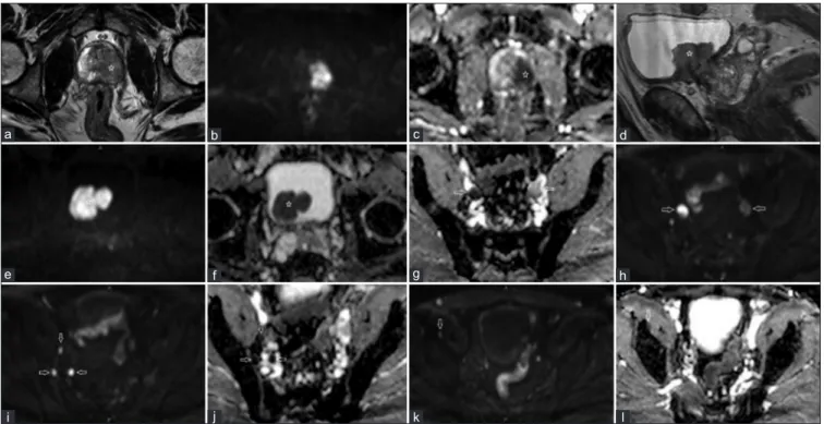
Figure 1: In transaxial images lesion located at the left side of the central zone was hypointense in T2A (a) and apparent diffusion coefficient
(c), while hyperintense in diffuse weighted imaging (b). The second lesion was located at the basis of central lobe and shows polypoid protrusion to the lumen of bladder (d‑f). Lymph nodes (arrows) larger than 1 cm in diameter in the bilateral parailiac region (g, h) and in the right obturator region (i, j) Bilateral parailiac and right obturator lymph nodes had restriction of diffusion. Retrospective evaluation of magnetic resonance imaging images following 18F‑labeled fluoro‑2‑deoxyglucose positron emission tomography/computed tomography scan pointed a focal cortical lesion showing restriction of diffusion at the anterior cortical edge of the right iliac bone (k and l)
k i j l a d h c g b f e
Cakir, et al.: Intense FDG uptake in high‑grade prostatic adenocarcinoma with negligible PSMA expression
Urology Annals | Volume 11 | Issue 4 | October-December 2019 441
the rare ductal carcinoma subtype can show poor PSMA avidity while exhibiting better FDG avidity. In our case, the tumor was entirely composed with Gleason pattern 5 highgrade tumor, it was not possible to recognize a specific subtype (acinar vs. ductal).
In this case, which was diagnosed as prostate cancer, the primary bulks, lymph node metastases, and iliac bone metastasis showed low PSMA expression while high F18-FDG PET uptake. On the other hand, the detection of primary bulk in mpMRI is not affected by PSA level while showing lymph node and bone metastases. There is an inverse correlation between Gleason score and apparent diffusion coefficient (ADC) values, and as it is our case, it shows explicit diffusion limitation in aggressive and high-grade tumors.[8] The lowADC values, calculated both
in the primary bulk and lymph node-bone metastases, indicate that the tumor is aggressive in our case.
Although there have been very important advancements in molecular and radiologic imaging methods, we observe differences in the imaging of prostate cancer which is a heterogeneous disease with distinct clinical features. Especially for PSMA-negative patients in high-grade prostate cancers with high metabolic activity, FDG and other agents can be more avid. While 68Ga-PSMA PET/
CT has demonstrated increasing utility in the evaluation of prostatic carcinoma, it is essential to be aware of false‑negative findings. Further subtype analyses of prostate cancer will be helpful in the understanding of underlying reasons.
Declaration of patient consent
The authors certify that they have obtained all appropriate patient consent forms. In the form the patient(s) has/have given his/her/their consent for his/her/their images and other clinical information to be reported in the journal. The patients understand that their names and initials will not be published and due efforts will be made to conceal their identity, but anonymity cannot be guaranteed.
Financial support and sponsorship
Nil.
Conflicts of interest
There are no conflicts of interest.
REFERENCES
1. Kabasakal L, Demirci E, Nematyazar J, Akyel R, Razavi B, Ocak M,
et al. The role of PSMA PET/CT imaging in restaging of prostate
cancer patients with low prostate‑specific antigen levels. Nucl Med Commun 2017;38:149-55.
2. Maurer T, Gschwend JE, Rauscher I, Souvatzoglou M, Haller B,
Figure 2: Primary prostatic lesions, metastatic lymph nodes, and bone lesions were significantly noticeable at the 18F‑labeled fluoro‑2‑deoxyglucose positron emission tomography/computed tomography images
Cakir, et al.: Intense FDG uptake in high‑grade prostatic adenocarcinoma with negligible PSMA expression
442 Urology Annals | Volume 11 | Issue 4 | October-December 2019
Weirich G, et al. Diagnostic efficacy of (68)Gallium‑PSMA positron
emission tomography compared to conventional imaging for lymph node staging of 130 consecutive patients with intermediate to high risk prostate cancer. J Urol 2016;195:1436-43.
3. Öbek C, Doğanca T, Demirci E, Ocak M, Kural AR, Yıldırım A,
et al. The accuracy of 68Ga-PSMA PET/CT in primary lymph node
staging in high-risk prostate cancer. Eur J Nucl Med Mol Imaging 2017;44:1806-12.
4. Mannweiler S, Amersdorfer P, Trajanoski S, Terrett JA, King D, Mehes G, et al. Heterogeneity of prostate-specific membrane
antigen (PSMA) expression in prostate carcinoma with distant metastasis. Pathol Oncol Res 2009;15:167-72.
5. Ross JS, Sheehan CE, Fisher HA, Kaufman RP Jr., Kaur P, Gray K,
et al. Correlation of primary tumor prostate‑specific membrane antigen
expression with disease recurrence in prostate cancer. Clin Cancer Res 2003;9:6357-62.
6. Shetty D, Loh H, Bui C, Mansberg R, Hadjashrafi A, Do V, et al.
Non‑prostate‑specific membrane antigen‑avid metastatic lung nodule from primary prostatic adenocarcinoma. Clin Nucl Med 2016;41:776-8. 7. McEwan LM, Wong D, Yaxley J. Flourodeoxyglucose positron emission tomography scan may be helpful in the case of ductal variant prostate cancer when prostate specific membrane antigen ligand positron emission tomography scan is negative. J Med Imaging Radiat Oncol 2017;61:503-5.
8. Maurer MH, Härmä KH, Thoeny H. Diffusion-weighted genitourinary imaging. Radiol Clin North Am 2017;55:393-411.