Gene Therapy and Molecular Biology Vol 16, page 95
Gene Ther Mol Biol Vol 16, 95-104, 2014
The effect of hysteroscopic polypectomy on the
gene expression of endometrial receptivity
markers: HOXA10, HOXA11 and LIF
Research Article
Engin Yildirim, M.D
1*, Onder Celik, M.D
1, Muslum Akgoz, Ph.D
2, Ilknur
Aksoy, M.Sc.
3,4, Ali Türkan, Ph.D
4, Zeynal Mete Karaca, B.Sc.
5, Yilmaz
Cigremis, Ph.D
51 Inonu University, Medical Faculty, Department of Obstetricsand Gynecology, Malatya, Turkey, [email protected], [email protected], +90 422 3410660
2 TUBITAK Ulusal Metroloji Enstitüsü (UME), Biyoanalysis Laboratory, 41470, Gebze, Kocaeli, Turkey, [email protected], +90 262 679 5000 3 Current address: Istanbul Medipol University, Vocational School of Health Services, Medical Laboratory Techniques Program, Istanbul, Turkey, [email protected], +90216 68153 99
4 Department of Chemistry, Faculty of Science, Gebze Institute of Technology, Çayırova, 41400, Gebze, Kocaeli, Turkey, [email protected], +90 262 605 30 56
5 Inonu University, Medical Faculty, Department of Medical Biology and Genetics, Malatya, Turkey, [email protected], [email protected], +90 422 341 0660
________________________________________________________________________________________________________________________________
*Correspondence: Engin Yıldırım, PhD, Inonu University, Turgut Ozal Medical Center,
Department of Obstetrics and Gynecology, 44069, Malatya, Turkey, Tel: +90 286 2180018 / 2107, E-mail: [email protected]
Keywords: homeobox A10, homeobox A11, Endometrialpolyp, RT-qPCR, HOXA10, HOXA11 and LIF Received: 4 April 2014; Revised: 30 June 2014
Accepted: 2 July 2014; electronically published: 3 July 2014
Summary
The aim of this study is to determine the effect of hysteroscopic polypectomy on the mRNA expression levels of the endometrial receptivity markers, namely, homeobox A10 (HOXA10), homeobox A11 (HOXA11) and leukemia inhibitory factor (LIF). Twenty-five reproductive-aged women with endometrial polyps underwent hysteroscopy. Samples were taken at the mid-secretory phase using hysteroscopic polypectomy and 4 months after polypectomy, and the change in mRNA expression levels of normalized HOXA10, HOXA11 and LIF genes were determined using Reverse Transcription Quantitative Real Time–Polymerase Chain Reaction (RT-qPCR). The results show that mRNA levels of HOXA10 and HOXA11 taken prior to surgery and 4 months after the complete hysteroscopic removal of polyps were not significantly different (P=0.79 and P=0.14, respectively). Moreover, a marked difference could not be obtained between preoperative and postoperative endometrial LIF mRNA expression levels (P=0.86). As a conclusion, these results indicate that mRNA levels of HOXA10, HOXA11 and LIF genes, three of molecular markers of endometrial receptivity, are not affected by hysteroscopic
96
I. Introduction:
Successful embryo implantation requires a well-coordinated reciprocal action between hormones, cytokines and growth factors (Carson et al, 2000; Dey et al, 2004; Tabibzadeh and Babaknia, 1995). Endometrial polyps, which are benign, hyperplastic-localized overgrowth of glands and stroma have been reported in 15-25% of infertile women and in about 1.4% of women undergoing in vitro fertilization (IVF) (de Sa Rosa e de Silva et al, 2005; Salm, 1972; Sanders, 2006; Shokeir et al, 2004). The mechanism by which endometrial polyp influences fertility is not clearly understood yet; however, it has been suggested that endometrial polyps may be related to the mechanical interference of sperm transport, embryo implantation or aberrant expression of endometrial markers consisting of HOXA10 and HOXA11, insulin growth factor binding protein 1 (IGFBP1), tumor necrosis factor alpha (TNF alpha) (Elbehery
et al, 2011; Inagaki et al, 2003; Rackow et al, 2011). HOXA10 is essential for both endometrial development during menstrual cycle and endometrial receptivity (Aghajanova et al, 2008; Rackow and Taylor, 2010; Taylor, 2000; Taylor et al, 1998; Taylor et al, 2003). An in vitro study has demonstrated that decreased HOXA10 expression is associated with diminished implantation, which proposes that change in HOXA10 levels modulates the degree of endometrial receptivity (Bagot et al, 2000). Similarly, an altered expression of HOXA11 results in poor endometrial development and lower implantation rate (Gendron et al, 1997; Hsieh-Li et al, 1995).
Although evidence from previous reports has shown that HOXA10 and HOXA11 appeared to influence implantation, several other molecules are also needed in this highly coordinated process where hundreds of molecules are intertwining rather than only a single molecule (Altmae et al, 2014; Altmae et al, 2010; Giudice and Kao, 2004; Horcajadas et al, 2007; Koler et al, 2009).
We suggest that different mechanisms might be related with implantation failure.LIF, which is known to be a member of pleiotropic cytokine family, can exert proinflammatory and anti-inflammatory responses (Aghajanova et al, 2008; Giudice and Kao, 2004). In human, LIF is expressed in endometrial glands during menstrual cycles and its expression peaks in the middle-late secretory phase (Singh et al, 2011). It has also been reported that LIF might also be involved in embryonic implantation, and its expression level might decrease with unexplained infertility when compared to fertile controls during implantation window (Aghajanova, 2010; Aghajanova et al, 2009; Charnock-Jones et
al, 1994). It has been reported that LIF gene activity was lower with women who had unexplained infertility rates, thus LIF signaling pathway could possibly offer an explanation for increased infertility rates (Aghajanova et al, 2009).
Although endometrial polyps have been identified as a possible factor for infertility, their implication on endometrial molecular markers involved in implantation has not been studied extensively. It was postulated that the presence of endometrial polyps in uterine cavity may alter endometrial signaling pathways leading to impaired endometrial receptivity; therefore, removal of polyps will have beneficial impact on endometrial receptivity (Rackow
et al, 2011). Based on evidence from aforementioned studies, we attempted to evaluate the impact of hysteroscopic endometrial polypectomy on established endometrial receptivity markers including HOXA10, HOXA11 and LIF.
Gene Therapy and Molecular Biology Vol 16, page 97
II. Materials and Methods: A. Biopsy
This prospective interventional study was carried out at Inonu University, Turgut Ozal Medical Centre. From unselected population of non-pregnant, reproductive aged women, 25 consecutive subjects (Six of patients did not have any children and one of these women lost a baby by abortion) who were diagnosed of endometrial polyp during transvaginal ultrasound (TV-USG) were invited to be involved in this study. The study protocol was approved by Institutional Ethical Committee for Research on Human Subjects. Informed written consent form was obtained from all participants.
All participants meets following inclusion criteria: 1) ages ranging from 22 to 49 years; 2) regular menstrual cycles; 3) presence of endometrial polyp on TV-USG that was also confirmed on saline infusion sonohysterogram; 4) acceptance of second examination at follow-up screening; 5) not taking any hormonal medications or treatments at least 3 months before surgery. The exclusion criteria were determined as the following: 1) having any confounding medical conditions known to affect endometrial receptivity such as endometriosis, polycystic ovarian syndrome (PCOS) or hydrosalpinges; 2) presence of any other endometrial pathology including Asherman Syndrome, submucosal fibroids, endometrial cancer or endometrial hyperplasia; 3) diagnosis of pelvic inflammatory disease at the time of the study; 4) previous history of endometrial surgery; 5) history of habitual abortion.
All women had two-dimensional transvaginal ultrasound scan (Toshiba Xario, Japan) using high frequency transducers of 5-7.5 MHz, followed by saline infusion hysterosonography for the confirmation of endometrial polyps (Lee et al, 2006).
Subsequently, all the women recruited for the study had endometrial polyps removed through pipelle in the secretory phase of menstrual cycle during the implantation window. The endometrial specimens were divided into two groups for both pathological evaluation and biochemical-molecular analysis. Endometrial polyps were excised under hysteroscopic guidance. The secretory phase was calculated as 7 to 9 days after the ultrasonographic confirmation of ovulation. Subsequently,
after polypectomy. Biopsy samples were evaluated by an independent pathologist experienced in gynecological pathology and only patients who are having polyps were included in the study. All endometrial biopsy samples were taken 7-9 days after ovulation and also maturation of endometrium was evaluated by Noyes criteria. Age of the patients, time of the surgery, obstetric and gynecological history, medical conditions, last menstrual period, preoperative and postoperative diagnoses data were recorded for all patients. Follow-up hysteroscopy was performed after four months at mid-luteal phase of menstrual cycle, and endometrial samples were collected from all participants.
B. RT-qPCR
All samples were stored in 1 ml of RNA-later solution at -80 °C until they were analyzed for the expression levels of HOXA10,
HOXA11, LIF and β-actin mRNA.
Endometrial samples and polyps that were obtained prior to and after polypectomy were subjected to manufacturer’s total RNA isolation protocol (RNeasy Mini Kit, QIAGEN, Hilden, Germany). Total extracted RNA was run on 1% agarose gel and integrity of the mRNA was confirmed by visualization of ribosomal bands with ethidium bromide staining over a UV transilluminator (data not shown). The concentration of the total purified RNA was determined by NanoDrop® Spectrophotometer. SuperScript® III Reverse Transcriptase Kit (Invitrogen, Carlsbad, CA) was used for reverse transcription (RT) reactions with minor changes. Equal amounts of total RNA were added into each RT reaction and oligo dT-18 primer was used to extend all mRNAs in the first strand cDNA synthesis. Primers were designed by BLAST
primer designing software
(http://www.ncbi.nlm.nih.gov/tools/primer-blast/) using human gene as a template (Table 4). Quantitative PCR was carried out by using cDNA as template with 96 well qPCR platform (Roche LC480).
Briefly, PCR amplification mixture (20 μl) contained 1 μl of RT reaction mixture (cDNA), 1 μl of forward (10 pmol/ul), 1 μl of reverse primers (10 pmol/ul) and 10 μl of 2x SYBR
98
Master Mix, Roche 04707516001). Amplification was performed at 95°C for 10 min, followed by 45 cycles of 95°C for 20 sec, 60°C for 20 sec, and 70°C for 30 sec heating and cooling cycles. Melting analysis was also performed at the end of PCR. Ct values were determined by automatic setting of the software. All qPCR were performed in triplicates in the same plate with β-actin housekeeping gene. For each gene, relative mRNA expression levels were calculated according to housekeeping genes, using the 2−ΔΔCt method (Livak and Schmittgen, 2001).
C. Statistical Analysis
The data were analyzed using the Statistical Package for Social Sciences software 19.0 for Windows package software (SPSS, Inc., Chicago, IL). Power analysis revealed that 16 patients are required for HOXA11 gene with the following conditions: HOXA11 average: 0.045; standard deviation: 0.052; type I error (alpha): 0.05 and type II error (beta: power): 0.10.
The distribution normality of data was evaluated with the Kolmogorov–Smirnov test and gene expression levels was found to be non-normally distributed (P<0.05). The results were presented as median, minimum and maximum. Comparison of HOXA10, HOXA11 and LIF concentrations in endometrial samples prior to and after polypectomy was performed using Wilcoxon Signed-Rank Sum Test. Correlation of variables was assessed using Spearman’s correlation test. For all comparisons, probability of < 0.05 was considered to be significant.
III. Results
The basal characteristics and serum hormone levels of patients are presented in Table 1. All endometrial tissue samples and hysteroscopically excised endometrial polyps underwent histological evaluation and endometrial polyps were identified as polyps and biopsy samples revealed normal secretory endometrium. The mean time interval between two endometrial samplings was 105.4 ±53.6 days.
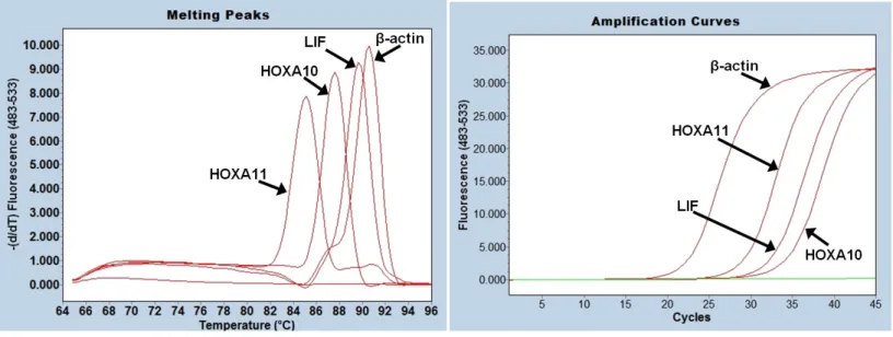
To compare HOXA10, HOXA11 and LIF mRNA levels prior and after polypectomy, total RNA was isolated from all samples and cDNA was transcribed from all mRNA. mRNA levels of HOXA10, HOXA11, LIF and β-actin mRNA levels were measured by qPCR method and Ct values were calculated form SYBR Green fluorescence amplification graphs (Figure 1.A). Melting analysis of PCR products revealed a single peak (Figure1.B) which proves that SYBR Green fluorescence signal belongs to only one kind of PCR product. Since melting analysis does not give information about the PCR product size, PCR products were also run in DNA agarose gels and correct sized PCR products were obtained for HOXA10, HOXA11 and LIF genes as well as for the control β-actin housekeeping gene, 110 bp, 128 bp, 113 bp and 112 bp, respectively (Figure 2, Table 2).
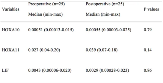
Expression levels HOXA10, HOXA11 and LIF mRNA were normalized according to β-actin housekeeping gene and they were presented in Table 3. The expression levels of HOXA10, HOXA11 and LIF mRNA of endometrial samples taken prior to polypectomy were not significantly different than endometrium samples obtained 4 months after removal of whole endometrial polyp (P=0.79, P=0.14 and P=0.86, respectively). The size of polyps and expression levels of HOXA10, HOXA11 and LIF (r=0.252, P=0.29; r=0.391, P=0.88; r=0.152, P=0.52, respectively) were compared but a significant correlation was not observed. The comparison of patient age with expression levels of HOXA10, HOXA11 and LIF also did not correlate significantly (0.307, P=0.19; r=0.157, P=0.51; r=-0.197, P=0.41, respectively).
Gene Therapy and Molecular Biology Vol 16, page 99
Figure 1: RT-qPCR amplification and melting analysis of HOXA10, HOXA11, LIF and β-actin mRNA. HOXA10, HOXA11LIF and β-actin mRNA samples were subjected to reverse transcription and SYBR Green based Real Time PCR analysis (A). After RT-qPCR, products were subjected to melting analysis (B).
Figure 2: DNA gel electrophoresis of RT-qPCR of HOXA10, HOXA11, LIF and β-actin mRNA. PCR products were loaded on 1.5% DNA agarose gel and the size of each mRNA was determined by using of DNA marker (100 bp, Fermentas). The expected sizes of the PCR products were given in Table 4.
100
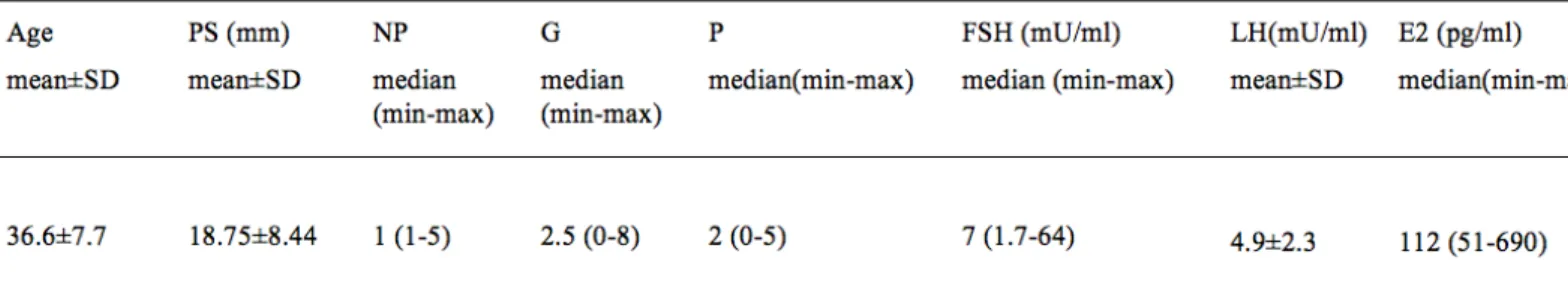
Table 1: The basal characteristic of patients and serum level of FSH, LH and E2.
PS; Polyp size, NP; Number of polyps, G; Gravida, P; Para, FSH; Follicle stimulating hormone, LH; Luteinizing hormone, E2; Estradiol
Table 2: Primer sequence and expected PCR product size for endometrial receptivity markers and internal standard β-actin mRNA.
a
β-actin: Beta-actin gene, HOXA10: Homeobox A10 gene, HOXA11: Homeobox A11 gene, LIF: Leukemia Inhibitory Factor gene.
b
F: forward primer, R: reverse primer
Primer sequences were designed by BLAST primer designing tool (http://www.ncbi.nlm.nih.gov/tools/primer-blast/) using human genes as template.
Gene Therapy and Molecular Biology Vol 16, page 101
Table 3: Comparison of expression of endometrial receptivity markers including HOXA10, HOXA11 and LIF between pre- and post-polypectomy in the whole group. Values are presented as “median (min-max)”. Comparison between groups was performed using “Wilcoxon Signed-Rank Sum Test.
IV. Discussion
In this study, the expression levels of HOXA10, HOXA11 and LIF genes were investigated before and after endometrial polyp removal and it was found that endometrial polyp removal does not change the expression levels of HOXA10, HOXA11 and LIF genes. Any significant correlation was not also noted with the polyp size and age of the patients. As a conclusion, we suggest that several other genes may also be involved in the regulation of endometrial polyp removal. In literature, several observational studies stated that the removal of endometrial polyp with infertility resulted in a significant improvement in pregnancy rate in subfertile or infertile women who underwent hysteroscopic endometrial polyp removal (Delage et al, 1995; Shokeir et al, 2004; Spiewankiewicz et al, 2003; Varasteh
et al, 1999). The mechanism of endometrial polyp infertility has not been determined yet, but several hypotheses were proposed including mechanical inhibition of sperm and embryo transport, altered endometrial receptivity or impairment of endometrial
implantation (Rackow et al, 2011). The result of one randomized controlled trial concerning the effect of endometrial polypectomy on subfertility has demonstrated that pregnancy rate considerably increased in women who underwent endometrial polypectomy (Perez-Medina et al, 2005).
Another prospective study comparing concentrations of endometrial markers in consecutive endometrial flushing fluid prior to post hysteroscopic polypectomy has shown a significant increase in IGFBP1, TNF alpha and osteopontin concentrations after hysteroscopic polypectomy (Ben-Nagi
et al, 2009).
Further, a case-controlled study has reported significantly lower mRNA expression of HOXA10 and HOXA11 in endometrium from women with polyps than controls in the proliferative phase with infertile subjects (Rackow et al, 2011). The endometrial polyp is type-a benign tumor and it is the result of monoclonal neoplastic proliferation of stromal cells with a non-neoplastic glandular component (Christopher P. Crum, 2011).
102 The endometrial polyps are
characterized with the following features: thick-walled vessels, fibrous or collagenous stroma or irregular gland structure. The possible explanation of this discrepancy between the previous reports with our result may be the paucity of pathological discrimination of endometrial polyps. It has been demonstrated that endometrial polyps can be divided into two patterns including proliferative/hyperplastic, atrophic or functional, and these patterns frequently overlap (Robert J. Kurman, 2011). The other explanation for this difference may be the variation of methodology that other studies did not re-evaluate these endometrial receptivity markers postoperatively.
Despite an obvious improvement in fertility rate after removal of endometrial polyps reported previously, the significance of the location and the size of polyp still remain controversial. Evidence from retrospective data suggests that removal of endometrial polyp improves the pregnancy rate in infertile women, irrespective of the number or size of present polyps (Livak and Schmittgen, 2001; Preutthipan and Herabutya, 2005; Rackow and Taylor, 2010). A randomized controlled study on the effect of size of endometrial polyp reported that significant differences were not observed in the pregnancy rate after polypectomy between the groups with different size of polyps; polyp diameter ranged <5, 5-10, 11-20 and >11-20 mm (Perez-Medina et al, 2005). In a case-controlled study, Rackow et al. (Rackow et al, 2011) demonstrated that expression levels of endometrial receptivity markers (HOXA10 and HOXA11) did not change significantly between a single polyp and multiple polyps. The same study also suggests that the size of polyps was not associated with the endometrial receptivity markers; HOXA10 and HOXA11 (Rackow et
al, 2011). LIF gene, also considered as endometrial receptivity marker, was also investigated in another study and it was suggested that the expression levels of LIF mRNA did not significantly differ between
submucosal myoma carrying women and control subjects (Rackow and Taylor, 2010). On the contrary, a recent report presented lower LIF expression in patients with an endometrial polyp than the controls at the midsecretory phase (Hasegawa et al, 2012). The development and characteristics of endometrial polyps and fibroids varies from the onset of disease. Moreover, the finding of this study demonstrated that LIF expression did not alter after removal endometrial polyp. The plausible reason for this controversial result may be due to the small number of cases (n=5), which was analyzed in the previous study (Hasegawa et
al, 2012). Since LIF expression was also assessed in the endometrium of different individuals, rather than the same patients, the discrepancy between the results of our study with Hasegawa’s study might be explained with grouping bias.
In summary, the present findings suggest that endometrial polyps may not alter the expression of established implantation factors during midsecretory implantation window. Earlier studies reported that removal of endometrial polyps improves fertility (Perez-Medina et al, 2005; Shokeir
et al, 2004; Spiewankiewicz et al, 2003;
Varasteh et al, 1999). Based on our study, it is not possible to comment on the implication of endometrial polypectomy on the fertility rate.
The other reason for this discrepancy may be explained with the short period when the expressions of implantation genes were up-regulated to normal levels after the removal of endometrial polyp; however, evidence from previous study has shown that the HOXA10 expression improved to normal levels 4 months after salpingectomy (Daftary et al, 2007). This study is the first report demonstrated that the endometrial polyp does not adversely affect the expressions of three implantation genes, HOXA10, HOXA11 and LIF genes. It is also possible that several other implementation genes other than HOXA10, HOXA11 and LIF genes may be regulated by the removal of
Gene Therapy and Molecular Biology Vol 16, page 103
endometrial polyp. In addition to RT-PCR analysis of gene expression, microarray technology can also be used to scan several genes at the same time and endometrial receptivity array (ERA) was developed to determine transcriptomic signature of potential endometrial receptivity biomarkers cluster (Diaz-Gimeno et al, 2011) and to treat patients with repeated implantation failure (Ruiz-Alonso et al, 2013).
Declaration of interest: The authors declare that they have no conflict of interest.
Funding: This work was supported by İnönü University Internal Project, (Grant number: 2011/174)
Acknowledgement: Authors thank to Dr. Engin Nasuhi Aydın for pathological evaluation.
References:
Aghajanova L (2010). Update on the role of leukemia inhibitory factor in assisted reproduction. Current Opinion in Obstetrics & Gynecology, 22: 213-219.
Aghajanova L, Altmae S, Bjuresten K, Hovatta O, Landgren BM and Stavreus-Evers A (2009). Disturbances in the LIF pathway in the endometrium among women with unexplained infertility. Fertility and sterility, 91: 2602-2610.
Aghajanova L, Simón C and Horcajadas JA (2008). are favorite molecules of endometrial receptivity still in favor? Expert Review of Obstetrics and Gynecology, 3: 487-501. Altmae S, Esteban FJ, Stavreus-Evers A, Simon C,
Giudice L, Lessey BA, Horcajadas JA, Macklon NS, D'Hooghe T, Campoy C, Fauser BC, Salamonsen LA and Salumets A (2014). Guidelines for the design, analysis and interpretation of 'omics' data: focus on human endometrium. Human Reproduction Update, 20: 12-28.
Altmae S, Martinez-Conejero JA, Salumets A, Simon C, Horcajadas JA and Stavreus-Evers A (2010). Endometrial gene expression analysis at the time of embryo implantation in women with unexplained infertility. Molecular Human Reproduction, 16:
178-Alteration of maternal Hoxa10 expression by in vivo gene transfection affects implantation. Gene Ther, 7: 1378-1384.
Ben-Nagi J, Miell J, Yazbek J, Holland T and Jurkovic D (2009). The effect of hysteroscopic polypectomy on the concentrations of endometrial implantation factors in uterine flushings. Reprod Biomed Online, 19: 737-744.
Carson DD, Bagchi I, Dey SK, Enders AC, Fazleabas AT, Lessey BA and Yoshinaga K (2000). Embryo implantation. Dev Biol, 223: 217-237.
Charnock-Jones DS, Sharkey AM, Fenwick P and Smith SK (1994). Leukaemia inhibitory factor mRNA concentration peaks in human endometrium at the time of implantation and the blastocyst contains mRNA for the receptor at this time. J Reprod Fertil, 101: 421-426.
Christopher P. Crum MRN, and Kenneth R. Lee (2011) Diagnostic Gynecologic and Obstetric Pathology, 2nd Edition: Elsevier.
Daftary GS, Kayisli U, Seli E, Bukulmez O, Arici A and Taylor HS (2007). Salpingectomy increases peri-implantation endometrial HOXA10 expression in women with hydrosalpinx. Fertility and sterility, 87: 367-372.
de Sa Rosa e de Silva AC, Rosa e Silva JC, Candido dos Reis FJ, Nogueira AA and Ferriani RA (2005). Routine office hysteroscopy in the investigation of infertile couples before assisted reproduction. J Reprod Med, 50: 501-506.
Delage G, Moreau JF, Taupin JL, Freitas S, Hambartsoumian E, Olivennes F, Fanchin R, Letur-Konirsch H, Frydman R and Chaouat G (1995). In-vitro endometrial secretion of human interleukin for DA cells/leukaemia inhibitory factor by explant cultures from fertile and infertile women. Hum Reprod, 10: 2483-2488.
Dey SK, Lim H, Das SK, Reese J, Paria BC, Daikoku T and Wang H (2004). Molecular cues to implantation. Endocr Rev, 25: 341-373.
Diaz-Gimeno P, Horcajadas JA, Martinez-Conejero JA, Esteban FJ, Alama P, Pellicer A and Simon C (2011). A genomic diagnostic tool for human endometrial receptivity based on the transcriptomic signature. Fertility and sterility, 95: 50-60, 60 e51-15.
Elbehery MM, Nouh AA, Mohamed ML, Alanwar AA, Abd-Allah SH and Shalaby SM (2011). Insulin-like growth factor binding protein-1 and glycodelin levels in uterine flushing before and after hysteroscopic polypectomy.
104 Gendron RL, Paradis H, Hsieh-Li HM,
Lee DW, Potter SS and Markoff E (1997). Abnormal uterine stromal and glandular function associated with maternal reproductive defects in Hoxa-11 null mice. Biol Reprod, 56: 1097-1105.
Giudice LC and Kao LC (2004). Endometriosis. Lancet, 364: 1789-1799.
Hasegawa E, Ito H, Hasegawa F, Hatano K, Kazuka M, Usuda S and Isaka K (2012). Expression of leukemia inhibitory factor in the endometrium in abnormal uterine cavities during the implantation window. Fertility and sterility, 97: 953-958.
Horcajadas JA, Pellicer A and Simon C (2007). Wide genomic analysis of human endometrial receptivity: new times, new opportunities. Human Reproduction Update, 13: 77-86. Hsieh-Li HM, Witte DP, Weinstein M, Branford W,
Li H, Small K and Potter SS (1995). Hoxa 11 structure, extensive antisense transcription, and function in male and female fertility. Development, 121: 1373-1385.
Inagaki N, Ung L, Otani T, Wilkinson D and Lopata A (2003). Uterine cavity matrix metalloproteinases and cytokines in patients with leiomyoma, adenomyosis or endometrial polyp. Eur J Obstet Gynecol Reprod Biol, 111: 197-203.
Koler M, Achache H, Tsafrir A, Smith Y, Revel A and Reich R (2009). Disrupted gene pattern in patients with repeated in vitro fertilization (IVF) failure. Hum Reprod, 24: 2541-2548. Lee C, Ben-Nagi J, Ofili-Yebovi D, Yazbek J,
Davies A and Jurkovic D (2006). A new method of transvaginal ultrasound-guided polypectomy: a feasibility study. Ultrasound Obstet Gynecol, 27: 198-201.
Livak KJ and Schmittgen TD (2001). Analysis of relative gene expression data using real-time quantitative PCR and the 2(-Delta Delta C(T)) Method. Methods, 25: 402-408.
Perez-Medina T, Bajo-Arenas J, Salazar F, Redondo T, Sanfrutos L, Alvarez P and Engels V (2005). Endometrial polyps and their implication in the pregnancy rates of patients undergoing intrauterine insemination: a prospective, randomized study. Hum Reprod, 20: 1632-1635.
Preutthipan S and Herabutya Y (2005). Hysteroscopic polypectomy in 240 premenopausal and postmenopausal women. Fertility and sterility, 83: 705-709.
Rackow BW, Jorgensen E and Taylor HS (2011). Endometrial polyps affect uterine receptivity. Fertility and sterility, 95: 2690-2692.
Rackow BW and Taylor HS (2010). Submucosal uterine leiomyomas have a global effect on
molecular determinants of endometrial receptivity. Fertility and sterility, 93: 2027-2034.
Robert J. Kurman LhE, Brigitte M. Ronnett (2011) Blaustein’s Pathology of the Female Genital Tract, 6th ed. 344. Springer
Ruiz-Alonso M, Blesa D, Diaz-Gimeno P, Gomez E, Fernandez-Sanchez M, Carranza F, Carrera J, Vilella F, Pellicer A and Simon C (2013). The endometrial receptivity array for diagnosis and personalized embryo transfer as a treatment for patients with repeated implantation failure. Fertility and sterility, 100: 818-824.
Salm R (1972). The incidence and significance of early carcinomas in endometrial polyps. J Pathol, 108: 47-53.
Sanders B (2006). Uterine factors and infertility. J Reprod Med, 51: 169-176.
Shokeir TA, Shalan HM and El-Shafei MM (2004). Significance of endometrial polyps detected hysteroscopically in eumenorrheic infertile women. J Obstet Gynaecol Res, 30: 84-89. Singh M, Chaudhry P and Asselin E (2011).
Bridging endometrial receptivity and implantation: network of hormones, cytokines, and growth factors. J Endocrinol, 210: 5-14. Spiewankiewicz B, Stelmachow J, Sawicki W,
Cendrowski K, Wypych P and Swiderska K (2003). The effectiveness of hysteroscopic polypectomy in cases of female infertility. Clin Exp Obstet Gynecol, 30: 23-25.
Tabibzadeh S and Babaknia A (1995). The signals and molecular pathways involved in implantation, a symbiotic interaction between blastocyst and endometrium involving adhesion and tissue invasion. Hum Reprod, 10: 1579-1602.
Taylor HS (2000). The role of HOX genes in human implantation. Hum Reprod Update, 6: 75-79.
Taylor HS, Arici A, Olive D and Igarashi P (1998). HOXA10 is expressed in response to sex steroids at the time of implantation in the human endometrium. J Clin Invest, 101: 1379-1384.
Taylor HS, Daftary GS and Selam B (2003). Endometrial HOXA10 expression after controlled ovarian hyperstimulation with recombinant follicle-stimulating hormone. Fertility and sterility, 80 Suppl 2: 839-843. Varasteh NN, Neuwirth RS, Levin B and Keltz MD
(1999). Pregnancy rates after hysteroscopic polypectomy and myomectomy in infertile women. Obstet Gynecol, 94: 168-171.