1 JOURNAL OF HEALTH SCIENCES
A. J. Health Sci. Volume 2 No 1 | January 2020, 1-10
Research Article
A Comparative Analysis of Biofilm Characteristics of Dual-SpeciesPeriodontopathogenic Biofilm based on Fusobacterium nucleatum and the Dual-Species Biofilm Response in the Presence of Antimicrobial Peptide
Mutlu Keskin1, 2 ORCID: 0000-0002-4929-800X
1Vocational School of Health Services, Altınbaş University, Istanbul, Turkey. 2Institute of Dentistry, University of Turku, Turku, Finland
Submitted: October 7, 2019; Accepted: December 3, 2019
Abstract: Fusobacterium nucleatum (F.nucleatum), which acts as a linking periodontopathogenic bacteria, has the
ability of coaggregation with early and late colonization species. It is the most intense gram-negative bacteria found in periodontal health and increases in periodontal diseased areas numerically. In this study, comparative analyses of dual and mono-biofilm structural characteristics formed by Porphyromonas gingivalis and Prevotella intermedia with F.
nucleatum have been evaluated. In all results, more biofilm formation was observed in dual-species compared to
mono-species biofilms. Biofilm formation of F. nucleatum-P.intermedia has increased in the presence of Human Neutrophilic Peptide (HNP-1). In this study it is concluded that dual-periodontopathogenic coaggregations based on F. nucleatum could produce more intense biofilm structure than single-species ones, and the increase in dual-species biofilm mass in the presence of low doses of HNP-1 may be a defense mechanism of dual periodontopathogenic biofilm.
Keywords: Periodontitis; subgingival; biofilm; antimicrobial; peptide; gram-negative bacteria; defensin; inflammation
Address of Correspondence: Mutlu Keskin- [email protected] Tel: +90 (212)7094528,
Department of Oral and Dental Health, Vocational School of Health Services, Altınbaş University, Zuhuratbaba, İncirli Cd. No:11-A, 34147 Bakırköy, İstanbul, Turkey
1. Introduction
Bacteria within the oral cavity are organized as multi-species biofilms. These structures containing around 500-700 species play an important role in the development of caries and periodontal diseases. Periodontitis is a biofilm induced bacterial inflammatory disease and characterized by progressive destruction of tooth supporting tissues. It is known that bacteria in biofilm have different characteristics compared to their planktonic life (Walker et al., 2007). There are studies showing that bacteria in biofilm can possess a more resistant structure against variables such as antimicrobial agents, physical forces, and pH changes (Shaddox et al., 2010). It has also been reported that multi-species biofilm structure may exhibit more
2
pathogenicity than single-species bacterial infections (Ebersole et al., 2017; Lamont et al., 2015; Lamont et al., 2018). Gram-negative bacteria are etiologically the most common species in the development of periodontitis. Among these, F. nucleatum plays a key role in the formation of dental biofilms and is involved in the association of early and late colonization types, including P. gingivalis and P. intermedia, which play important role in the progression of periodontitis (Bolstad et al., 1996; Kolenbrander et al., 1993). The Cationic Antimicrobial Peptides (CAMPs), important elements of the host defense system against the presence of infectious agents, belong to two families in human, namely defensins and cathelicidins. CAMPs act as a broad spectrum antimicrobial and also involve in immune system stimulation. HNP-1 has been shown to be the most abundant one among the 4 types of defensins isolated from neutrophilic granules (Keskin et al., 2014; Lundy et al., 2008). Bacteria and defensins are constantly present in the mouth. According to study of Keskin M. et al. (2014) it was concluded that F. nucleatum could develop some adaptation responses to the presence of defensins. The aim of this study is to investigate the dual-species biofilm characteristics of F. nucleatum with P. gingivalis and P. intermedia and also the dual-dual-species biofilm characteristics in the presence of low concentration of HNP-1.
2. Materials and Methods
2.1. Bacterial Species and Cultures
In all laboratory experiments, type strains of F. nucleatum, P. gingivalis and P. intermedia (F. nucleatum ATCC 25586, P. gingivalis ATCC 33277, P. intermedia ATCC 25611) were used. All bacteria were incubated in hemin (5 mg/l) and vitamin K1 (10 mg/l) supplemented Brucella agar at 37°C in 10% H2, 5% CO2, and 85% N2 anaerobic atmosphere (Whitley A35 Anaerobic Workstation, Don Whitely Scientific Ltd., West Yorkshire, UK) for 72 hours. Mature colonies were collected with sterile cotton swabs and transferred to KH2PO4 (final concentration: 25 mM), MgSO4 (final concentration: 4 mM) and saccharose (final concentration: 1%) supplemented Bactoetryptone specific broths and incubated for 48 hours under anaerobic conditions. Optical densities were standardized to 0.6OD at 490 nm just before the experiments.
2.2. Experimental Groups
The following seven different groups were designed;
Group A: F. nucleatum alone: 180 μl bacterial growth media + 20 μl F.nucleatum Group B: P. gingivalis alone: 180 μl bacterial growth media + 20 μl P.gingivalis Group C: P. intermedia alone: 180 μl media + 20 μl P.intermedia
Group D: F.nucleatum +P.gingivalis (Dual): 180 μl media + 10 μl F.nucleatum + 10 μl P.gingivalis Group E: F.nucleatum + P. intermedia (Dual): 180 μl media + 10 μl F.nucleatum + 10 μl P.intermedia
3 JOURNAL OF HEALTH SCIENCES
A. J. Health Sci.
Group F: F.nucleatum + P. gingivalis. (Dual+HNP): 180 μl media + 10 μl F.nucleatum + 10 μl P.gingivalis. + 5 μg/ml HNP-1)
Group G: F.nucleatum + P. intermedia (Dual+HNP): 180 μl media + 10 μl F.nucleatum + 10 μl P.gingivalis + 5 μg/ml HNP-1)
All experiments were performed in triplicate wells for each condition and repeated at least twice. Samples were taken from all the wells and placed on Brucella agar for contamination control.
2.3. Preparation of Defensin Suspension
HNP-1 was purchased commercially (HNP-1/Code: 4271-s Lot: 610505, Peptide institute, Japan) and stored at -20°C until use. HNP-1 was dissolved in bacterial growth media on the day of the experiment and prepared as 5 μg/ml concentration.
2.4. Preparation of Saliva Coated 96-well Plates
Clarified Saliva were prepared and stocked before all experiments. Salivas were collected from healthy male and female volunteers to 50 ml propylene tubes and centrifuged at 12,000 g at 4°C for 40 minutes. The supernatants were collected and divided into sterile glass tubes. The tubes were incubated for 30 minutes at 60°C for pasteurization and then centrifuged again at 12,000 g and stored at +4°C until use. 20 μl of each supernatant was collected and placed on Brucella agar, then incubated under aerobic and anaerobic conditions for 4 days and sterilization controls were performed. 50 μl of saliva was added to all the wells and plates were incubated for 1 hour at 37°C. All wells were then dried without washing and prepared for the experiment.
2.5. Analysis of Biofilm Mass Formation
Biofilm mass formation tests were performed by Crystal Violet Staining Method in all groups (Keskin et al., 2014). All wells were stained with 0.1% Crystal Violet. After incubation at room conditions for 15 minutes, the wells were washed twice with PBS (Phosphate Buffer Saline) to remove excess dye. 200 ml of acetic acid was added to each well and incubated for 10 minutes to allow homogenous distribution of Crystal Violet dye. Afterwards, spectrometric analysis (570 nm) was performed on all plates.
2.6. Analysis of Biofilm Protein Content
The incubated plates were washed with PBS and dried. At the end of these washing and drying processes, wells free from planktonic bacteria and liquid medium were filled with 100 ll of 0.2 N NaOH. 2 sec/ 80 W sonication (dr hielscher UP50H homogenizer) was applied to each well to ensure homogeneous distribution of the present biofilms at the bottom (Lu et al., 2007). Afterwards, the plates were transferred to a microwave oven operated at 600 W for 20 seconds (Akins et al., 1995). Then, BIO-RAD protein assay dye was added to each well at a rate of 1/4 of the total volume (25 μl). Following the instructions for the
4
FITC-labeled Concanavalin A (FITC-Con A) fluorescent dye was used for fluorometric analysis of biofilm polysaccharide content. Washing and drying of 96-well plates was carried out for twice with PBS. Each well was filled with 100 μl of FITC-Con A fluorescent dye (concentration of 50 μg/ml) and incubated at room temperature in a dark room for 5 minutes. After incubation, plates were washed twice with PBS and dried. The 96-well plates were analyzed and recorded by using the BIOTEK Synergy HT Fluorometer Instrument (Excitation=485, Emission=528).
2.8. Statistical Analysis
In species biofilm tests, single bacterial values were accepted as constant, and each of different dual-species biofilm structures was accepted as a variable. Paired-sample parametric t-test was used to evaluate whether there was a difference in the biofilm structures of any group. P values <0.05 were considered statistically significant. In defensin tests, dual-species biofilms with HNP-1 free media were accepted as constant value and the wells with HNP-1 were accepted as variable and paired-sample parametric t test was applied for statistical analysis.
3. Results
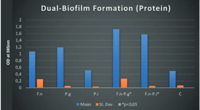
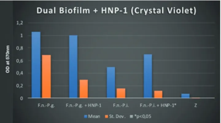
P. gingivalis- F. nucleatum dual-species biofilms produced more biofilm mass, protein and polysaccharide synthesis compared to all mono-species biofilms (Figure 1, 2 and 3) (p <0.05). Similar results have been observed in the dual-species biofilms of P. intermedia and F. nucleatum (Figure 1, 2 and 3) (p <0.05). Statistically significant polysaccharide synthesis was found in P. gingivalis- F. nucleatum and P. intermedia - F. nucleatum dual-species biofilm structures compared to mono-species biofilms (Figure 3) (p <0.05). It was observed that F. nucleatum- P. intermedia dual-species biofilms were able to produce statistically significant amounts of biofilms in the presence of HNP-1 compared to HNP-1 free media (Figure 4) (p <0.05). All other dual-species biofilm tests with HNP-1 showed no statistically significant difference (Figure 5 and 6).
5 JOURNAL OF HEALTH SCIENCES
A. J. Health Sci.
Figure 1. Dual-Biofilm Formation (Crystal Violet)
Figure 2. Dual-Biofilm Formation (Protein)
6
Figure 5. Dual-Biofilm + HNP-1 (Protein)
7 JOURNAL OF HEALTH SCIENCES
A. J. Health Sci.
4. Discussion
Gram-negative, anaerobic bacilli types are most closely associated with periodontitis. Among these, F. nucleatum, P. intermedia, P. gingivalis are the most common bacterial species and play a key role in the progression of the disease (Socransky et al., 1998; Teles et al., 2010).
In this study, characteristics of dual-species biofilms formed by P. gingivalis and P. intermedia with F. nucleatum were investigated. In order to grow them, incubation time of the bacteria in anaerobic conditions was determined as 48 hours for biofilm formation experiments. In previous studies it was shown that anaerobic incubation period of 48-72 hours could provide the appropriate time for the biofilm formation of F. nucleatum (Jang et al., 2013; Saito et al., 2008). The Crystal Violet technique was used in the analysis of biofilm mass after incubation. Because it is known as a reliable method and has been used in many studies (Merritt et al., 1998; Pitts et al., 2003; Stepanovic et al., 2000). Bradford Protein Analysis was also included for comparative analysis of total biofilm mass and biofilm protein values. While performing Bradford Protein Analysis, sonication was applied to all the wells to ensure homogeneous distribution of the biofilm mass (Bradford et al., 1976; Lu et al., 2007). FITC - Con A fluorescent dye was used to determine the amount of biofilm polysaccharide. FITC labeled Concanavalin A is a fluorescent fluorophore substance that binds with polysaccharide and is frequently used in in vitro polysaccharide studies (Roth et al., 1978; Sato et al., 2006; Yang et al., 2006).
The P. gingivalis and P. intermedia dual-species biofilms formed by F. nucleatum showed statistically significant increase in biofilm mass, polysaccharide synthesis and protein ratios when compared with mono-species biofilms. All these results show that P. gingivalis and P. intermedia dual-species biofilms formed with F. nucleatum are able to produce intense biofilms compared to single-species biofilms synergistically. Based on previous studies, it was observed that bacterial coaggregation plays an important role in biofilm formation. Similar to the results of this study, there are many other studies showing that F. nucleatum could play an important role in binding of late colonization species within the biofilm (Kolenbrander et al., 1989; Kolenbrander et al., 1993; Rickard et al., 2003).
In this study, dual periodontopathogenic biofilm characteristics were analyzed in the presence of HNP-1 comperatively. Other studies show that the HNP-1 concentration in the oral cavity can be found in a wide range of values from 0.40 μg/ml to 100 μg/ml (Dale et al., 2006; Fanali et al., 2008; Goebel et al., 2000; Puklo et al., 2008). However, there is a finding which indicates no lethal effect at low concentration levels of HNP-1 on F. nucleatum (Miyasaki et al., 1998). Therefore, the concentration of HNP-1 (5 μg/ml), which can be found in oral cavity continously, was studied to evaluate the defensive response of dual-species biofilms to HNP-1 challenge. It was observed that F. nucleatum - P. gingivalis dual-species biofilm did not show any significant change of biofilm production in the presence of HNP-1 compared to defensin-free media. Additionally, it was seen that the presence of HNP-1 did not lead to statistically significant difference in polysaccharide production at any biofilm group. It was formerly reported that dual-bacterial biofilms form an intense biofilm structure than single bacteria biofilms (Okuda et al., 2012; Saito et al., 2007). Although P. intermedia - F. nucleatum dual-species biofilm showed a statistically significant increase in biofilm mass in the presence of HNP-1, the same increase was not observed in polysaccharide
8
doses of HNP-1 could act as a defense mechanism of F. nucleatum- P. intermedia dual-species biofilm. More enlightening studies are needed over this subject.
Conflict of interest
Author declares no conflict of interests.
References
Akins, R. E., Tuan, R. S. (1995). Ultrafast protein determinations using microwave enhancement. Molecular Biotechnology, 4(1), 17–24. Doi: 10.1007/bf02907468
Bolstad, A. I., Jensen, H. B., Bakken, V. (1996). Taxonomy, biology, and periodontal aspects of Fusobacterium nucleatum. Clinical Microbiology Reviews, 9(1), 55–71. Doi: 10.1128/cmr.9.1.55
Bradford, M. (1976). A rapid and sensitive method for the quantitation of microgram quantities of protein utilizing the principle of protein-dye binding. Analytical Biochemistry, 72(1-2), 248–254. Doi: 10.1006/ abio.1976.9999
Dale, B. A., Tao, R., Kimball, J. R., Jurevic, R. J. (2006). Oral antimicrobial peptides and biological control of caries. BMC Oral Health, 6(S1). Doi: 10.1186/1472-6831-6-s1-s13
Ebersole, J. L., Dawson, D., Emecen-Huja, P., Nagarajan, R., Howard, K., Grady, M. E., Gonzalez, O. A. (2017). The periodontal war: microbes and immunity. Periodontology 2000, 75(1), 52–115. Doi: 10.1111/prd.12222 Fanali, C., Inzitari, R., Cabras, T., Pisano, E., Castagnola, M., Celletti, R., Messana, I. (2008). α-Defensin levels in whole saliva of totally edentulous subjects. International Journal of Immunopathology and Pharmacology, 21(4), 845–849. Doi: 10.1177/039463200802100409
Flemming, H. C., Wingender, J. (2010). The biofilm matrix. Nature Reviews Microbiology, 8(9), 623–633. Doi: 10.1038/nrmicro2415
Goebel, C., Mackay, L., Vickers, E., Mather, L. (2000). Determination of defensin HNP-1, HNP-2, and HNP-3 in human saliva by using LC/MS. Peptides, 21(6), 757–765. Doi: 10.1016/s0196-9781(00)00205-9
Jang, Y. J., Choi, Y. J., Lee, S. H., Jun, H. K., Choi, B. K. (2013). Autoinducer 2 of Fusobacterium nucleatum as a target molecule to inhibit biofilm formation of periodontopathogens. Archives of Oral Biology, 58(1), 17–27. Doi: 10.1016/j.archoralbio.2012.04.016
9 JOURNAL OF HEALTH SCIENCES
A. J. Health Sci.
Keskin, M., Könönen, E., Söderling, E., Isik, G., Firatli, E., Uitto, V.J., Gürsoy, U.K. (2014). Increased proliferation and decreased membrane permeability as defense mechanisms of Fusobacterium nucleatum against human neutrophilic peptide-1. Anaerobe, 30, 35–40. Doi: 10.1016/j.anaerobe.2014.08.001
Kolenbrander, P. E., Andersen, R. N. (1989). Inhibition of coaggregation between Fusobacterium nucleatum and Porphyromonas (Bacteroides) gingivalis by lactose and related sugars. Infect Immun, 57, 3204-3209. Kolenbrander, P. E., Andersen, R. N., Moore, L. V. (1989). Coaggregation of Fusobacterium nucleatum, Selenomonas flueggei, Selenomonas infelix, Selenomonas noxia, and Selenomonas sputigena with strains from 11 genera of oral bacteria. Infect Immun, 57, 3194-3203.
Kolenbrander, P. E., London, J. (1993). Adhere today, here tomorrow: oral bacterial adherence. J Bacteriol 175, 3247-3252
Lamont, R. J., Hajishengallis, G. (2015). Polymicrobial synergy and dysbiosis in inflammatory disease. Trends in Molecular Medicine, 21(3), 172–183. Doi: 10.1016/j.molmed.2014.11.004
Lamont, R. J., Koo, H., Hajishengallis, G. (2018). The oral microbiota: dynamic communities and host interactions. Nature Reviews Microbiology, 16(12), 745–759. Doi: 10.1038/s41579-018-0089-x
Lu, T. K., Collins, J. J. (2007). Dispersing biofilms with engineered enzymatic bacteriophage. Proceedings of the National Academy of Sciences, 104(27), 11197–11202. Doi: 10.1073/pnas.0704624104
Lundy, F. T., Nelson, J., Lockhart, D., Greer, B., Harriott, P., Marley, J. J. (2008). Antimicrobial activity of truncated α-defensin (human neutrophil peptide (HNP)-1) analogues without disulphide bridges. Molecular Immunology, 45(1), 190–193. Doi: 10.1016/j.molimm.2007.04.018
Merritt, K., Gaind, A., Anderson, J. M. (1998). Detection of bacterial adherence on biomedical polymers. Journal of Biomedical Materials Research, 39(3), 415–422. Doi: 10.1002/(sici)1097-4636(19980305)39:3<415::aid-jbm10>3.0.co;2-9
Okuda, T., Kokubu, E., Kawana, T., Saito, A., Okuda, K., Ishihara, K. (2012). Synergy in biofilm formation between Fusobacterium nucleatum and Prevotella species. Anaerobe, 18(1), 110–116. Doi: 10.1016/j. anaerobe.2011.09.003
Pitts, B., Hamilton, M. A., Zelver, N., Stewart, P. S. (2003). A microtiter-plate screening method for biofilm disinfection and removal. Journal of Microbiological Methods, 54(2), 269–276. Doi: 10.1016/s0167-7012(03)00034-4
Puklo, M., Guentsch, A., Hiemstra, P. S., Eick, S., Potempa, J. (2008). Analysis of neutrophil-derived antimicrobial peptides in gingival crevicular fluid suggests importance of cathelicidin LL-37 in the innate immune response against periodontogenic bacteria. Oral Microbiology and Immunology, 23(4), 328–335. Doi: 10.1111/j.1399-302x.2008.00433.x
10
histochemical double labeling of carbohydrate components. Histochemistry, 56(3-4), 265–273. Doi: 10.1007/bf00495988
Saito, Y., Fujii, R., Nakagawa, K. I., Kuramitsu, H. K., Okuda, K., Ishihara, K. (2007). Stimulation of Fusobacterium nucleatum biofilm formation by Porphyromonas gingivalis. Oral Microbiology and Immunology, 23(1), 1–6. Doi: 10.1111/j.1399-302x.2007.00380.x
Sato, K., Anzai, J. I. (2006). Fluorometric determination of sugars using fluorescein-labeled concanavalin A–glycogen conjugates. Analytical and Bioanalytical Chemistry, 384(6), 1297–1301. Doi: 10.1007/s00216-005-0279-z
Shaddox, L., Alfant, B., Tobler, J., Walker, C. (2010). Perpetuation of subgingival biofilms in an in vitro model. Molecular Oral Microbiology, 25(1), 81–87. Doi: 10.1111/j.2041-1014.2009.00549.x
Socransky, S., Haffajee, A., Cugini, M., Smith, C., Kent, R. L. (1998). Microbial complexes in subgingival plaque. Journal of Clinical Periodontology, 25(2), 134–144. Doi: 10.1111/j.1600-051x.1998.tb02419.x
Stepanović, S., Vuković, D., Dakić, I., Savić, B., Švabić-Vlahović, M. (2000). A modified microtiter-plate test for quantification of staphylococcal biofilm formation. Journal of Microbiological Methods, 40(2), 175– 179. Doi: 10.1016/s0167-7012(00)00122-6
Teles, R., Sakellari, D., Teles, F., Konstantinidis, A., Kent, R., Socransky, S., Haffajee, A. (2010). Relationships among gingival crevicular fluid biomarkers, clinical parameters of periodontal disease, and the subgingival microbiota. Journal of Periodontology, 81(1), 89–98. Doi: 10.1902/jop.2009.090397
Walker, C., Sedlacek, M. J. (2007). An in vitro biofilm model of subgingival plaque. Oral Microbiology and Immunology, 22(3), 152–161. Doi: 10.1111/j.1399-302x.2007.00336.x
Yang, Y., Sreenivasan, P. K., Subramanyam, R., Cummins, D. (2006). Multiparameter assessments to determine the effects of sugars and antimicrobials on a polymicrobial oral biofilm. Applied and Environmental Microbiology, 72(10), 6734–6742. Doi: 10.1128/aem.01013-06