691 Case Report / Olgu Sunumu
Turkish Journal of Thoracic and Cardiovascular Surgery 2020;28(4):691-694
http://dx.doi.org/doi: 10.5606/tgkdc.dergisi.2020.19613
Left subclavian artery originating from left pulmonary artery in
DiGeorge syndrome
DiGeorge sendromunda sol pulmoner arterden köken alan sol subklavyen arter Nazlıcan Çivilibal Tang1, Nida Çelik Alaçam2, Helen Bornaun3, Abdullah Erdem2, Elif Yılmaz Güleç4
ÖZ
Sol pulmoner arterden köken alan sol subklavyen arter, nadir bir arkus aorta anomalisidir. Bu yazıda, Türkiye’de ilk kez, DiGeorge sendromunda duktus arteriyozus ile bağlantılı sol pulmoner arterden köken alan ve subklavyen çalma sendromuna neden olan sol subklavyen arter olgusu sunuldu.
Anah tar söz cük ler: Aberran subklavyen arter, DiGeorge sendromu,
subklavyen çalma sendromu.
ABSTRACT
Left subclavian artery originating from the left pulmonary artery is a rare aortic arch anomaly. Herein, we, for the first time in Turkey, present a case of left subclavian artery originating from the left pulmonary artery via ductus arteriosus in DiGeorge syndrome and causing subclavian steal syndrome.
Keywords: Aberrant subclavian artery, DiGeorge syndrome, subclavian
steal syndrome.
Institution where the research was done:
University of Health Sciences, Kanuni Sultan Süleyman Training and Research Hospital, Istanbul, Turkey
Author Affiliations:
1Department of Pediatrics, University of Health Sciences, Kanuni Sultan Süleyman Training and Research Hospital, Istanbul, Turkey 2Department of Pediatric Cardiology, Istanbul Medipol University, Istanbul, Turkey
3Department of Pediatric Cardiology, University of Health Sciences, Kanuni Sultan Süleyman Training and Research Hospital, Istanbul, Turkey 4Department of Medical Genetics, University of Health Sciences, Kanuni Sultan Süleyman Training and Research Hospital, Istanbul, Turkey
Received: March 05, 2020 Accepted: June 03, 2020 Published online: October 21, 2020
Correspondence: Nazlıcan Çivilibal Tang, MD. Saglık Bilimleri Üniversitesi, Kanuni Sultan Süleyman Eğitim ve Araştırma Hastanesi Çocuk Hastalıkları Kliniği, 34303 Küçükçekmece, İstanbul, Türkiye. Tel: +90 532 - 484 97 52 e-mail: [email protected]
©2020 All right reserved by the Turkish Society of Cardiovascular Surgery.
This is an open access article under the terms of the Creative Commons Attribution-NonCommercial License, which permits use, distribution and reproduction in any medium, provided the original work is properly cited and is not used for commercial purposes (http://creativecommons.org/licenses/by-nc/4.0/).
Çivilibal Tang N, Çelik Alaçam N, Bornaun H, Erdem A, Yılmaz Güleç E. Left subclavian artery originating from left pulmonary artery in DiGeorge syndrome. Turk Gogus Kalp Dama 2020;28(4):691-694
Cite this article as: It is well known that DiGeorge (22q11.2 deletion) syndrome is associated with conotruncal anomalies and abnormal branching or the laterality of the aortic arch, regardless of the primary cardiovascular defect.[1] The haploinsufficiency of three genes in
chromosome 22q11.2 (TBX1, CRKL, and ERK2) has been associated with the dysfunction of the neural crest cell and anterior heart field, leading to anomalies of this syndrome such as cardiac conotruncal, aortic arch, and pharyngeal structural
abnormalities.[1] The most common conotruncal
anomalies in DiGeorge syndrome are interrupted aortic arch type B (30%), truncus arteriosus (25%), and tetralogy of Fallot (20%) cases. The right aortic
arch, aberrant subclavian artery, cervical origin of the subclavian artery, crossing pulmonary arteries, and major aortopulmonary collateral arteries are frequently associated with 22q11.2 deletion syndrome.[1]
Although the isolation of the left subclavian artery (LSCA) with a right aortic arch is mostly associated with congenital heart disease, there are a few cases reported in the literature showing that it occurred with
a normal intracardiac anatomy, as well.[2,3]
In this report, we present a case of LSCA originating from the left pulmonary artery (LPA) in a normal cardiac anatomy with DiGeorge syndrome.
692
Turk Gogus Kalp Dama 2020;28(4):691-694
CASE REPORT
A baby girl was delivered by cesarean section at 39 weeks with Appearance, Pulse, Grimace, Activity, and Respiration (APGAR) scores of 6 and 8 at 1 and 5 min, respectively. Shortly after birth, she was
hospitalized in the neonatal intensive care unit due to hypoxia and newborn hyperbilirubinemia. On her physical examination, her weight was 2,400 g (<3rd percentile), her height was 46 cm (<3rd percentile),
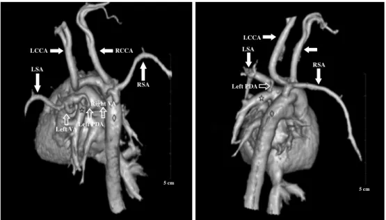
and her head circumference was 34 cm (<3rd percentile). Figure 1. Computed tomography angiography showing an isolated origin of LSA from LPA via a
left-sided PDA.
LCCA: Left common carotid artery; LSA: Left subclavian artery; RCCA: Right common carotid artery; RSA: Right subclavian artery; VA: Vertebral artery; PDA: Patent ductus arteriosus; LPA: Left pulmonary artery.
LCCA
LCCA RCCA
LSA
LSA
Left VALeft PDA Right VA RSA 5 cm 5 cm RSA Left PDA
Figure 2. Coronal and three-dimensional reconstruction slices showing absence of origin of isolated
left subclavian artery from aorta and relation between isolated left subclavian artery and left vertebral artery.
RCA: Right carotid artery; RSA: Right subclavian artery; LCA: Left carotid artery; ILSA: Isolated left subclavian artery; DAo: Descending aorta; RPA: Right pulmonary artery; LPA: Left pulmonary artery; LVA: Left vertebral artery; AAo: Ascending aortic artery; MPA: Main pulmonary artery.
693 Çivilibal Tang et al.
Aberrant left subclavian artery in DiGeorge syndrome
The heart rate was 146 bpm and the respiratory rate was 72 breaths/min. The left radial pulse was weak, while the right radial and femoral pulses were normal. The blood pressure was 90/40 mmHg in the right arm and 60/30 mmHg in the left arm. The oxygen saturation was 65 to 75% on room air due to transient tachypnea of the newborn. She had a dysmorphic appearance with hypertelorism, narrow palpebral fissures, small mouth, short philtrum, and low set ears. There was a systolic 2/6 murmur at the pulmonary area. The laboratory investigations showed hypocalcemia, hyperphosphatemia, and low parathyroid hormone levels. Absolute counts and percentages of lymphocyte subsets were within normal limits. The fluorescence in situ hybridization (FISH) test for 22q11.2 deletion was positive.
An electrocardiogram indicated sinus rhythm with a right QRS axis. The superior mediastinum appeared narrow on chest radiograph. Cervical ultrasonography (USG) indicated an absent thymus in the anterior mediastinum. Echocardiography revealed that there were secundum atrial septal defect (ASD) 7 mm in diameter, restrictive patent ductus arteriosus (PDA), and right positional aortic arch (RAA). There was a
continuous flow pattern at the atypical location toward the right pulmonary artery and a flow to the LPA with the same character. Thoracic CT angiography indicated a right aortic arch causing left common carotid artery, right common carotid artery, and right subclavian artery. The LSCA was originating from the LPA via PDA, instead of the aortic arch. The PDA on the right side was between the aortic arch and right pulmonary artery (Figures 1 and 2).
At the end of the third day, echocardiography showed that both PDAs were closed. Hypoxia responded well to nasal oxygen and tachypnea regressed, and the pathology causing hypoxia was transient tachypnea of the newborn. The baby was, then, discharged with normal oxygen saturation and well condition on the fourth day.
At eight months of follow-up, the patient presented with weakness in her left arm. On her neurological examination, there were 4/5 tone in her left arm and 5/5 tone in her right arm. On Doppler echocardiography, there were secundum ASD 4-mm in diameter and right aortic arch. Doppler USG indicated reversed flow direction on the left vertebral artery which was compatible with the subclavian steal syndrome. Thoracic CT angiography revealed the absence of origin of the LSCA from the aorta and the connection with LSCA and left vertebral artery.
A written informed consent was obtained from each parent.
DISCUSSION
Isolation of the subclavian artery is a rare aortic arch anomaly with fewer than 100 cases reported in the literature. Isolation of the LSCA is seen in 0.8% of the
right aortic arches.[4] In normal cardiac pathology, the
reported case is very rare and the first case similar to
this one was reported in 2006 by Madan et al.[3]
Edward suggested an embryological model for bilateral ductus arteriosus and isolation of the LSCA
with a right aortic arch.[5] Interruption in the double
aortic arch plan takes place at two levels; one between the left common carotid and LSCAs and the other one between the LSCA and the descending aorta. Then, this regression results in a right arch with three vessels arising from the arch in the following order: left common carotid, right common carotid, and right subclavian arteries (Figure 3). The LSCA is not attached to either the left common carotid artery or the arch, but rather is tethered to the LPA by a left ductus arteriosus which may be patent or closed. The blood supply to the LSCA may involve a mediastinal,
Figure 3. The view of Edwards hypothetical double aortic
arch. Arrows show the interruption of right aortic arch at two locations.[5]
Ao: Aorta; RPA: Right pulmonary artery; LPA: Left pulmonary artery; PT: Pulmonary trunk.
Right common carotid artery Left common carotid artery
Right vertebral artery
Left vertebral artery
Right subclavian artery
PT Ao
1 2
RPA LPA
694
Turk Gogus Kalp Dama 2020;28(4):691-694
thoracic anastomosis, or vertebral pathway. Therefore, pulmonary steal syndrome and/or subclavian steal syndrome may occur. When the left ductus arteriosus obliterates, the isolated LSCA is filled by retrograde flow from the left vertebral artery, a congenital left subclavian steal similar to our patient.[5]
The subclavian steal syndrome is characterized by a subclavian artery anomaly located proximal to the origin of the vertebral artery. However, subclavian artery lesions are usually asymptomatic due to the abundant collateral blood supply in the head, neck, and shoulder. It is important to note that muscle weakness is the most common symptom of the syndrome, after it becomes symptomatic, which was observed in our
patient, as well.[6] Other possible symptoms of ischemia
are pain, coldness, paresthesia in the upper extremity, and vertebrobasilar insufficiency (i.e., dizziness, vertigo, headache, tinnitus, disturbances of vision,
drop attacks, and syncope).[4,7] In another study of 39
patients, vertebrobasilar insufficiency was found in five patients and ischemic symptoms of the left arm
were observed in another five patients.[4]
For the diagnosis, several imaging tools including color Doppler USG, computed tomography (CT), magnetic resonance (MR) angiography, and invasively aortic arch aortography are used. The precise ultrasonic evaluation of both vertebral arteries is useful for early
diagnosis.[7] As a non-invasive method, however, color
Doppler USG is typically the first and sufficient choice for the diagnosis. Other methods are useful for further exploring the anatomy. It is diagnostic in the arteriography to visualize the retrograde filling of the vertebral artery and the passage of the contrast agent from the vertebral artery to the subclavian artery.[7] In
our patient, echocardiography findings were suspicious, and the diagnosis was confirmed with combined Doppler USG and CT. The first indicated reversed flow direction on the left vertebral artery, whereas the latter revealed the anatomy and determined the etiology.
There are some therapeutic options for isolation of the subclavian artery; device occlusion of the PDA, simple ligation of the LSCA, surgical reimplantation
and follow-up,[8] and treatment varies depending on
the presence of symptoms. Surgery is appropriate
for obliterated PDA and symptomatic patients. In our patient, both ductus arteriosus were patent at birth. However, the right ductus arteriosus regressed first and the left ductus arteriosus regressed three days later. Therefore, device occlusion of PDA was not considered.
In conclusion, in this report, we describe the first case of aberrant LSCA originating from the pulmonary artery in a normal cardiac anatomy with DiGeorge syndrome. Furthermore, the existence of congenital left subclavian steal syndrome presenting with the left arm weakness makes this case a more interesting one.
Declaration of conflicting interests
The authors declared no conflicts of interest with respect to the authorship and/or publication of this article.
Funding
The authors received no financial support for the research and/or authorship of this article.
REFERENCES
1. Momma K. Cardiovascular anomalies associated with chromosome 22q11.2 deletion syndrome. Am J Cardiol 2010;105:1617-24.
2. Abe M, Isobe T, Atsumi N. Right aortic arch with isolation of the left subclavian artery and bilateral patent ductus arterioses. Pediatr Cardiol 2000;21:497-9.
3. Madan N, Schneider DJ, Jacobs ML. Right aortic arch, isolated left subclavian artery and ductus arteriosus with normal intracardiac anatomy: rare manifestation of chromosome 22q11 deletion. Pediatr Cardiol 2006;27:781-3.
4. Luetmer PH, Miller GM. Right aortic arch with isolation of the left subclavian artery: case report and review of the literature. Mayo Clin Proc 1990;65:407-13.
5. Edwards JE. Anomalies of the derivatives of the aortic arch system. Med Clin North Am 1948;32:925-49.
6. Perler BA, Becker GJ, editors. Vascular intervention: A clinical approach. London: Thieme; 1998.
7. Boztosun B, Avcı A, Saglam M, Meriç M. Coronary subclavian steal syndrome: a case report. Turk Gogus Kalp Dama 2010;18:141-4.
8. Lee Y, Hong SW. Abnormal origin of the left subclavian artery from the left pulmonary artery in a patient with double outlet right ventricle. Korean J Thorac Cardiovasc Surg 2014;47:32-4