Case Report
Resembling Left Ventricular False Tendon in a Father and
His Daughter
Abdullah Kaplan
and Hac
ı Murat Gunes
Department of Cardiology, Istanbul Medipol University, Istanbul, Turkey Correspondence should be addressed to Abdullah Kaplan; [email protected]
Received 3 August 2018; Revised 7 October 2018; Accepted 1 November 2018; Published 9 December 2018 Academic Editor: Assad Movahed
Copyright © 2018 Abdullah Kaplan and Hacı Murat Gunes. This is an open access article distributed under the Creative Commons
Attribution License, which permits unrestricted use, distribution, and reproduction in any medium, provided the original work is properly cited.
Left ventricular false tendons (LVFTs) are linearfibrous or fibromuscular bands stretching across left ventricular cavity. Although
LVFTs have been associated with various heart pathologies and investigated embryologically and histologically, there is only one report in the literature connoting possible hereditary transmission of this entity. We reported a father and his daughter having similar types of LVFTs with regard to location and thickness. With this report, we will contribute in the literature in respect to potential genetic inherence of LVFTs.
1. Introduction
Left ventricular false tendons (LVFTs) are linearfibrous or fibromuscular bands that extend between ventricular septum and papillary muscle, left ventricular free wall, or apex [1, 2]. LVFTs were incidentalfindings at cadaver dissection in early days, but later technical improvement in the heart imaging such as echocardiography and magnetic resonance imaging let the percentage of LVFT visualization rise [2, 3]. LVFT prevalence in the literature spreads over a wide range, while in the earliest reports, it was 0.5%; current investigations have reported up to 78% [3].
Although there is no consensus on the implication of LVFTs in heart diseases, they have been associated with various clinical manifestations in otherwise structurally normal hearts such as functional murmur, preexcitation, idiopathic ventricular tachycardia, infective endocarditis, cavitary thrombi, regional myocardial hypertrophy, repolar-ization abnormalities, and genesis of J-waves [2–9].
Our knowledge about LVFT continuously grows. In order to understand clinical implication of LVFT, we need to evaluate this entity in all its aspects. The questions rose regarding its clinical implication let researches to extend histologic characteristic and embryologic development of
LVFT. The lacking information regarding genetic transi-tion of LVFT is the missing link of this chain. We offered an insight into genetic aspect of this entity by reporting these cases.
1.1. Case 1. An 18-year-old previously healthy girl presented with complaint of palpitation. She has experienced palpita-tion 2 or 3 times a month for 3 years. She did not declare other triggering factors for palpitation with the exception of caffeine. She stated that her palpitation episode sometime lasts one hour, and it is rarely accompanied by dizziness. On physical examination, the followings were noted: normal S1 and S2 without added sounds (S3 or S4) and murmur on cardiac auscultation, blood pressure 118/76 mmHg, heart rate 78/min, and normal breathing sounds. A 12-lead electro-cardiogram showed sinus rhythm with normal QRS mor-phology. Transthoracic echocardiography revealed a structurally normal heart with the exception of a broad false tendon within the left ventricle extending between apical lateral wall and basal septum (Figures 1(b)–4(b)). Ambulatory rhythm monitoring showed no isolated pre-mature ventricular complexes, ventricular couplets, or runs during 24 hours. She was asymptomatic during the 24 hours of ambulatory rhythm monitoring. She was advised
Volume 2018, Article ID 9543098, 4 pages https://doi.org/10.1155/2018/9543098
RV LA (a) RV LA (b)
Figure 1: mode echocardiographic image from the father (a) and his daughter (b). (a) and (b) Modified parasternal long axis view by B-mode recording. The arrow indicates the false tendon stretching from the left ventricular apical free wall to the ventricular septum. The star indicates the papillary muscles. LA: left atrium; RV: right ventricle.
(a) (b)
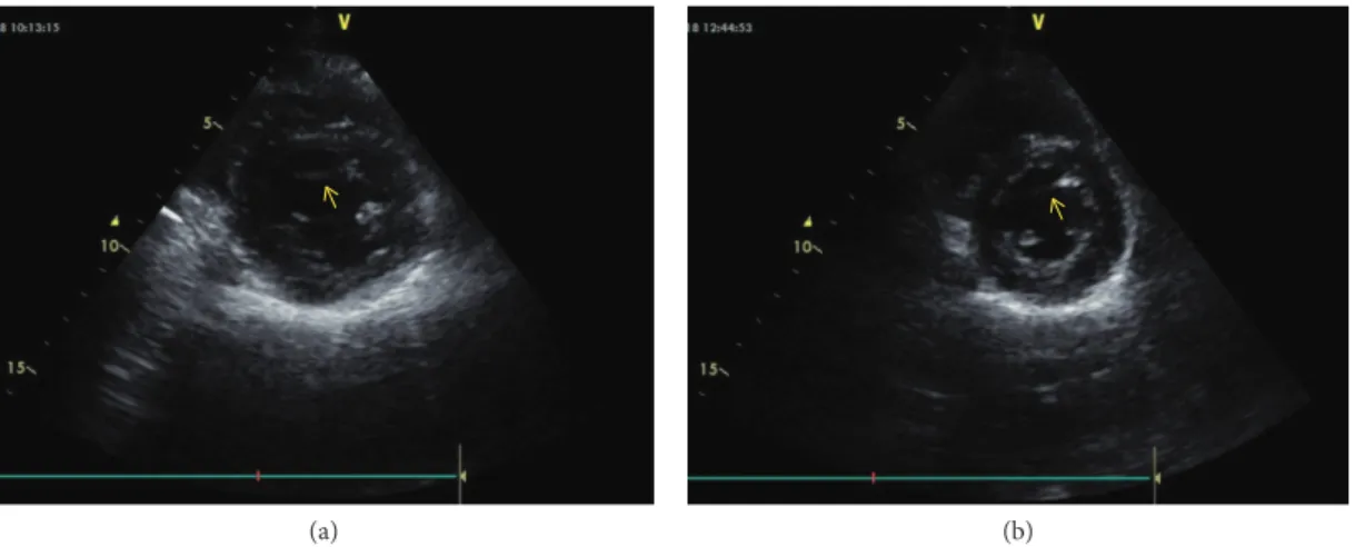
Figure 2: M-mode echocardiographic image from the father (a) and his daughter (b). (a) and (b) M-mode recording from the midsegment of the left ventricle. The arrow indicates the false tendon; The star indicates the papillary muscles.
LA RV (a) RV LA (b)
Figure 3: B-mode echocardiographic image from the father (a) and his daughter (b). (a) and (b) Modified apical four chamber view by B-mode recording. The arrow indicates the false tendon stretching from the left ventricular apical free wall to the ventricular septum. LA: left atrium; RV: right ventricle.
to obtain an electrocardiogram at the time of palpitation from the nearest medical center.
1.2. Case 2. The father of the girl in case 1, 52 years old, was admitted in our outpatient clinic due to a periodic examina-tion of coronary artery disease. He had coronary bypass sur-gery history. At the time of admission, he was asymptomatic with regard to coronary artery disease. On physical examina-tion, the followings were noted: normal S1 and S2 without added sounds (S3 or S4) and murmur on cardiac ausculta-tion, blood pressure 132/82 mmHg, heart rate 72/min, nor-mal breathing sounds, and no peripheral edema.
A 12-lead electrocardiogram revealed nonspecific ST changes in precordial leads. Transthoracic echocardiography showed normal systolic contraction in all left ventricular wall segments. There was no evidence of left ventricular cavity enlargement or hypertrophy according to the measurements suggested by chamber quantification guideline [10]. During the evaluation, a left ventricular false tendon extending between apical lateral wall and basal septum was noticed (Figures 1(a)–4(a)).
2. Discussion
Although there are several reports in the literature regarding embryologic development, histologic evaluation, and its clin-ical association of LVFT, to date, genetic aspect of LVFT has not been scrutinized thoroughly. Many years ago, genetic transmission of LVFT became the main topic of conversation upon a presentation of resembling LVFTs in a mother and her son in Russian literature [11]. Unlike the previous one,
we reported LVFT in a father and his daughter with a similar point of attachment and thickness in the left ventricle. To our best knowledge, this is the second report to point out the potential genetic inheritance of LVFT in the literature.
Clinical implication of LVFTs is still not well understood although several reports have showed their association with some heart diseases [3]. In order to disclose a potential link between LVFT and heart disease, histologic feature and embryologic development of this entity have been studied. Although embryologic origin of LVFT has not been fully elucidated, they are thought to be derived from inner muscle layer of primitive heart [2, 12, 13]. Histologic analyses of LVFTs revealed that they can containfibrous tissues, myo-cardial fibers, elastic fibers, and blood vessels [2, 14, 15]. LVFTs can be classified into three types based on their thick-ness although their functional differences remain unknown. Fibrous type is less than 1.4 mm in diameter,fibromuscular type is 1.5–2.4 mm, and muscular type is larger than 2.5 mm [2]. Our both cases had muscular type false tendon based on their thickness (Table 1).
LVFTs are classified into 5 types with varying percent-ages based on the points of attachment. Type 1 (66%) extends between posteromedial papillary muscle and ven-tricular septum, type 2 (12%) between anterolateral papillary muscle and posteromedial papillary muscle, type 3 (11%) between anterolateral papillary muscle and ventricular sep-tum, type 4 (9%) between left ventricular free wall and ven-tricular septum, and type 5 (1%) between one left venven-tricular free wall and another left ventricular free wall [16]. Our both cases had type 4 LVFT based on the points of attachment where the tendons extended between apical lateral wall and basal septum (Figures 1–4).
Although there is no consensus on clinical importance of LVFTs [3], it might be important to visualize the false tendon by cardiac imaging in some cases. In those who undergo heart surgery or left ventricular catheter ablation, the presence of LVFT must be known by the operator to avoid damage to these tendons, in view of the possible pres-ence of the conduction system and coronary artery branches in the tendons [2]. As well as, because LVFTs can be the
(a) (b)
Figure 4: B-mode echocardiographic image from the father (a) and his daughter (b). (a) and (b) B-mode image from the short axis. The arrow indicates the false tendon stretching from the left ventricular free wall to the ventricular septum.
Table 1: Maximum thickness of the left ventricular false tendon in both cases.
Subjects PSLAX (mm) SAX (mm) A4C (mm) Average (mm)
Father 8 3 9 6.6
Daughter 6 2 7 5
origin of the infective endocarditis [8, 17] and cavitary thrombus [18, 19], clinician must be vigilant in those who have unknown source of fever or systemic embolism.
Several heart diseases and electrocardiographic abnor-malities have been associated with LVFTs [2–4, 20–22]. However, in our both cases, we failed to show any heart pathology linked to LVFT as reported in the literature. Even though the girl presented with complaint of palpitation, ambulatory rhythm monitoring showed no arrhythmia during 24 hours. It is worth noting that she was asymptom-atic during the recording.
By reporting these cases, we draw attention of clinicians on the potential genetic inheritance of LVFTs. Clinical studies are warranted to confirm the genetic inheritance and reveal the pattern of genetic transmission of LVFTs.
Conflicts of Interest
The authors declare that they have no conflicts of interest.
References
[1] W. Turner,“Another heart with moderator band in left
ventri-cle,” Journal of Anatomy and Physiology, vol. 30, Part 4,
pp. 568-569, 1896.
[2] S. Philip, K. M. Cherian, M. H. Wu, and H. C. Lue, “Left
ventricular false tendons: echocardiographic, morphologic, and histopathologic studies and review of the literature,”
Pediatrics and Neonatology, vol. 52, no. 5, pp. 279–286, 2011.
[3] F. S. Ferrer, M. L. S. Ferrer, M. D. G. Murcia, M. S. Ferrer, and
F. S. del Campo,“Basic study and clinical implications of left
ventricular false tendon. Is it associated with innocent murmur in children or heart disease?,” Revista Española de Cardiología
(English Edition), vol. 68, no. 8, pp. 700–705, 2015.
[4] M. Nakagawa, K. Ezaki, H. Miyazaki et al.,“False tendons may
be associated with the genesis of J-waves: prospective study in
young healthy male,” International Journal of Cardiology,
vol. 172, no. 2, pp. 428–433, 2014.
[5] S. Kenchaiah, E. J. Benjamin, J. C. Evans, J. Aragam, and R. S.
Vasan,“Epidemiology of left ventricular false tendons: clinical
correlates in the Framingham Heart Study,” Journal of the
American Society of Echocardiography, vol. 22, no. 6, pp. 739–745, 2009.
[6] M. Kervancioğlu, D. Ozbağ, P. Kervancioğlu et al., “Echocar-diographic and morphologic examination of left ventricular
false tendons in human and animal hearts,” Clinical Anatomy,
vol. 16, no. 5, pp. 389–395, 2003.
[7] M. Loukas, C. T. Wartmann, R. S. Tubbs et al., “Right
ventricular false tendons, a cadaveric approach,” Surgical and
Radiologic Anatomy, vol. 30, no. 4, pp. 317–322, 2008.
[8] E. Aksakal, S. Sevimli, Y. Gürlertop, and H. Taş, “An
intracar-diac mobile mass: ruptured left-ventricular false tendon
with big vegetation due to Brucella endocarditis,” Anadolu
Kardiyoloji Dergisi, vol. 10, no. 6, pp. 557-558, 2010. [9] S. Pisiak, K. Dorniak, M. Hellmann, D. Rawicz-Zegrzda,
M. Węsierska, and M. Dudziak, “Left ventricular false tendons:
echocardiographic characteristics in the Polish population,”
Folia Morphologica, vol. 74, no. 2, pp. 225–228, 2015.
[10] R. M. Lang, L. P. Badano, V. Mor-Avi et al.,
“Recommenda-tions for cardiac chamber quantification by echocardiography
in adults: an update from the American Society of Echocardi-ography and the European Association of Cardiovascular
Imaging,” Journal of the American Society of
Echocardiogra-phy, vol. 28, no. 1, pp. 1–39.e14, 2015.
[11] V. A. Kuznetsov and A. A. Korzhenkov, False Tendons in the
Heart. Diagnostics and Clinical Significance: Guidelines for
Practitioners, Meditsinskaya kniga, Moscow, Russia, 2011. [12] L. W. Perry, R. N. Ruckman, S. R. Shapiro, K. S. Kuehl, F. M.
Galioto Jr., and L. P. Scott,“Left ventricular false tendons in
children: prevalence as detected by 2-dimensional
echocardi-ography and clinical significance,” The American Journal of
Cardiology, vol. 52, no. 10, pp. 1264–1266, 1983.
[13] M. Grzybiak, D. Lotkowski, and D. Kozłowski, “False tendons
in the left ventricle of the heart in humans during pre- and postnatal periods,” Folia Morphologica, vol. 55, no. 2,
pp. 89–99, 1996.
[14] L. M. Gerlis, H. M. Wright, N. Wilson, F. Erzengin, and D. F.
Dickinson, “Left ventricular bands. A normal anatomical
feature,” British Heart Journal, vol. 52, no. 6, pp. 641–647,
1984.
[15] J. I. Brenner, K. Baker, R. E. Ringel, and M. A. Berman,
“Echo-cardiographic evidence of left ventricular bands in infants and
children,” Journal of the American College of Cardiology, vol. 3,
no. 6, pp. 1515–1520, 1984.
[16] J. J. Silbiger,“Left ventricular false tendons: anatomic,
echocar-diographic, and pathophysiologic insights,” Journal of the
American Society of Echocardiography, vol. 26, no. 6, pp. 582–588, 2013.
[17] S. K. Chhabra, L. J. Bogar, M. V. DeCaro, and I. S. Cohen, “Complex mitral valve endocarditis involving a left atrial false tendon,” Journal of the American College of Cardiology, vol. 60, no. 22, p. 2330, 2012.
[18] A. Hosseinsabet, “A large thrombus on false tendon in a
cardiomyopathic patient,” Türk Kardiyoloji Derneği Arşivi,
vol. 42, no. 7, p. 689, 2014.
[19] S. Mukai, H. Fuseno, M. Nakamura, J. Yoshikawa, and
T. Shomura, “Dilated cardiomyopathy complicated by a
pedunculated and mobile left ventricular thrombus on
rup-tured false tendons,” Chest, vol. 99, no. 4, pp. 1042-1043, 1991.
[20] V. J. R. Reddy, A. Ali, and C. N. Manjunath, “A case of left
ventricular false tendon with ventricular tachycardia,” Heart
Views, vol. 18, no. 1, pp. 30-31, 2017.
[21] T. Betsuyaku, H. Muto, E. Sugiyama, A. Minoshima, and
M. Sato, “False tendon-related polymorphic ventricular
tachycardia,” Pacing and Clinical Electrophysiology, vol. 35,
no. 12, pp. e341–e344, 2012.
[22] A. Hussain, R. H. Anderson, and S. S. Dhillon,“Taut and click:
an unusual left ventricular false tendon,” Cardiology in the
Young, vol. 26, no. 7, pp. 1435–1437, 2016.
Stem Cells
International
Hindawi www.hindawi.com Volume 2018 Hindawi www.hindawi.com Volume 2018 INFLAMMATIONEndocrinology
International Journal ofHindawi www.hindawi.com Volume 2018 Hindawi www.hindawi.com Volume 2018
Disease Markers
Hindawi www.hindawi.com Volume 2018 BioMed Research InternationalOncology
Journal of Hindawi www.hindawi.com Volume 2013 Hindawi www.hindawi.com Volume 2018Oxidative Medicine and Cellular Longevity
Hindawi
www.hindawi.com Volume 2018
PPAR Research
Hindawi Publishing Corporation
http://www.hindawi.com Volume 2013 Hindawi www.hindawi.com
The Scientific
World Journal
Volume 2018 Immunology Research Hindawi www.hindawi.com Volume 2018 Journal ofObesity
Journal of Hindawi www.hindawi.com Volume 2018 Hindawi www.hindawi.com Volume 2018 Computational and Mathematical Methods in Medicine Hindawi www.hindawi.com Volume 2018Behavioural
Neurology
Ophthalmology
Journal of Hindawi www.hindawi.com Volume 2018Diabetes Research
Journal of Hindawiwww.hindawi.com Volume 2018
Hindawi
www.hindawi.com Volume 2018 Research and Treatment
AIDS
Hindawiwww.hindawi.com Volume 2018 Gastroenterology Research and Practice
Hindawi www.hindawi.com Volume 2018