Received: 29 August 2015 Revised: 9 February 2016 Accepted: 16 February 2016 http://dx.doi.org/10.1259/bjr.20150716
Cite this article as:
Karaman A, Diyarbakir B, Durur-Subasi I, Kose D, ¨Ozbek-Bilgin A, Topcu A, et al. A novel approach to contrast-induced nephrotoxicity: the melatonergic agent agomelatine. Br J Radiol 2016; 89: 20150716.
FULL PAPER
A novel approach to contrast-induced nephrotoxicity: the
melatonergic agent agomelatine
1ADEM KARAMAN,MD,2BUSRA DIYARBAKIR,MD,3IRMAK DURUR-SUBASI,MD,2DUYGU KOSE,MD,
2ASLI ¨OZBEK-BILGIN,MD,4ATILLA TOPCU,MD,5CEMAL GUNDOGDU,MD,Prof,6AFAK DURUR-KARAKAYA,MD, 7ZAFER BAYRAKTUTAN and1FATIH ALPER,MD,PhD
1Department of Radiology, Faculty of Medicine, Ataturk University, Erzurum, Turkey 2Department of Pharmacology, Faculty of Medicine, Ataturk University, Erzurum, Turkey
3Department of Radiology, Diskapi Yildirim Beyazit Training and Research Hospital, Ankara, Turkey 4Department of Pharmacology, Faculty of Medicine, Recep Tayyip Erdo˘gan University, Rize, Turkey 5
Department of Pathology, Faculty of Medicine, Ataturk University, Erzurum, Turkey
6Department of Radiology, Istanbul Medipol University, Istanbul, Turkey 7
Department of Biochemistry, Erzurum Regional Training and Research Hospital, Erzurum, Turkey Address correspondence to: Associate Professor Irmak Durur-Subasi
E-mail:[email protected]
Objective: To study the potential nephroprotective role of agomelatine in rat renal tissue in cases of contrast-induced nephrotoxicity (CIN). The drug’s action on the antioxidant system and proinflammatory cytokines, su-peroxide dismutase (SOD) activity, levels of glutathione (GSH) and malondialdehyde (MDA) and the gene expres-sion of interleukin-6 (IL-6), tumour necrosis factor (TNF)-a and nuclear factor kappa B (NF-kB) was measured. Tubular necrosis and hyaline and haemorrhagic casts were also histopathologically evaluated.
Methods: The institutional ethics and local animal care committees approved the study. Eight groups of six rats were put on the following drug regimens: Group 1: healthy controls, Group 2: GLY (glycerol), Group 3: CM (contrast media—iohexol 10 ml kg21), Group 4: GLY1CM, Group 5: CM1AGO20 (agomelatine 20 mg kg21), Group 6: GLY1 CM1AGO20, Group 7: CM1AGO40 (agomelatine 40 mg kg21) and Group 8: GLY1CM1AGO40. The groups were evaluated by one-way analysis of variance and Duncan’s multiple comparison test.
Results: Agomelatine administration significantly improved the serum levels of blood urea nitrogen (BUN) and creatinine, SOD activity, GSH and MDA. The use of agomelatine had
substantial downregulatory consequences on TNF-a, NF-kB and IL-6 messenger RNA levels. Mild-to-severe hyaline and haemorrhagic casts and tubular necrosis were observed in all groups, except in the healthy group. The histopathological scores were better in the agome-latine treatment groups.
Conclusion: Agomelatine has nephroprotective effects against CIN in rats. This effect can be attributed to its properties of reducing oxidative stress and inhibiting the secretion of proinflammatory cytokines (NF-kB, TNF-a and IL-6).
Advances in knowledge: CIN is one of the most important adverse effects of radiological procedures. Renal failure, diabetes, malignancy, old age and non-steroidal anti-inflammatory drug use pose the risk of CIN in patients. Several clinical studies have investigated ways to avoid CIN. Theophylline/aminophylline, statins, ascor-bic acid and iloprost have been suggested for this purpose. Agomelatine is one of the melatonin ligands and is used for affective disorders and has antioxidant features. In this study, we hypothesized that agomelatine could have nephroprotective, antioxidant and anti-inflammatory effects against CIN in rats.
INTRODUCTION
Contrast-induced nephrotoxicity (CIN) is the worsening of renal function (an increase in levels of creatinine by more than one-fourth or 44mmol l21) that occurs within 72 h after the administration of a contrast material in the ab-sence of any other aetiology.1 Although its incidence has
been reported to be low,2it is one of the most important
3
complication following the administration of contrast mate-rials is a common cause of hospital-acquired renal in-sufficiency and accounts for about 11% of cases,5
occurring during diagnostic and interventional procedures.6 Renal fail-ure, diabetes, malignancy, old age and non-steroidal anti-inflammatory drug use have all been found to be associated with a higher risk of CIN.2CIN occurs more commonly in
acute renal failure with the increasing use of contrast media (CM) for diagnostic and interventional procedures. In this regard, it is important to thoroughly investigate this issue in animal models. Pre-treatment with glycerol can be used to induce pre-existing renal damage in rats.7 By administering glycerol, we induced CIN in the rats to mimic any underlying pathology. Iohexol is a low-osmolar non-ionic radiocontrast agent that has improved safety and tolerability compared with classic radiocontrast agents. For these reasons, iohexol became the most common radiocontrast agent used in clinical and experimental “contrast-induced nephropathy” research.8,9
We therefore chose to use this contrast agent in our experimental model.
Several clinical studies have investigated ways to avoid CIN. Drugs such as theophylline/aminophylline, statins, ascorbic acid and iloprost have been suggested to be helpful in reducing the occurrence of this condition; nevertheless, a detailed evaluation of their impact is needed.10–13
Agomelatine is a melatonergic receptor agonist and a selective serotonin 5HT2Creceptor antagonist that has shown
antidepres-sant efficacy. Melatonin receptor type (MT)1 and MT2 receptors are predominating in the human foetal kidney cortex area and have been disclosed as renoprotective agents, as they are generally attributed to have the debated antioxidant capability.14,15 Mela-tonin has several applications related to the renal system and its production is impaired in chronic renal failure and it cannot protect the kidneys from inflammation which can cause renal damage.16,17In this context, to our knowledge, there has not been a study to date on the nephroprotective role of agomelatine for CIN aggravated by glycerol in rats.
In this study, we aimed to investigate the possible nephroprotective role of agomelatine. To appreciate the relationship between the nephroprotective action of agomelatine, the antioxidant system and proinflammatory cytokines, we measured the superoxide dismutase (SOD) activity, levels of glutathione (GSH) and malondialdehyde (MDA) and the gene expression of interleukin-6 (IL-6), tumour necrosis factor (TNF)-a and nuclear factor kappa B (NF-kB) in the renal tissue of rats with CIN.
METHODS AND MATERIALS
The study protocol was approved by the institutional ethics and local animal care committees. Animal experimentations and trials were implemented in a manner that was consistent with the na-tional guiding principles.
Animals
In total, 48 female albino Wistar rats (250–300 g) were used in the study. They were obtained from our university experimental animal laboratory. The rats were stored in typical plastic cages on sawdust bedding in a well-ventilated room at 22°C under precise light conditions (14/10 h light/dark cycle). Standard rat chow and tap water were provided ad libitum.
Chemicals
Clinical substances were obtained from Sigma Chemical Com-pany (Germany), agomelatine (Valdoxan® 25 mg, 28 tablets) was
obtained from Les Laboratories Servier (Istanbul, Turkey), iohexol (Omnipaque™ 300 mgI ml21, 100 ml) from Opakim Medical Products (Istanbul, Turkey) and 100% glycerol from Bikar (Istanbul, Turkey).
Experimental design
Eight sets of six rats in separate cages were administered the following drugs:
(1) Group 1: healthy control group (2) Group 2 (GLY): glycerol only
(3) Group 3 (CM): contrast media (iohexol 10 ml kg21) (4) Group 4 (GLY1CM): glycerol 1 contrast media (iohexol
10 ml kg21)
(5) Group 5 (CM1AGO20): contrast media (iohexol 10 ml kg21
) 1 agomelatine 20 mg kg21(po)
(6) Group 6 (GLY1CM1AGO20): glycerol 1 contrast media (iohexol 10 ml kg21)1 agomelatine 20 mg kg21(po) (7) Group 7 (CM1AGO40): contrast media (iohexol 10 ml kg21)
1 agomelatine 40 mg kg21
(po)
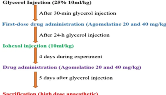
(8) Group 8 (GLY1CM1AGO40): glycerol 1 contrast media (iohexol 10 ml kg21)1 agomelatine 40 mg kg21(po). The contrast media-induced nephrotoxicity model Kidney injury was induced in Groups 2, 4, 6 and 8 by in-tramuscular administration of 25% glycerol as a single dose of 10 ml kg21body weight after 24-h water deprivation. The dose was divided among the legs. After glycerol injection, drinking water was resumed ad libitum.
24 h after glycerol injection, nephrotoxicity was induced with intravenous administration of iohexol (contrast media, CM) as a single dose of 10 ml kg21body weight over a period of 2 min to the animals in Groups 3–8 via the tail vein.8
30 min after glycerol injection, agomelatine (dissolved in iso-tonic sodium chloride solution) was administered orally to the animals in Groups 5–8. Agomelatine administration was re-peated on the second, third and fourth days (Figure 1). Following the drug administration period, the study wasfinished on the fifth day after glycerol injection by an overdose of a general anaesthetic (thiopental sodium, 50 mg kg21). Under anaesthetic conditions, blood samples were obtained by
intracardiac method. Kidney tissues were dissected out imme-diately. Half of one kidney was kept at280°C for biochemical evaluation; the rest was kept in an RNA stabilization reagent for molecular analysis, and the other kidney wasfixed in formalin for pathological examination. Serum was instantly separated by centrifugation at 4000 rpm for 10 min at 4°C and stored at280°C until it was analysed.
Determination of serum BUN and creatinine concentrations
Serum blood urea nitrogen (BUN) (Lot No: B0382A, S.R.I, Italy) and creatinine (Lot No: B0914A, S.R.I, Italy) levels were detected using commercially available kits. All analyses were performed in a ChemWell® 2910 Automated EIA and Chemistry analyzer (Awareness Technology®, Inc., Palm City, FL).
Biochemical examination of kidneys
After surgery, the tissues were stored at280°C. All samples were first perfused with PBS/heparin and then pulverized in liquid nitrogen using a TissueLyser II (Qiagen, Ankara, Turkey) grinding jar set. Nearly 100 mg of the pulverized tissue was homogenized in 1 ml of PBS homogenate buffer in an Eppen-dorf tube® using the TissueLyser II. Samples were then centri-fuged. SOD,18GSH19and MDA20levels from the supernatant of each sample and standard were measured at room temperature in duplicate via modified methods with an enzyme-linked im-munosorbent assay reader. The average absorbance of each sample and standard was calculated. A standard curve was plotted, and the equation was obtained from the absorbance of the standards. The linear SOD, GSH and MDA concentrations were calculated according to this equation. The SOD, GSH and MDA levels in the tissues were expressed as U mg21 protein, nmol mg21protein and nmol mg21protein, respectively. Obtained data were presented as mean6 standard deviation from 1 mg of protein.
Molecular investigations of kidney tissues Total RNA extraction and cDNA synthesis
Tissues (20 mg) were briefly stabilized in an RNA stabilization reagent (RNAlater®; Qiagen, Hilden, Germany) and then dis-rupted using the TissueLyser II (23 2 min for kidney tissue; 23 5 min, Qiagen). Total RNA was purified in Qiacube using the RNeasy Mini Kit (Qiagen) according to manufacturer instruc-tions. The RNA samples were reverse transcribed into comple-mentary DNA (cDNA) using a high-capacity cDNA reverse transcription kit (Applied Biosystem®; Hernakim, Istanbul, Turkey). From 10ml, total RNA was treated with 2 ml 103 RT buffer, 0.8ml 253 dNTPs mix, 2 ml 103 RT Random Primers, 1ml MultiScribe® reverse transcriptase and 4.2 ml DEPC-H2O. Reverse transcription was carried out at 25°C for 10 min, fol-lowed by 120 min at 37°C and finally 5 min at 85°C using a Veriti® 96-well thermal cycler (Applied Biosystem). The cDNA concentration and quality were assessed and quantified using the Epoch™ Spectrophotometer System and Take3 plate (Bio Tek®; Pera Medikal, Istanbul, Turkey).
Relative quantification of gene expression
Relative TNF-a, IL-6 and NF-kB expression analyses were performed with StepOne Plus™ Real Time PCR System
technology (Applied Biosystems) using synthesized cDNA from rat kidney RNAs. A quantitative polymerase chain re-action (qPCR) was run using a TaqMan® probe mix based on the TaqMan probe-based technology (Applied Biosystems). A real-time PCR was performed using primers generated for rat TNF-a (Rn00562055_m1), rat IL-6 (Rn01410330_m1), rat NF-kB (Rn01399583_m1) and rat b-actin (Rn00667869_m1). The results are expressed as relative-fold, compared with control groups. Expression data forb-actin in each tissue were used as endogenous controls. For each tissue, triplicate determinations were performed in a 96-well optical plate for both targets using 9ml of cDNA (100 ng), 1 ml of Primer Perfect Probe mix and 10ml of QuantiTect® Probe polymerase chain reaction (PCR) Master mix (Qiagen) in each 20-ml reaction. The plates were heated for 2 min at 50°C and for 10 min at 95°C. Subsequently, 40 cycles of 15 s at 94°C and cycles of 60 s at 60°C were applied. All data are expressed as fold change in expression, compared with the expression in other animal groups, using the 22DDCt method.21
Histological procedure
Kidneys from the rats in all groups were obtained, sectioned frontally andfixed in 10% neutral formalin for 48–72 h. Tissues were then processed routinely and embedded in paraffin wax, from which 4–5-mm-thick serial sections were cut. All tissue sections were stained with haematoxylin and eosin for histopa-thology and periodic acid–Schiff (PAS) stain for basement membrane assessment and examined under a light microscope (BX51, Olympus, Japan). For histopathological assessment, tu-bular necrosis, hyaline casts and haemorrhagic casts were eval-uated and scored independently by a pathologist blinded to the experiment. A minimum offive fields for each kidney slide at 1003 magnification was evaluated and assigned scores for se-verity of changes as follows: Grade 0:2 (negative), Grade 1: 11 (mild), Grade 2: 12 (moderate), Grade 3: 13 (severe) and Grade 4:14 (most severe). The other sections were stored for assessing the immunohistochemistry.
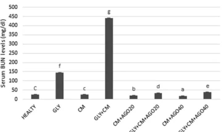
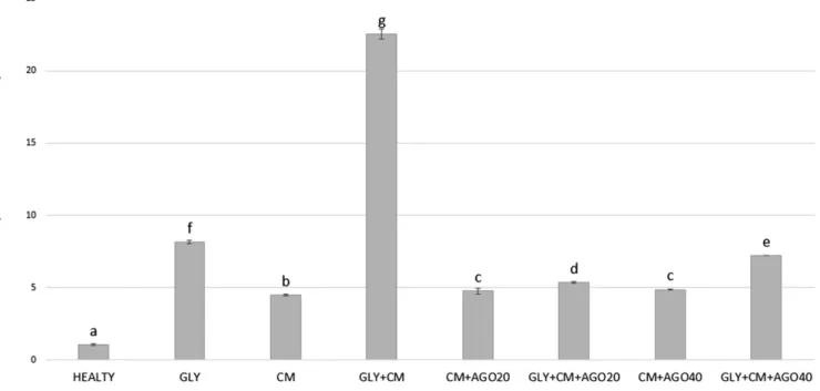
Figure 2. The serum blood urea nitrogen (BUN) levels (mg dl21) of all the experimental groups. Means in the same column with the same letter are not significantly different; means in the same column with different letters indicate significant differ-ences between the groups. AGO20 (agomelatine 20 mg kg21); AGO40 (agomelatine 40 mg kg21); CM, contrast media; GLY (glycerol).
Statistical analysis
For statistical analysis, we used SPSS® v. 20.0 (IBM Corp., New York, NY; formerly SPSS Inc., Chicago, IL). Results are presented as means6 standard deviation. Comparisons between groups were performed using one-way analysis of variance and Duncan’s multiple comparison test. Significance was accepted at p , 0.05. RESULTS
Serum concentrations of BUN and creatinine It was observed that BUN and creatinine serum levels significantly increased in the rats with GLY1CM-induced nephrotoxicity compared with the other groups (p, 0.05). Agomelatine ad-ministration in groups of 5–8 significantly decreased the activity of the serum enzymes compared with the GLY1CM group
(p, 0.05). The changes in the serum BUN and creatinine levels are shown inFigures 2and3, respectively.
Biochemical results for kidney tissue
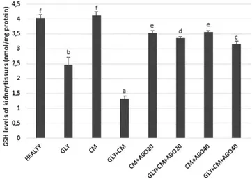
In the present study, SOD activity and levels of GSH and lipid peroxidation (MDA) were evaluated in all rat kidneys. SOD activity was significantly decreased in the GLY1CM group compared with that in the healthy group (p, 0.05). In addition, there were significant differences between the GLY and GLY1 CM groups (p, 0.05). In the treatment groups, SOD activity was significantly improved compared with the GLY1CM group, as shown in Figure 4 (p, 0.05). GSH levels significantly de-creased in the GLY1CM group compared with that in the healthy group (p, 0.05). There were also significant differences between the GLY and GLY1CM groups (p , 0.05). As seen in
Figure 5, GSH levels were significantly ameliorated in the drug
treatment groups when compared with that in the GLY1CM group (p, 0.05).
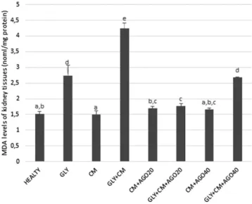
As shown inFigure 6, the MDA levels were significantly
in-creased in the GLY1CM group compared with the healthy group (p, 0.05). In addition, there were significant differ-ences between the GLY and GLY1CM groups (p , 0.05). The MDA levels in groups of 5–8 were significantly improved with the administration of agomelatine compared to the GLY1CM group (p , 0.05).
Molecular results
We investigated TNF-a, IL-6 and NF-kB messenger RNA (mRNA) expressions in the kidney tissue of rats using real-time PCR. As shown inFigure 7, TNF-a gene expression increased in
the GLY1CM groups (30.46-fold) compared with that in the healthy group (p, 0.05). Agomelatine had a significant down-regulatory effect on TNF-a mRNA expression, by 6.95-fold after 20 mg kg21doses and 7.40-fold for 40 mg kg21doses (p, 0.05).
Figure 3. The serum creatinine levels of all the experimental groups. Means in the same column with the same letter are not significantly different; means in the same column with different letters indicate significant differences between the groups. AGO20 (agomelatine 20 mg kg21); AGO40 (agomelatine 40 mg kg21); CM, contrast media; GLY (glycerol).
Figure 4. The tissue superoxide dismutase (SOD) activities of all the experimental groups. Means in the same column with the same letter are not significantly different; means in the same column with different letters indicate significant differ-ences between the groups. AGO20 (agomelatine 20 mg kg21); AGO40 (agomelatine 40 mg kg21); CM, contrast media; GLY (glycerol).
Figure 5. The tissue glutathione (GSH) levels of all the experimental groups. Means in the same column with the same letter are not significantly different; means in the same column with different letters indicate significant differences between the groups. AGO20 (agomelatine 20 mg kg21); AGO40 (agomelatine 40 mg kg21); CM, contrast media; GLY (glycerol).
As shown inFigure 8, compared with the control group, the NF-kB mRNA level was significantly higher in the GLY1CM group, by 22.37-fold (p, 0.05). NF-kB expression in the rat kidney tissues decreased by 5.82-fold and 7.45-fold in the treatment groups compared with that in the control group to nearly healthy levels (p, 0.05).
Compared with the control group, the IL-6 mRNA level was significantly higher (22.55-fold; p , 0.05) in the GLY1CM groups (Figure 9). Agomelatine had a significant downregulatory
effect on IL-6 mRNA expression in the treatment groups when compared with the GLY1CM group (5.36- and 7.21-fold, re-spectively; p, 0.05).
Histopathological results
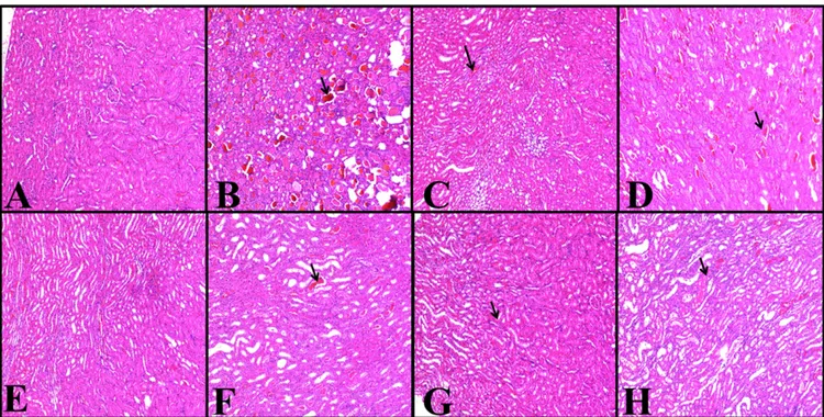
The histopathology of the kidneys of the animals in all groups was examined and scored; the results are given in Figure 10. Rats in the healthy group did not have any histopathological lesions (Figure 11), but severe lesions were seen in both the cortex and medulla of the glycerol-treated rats. Mild-to-severe hyaline and haemorrhagic casts and tubular necrosis were observed in all groups, except in the healthy group (Figure 11). The histopathological scores were better in the agomelatine treatment groups when compared with that in the GLY1 CM groups.
DISCUSSION
In our study, we focused on the restorative effects of ago-melatine for rats with contrast-induced nephropathy; we found that agomelatine administration significantly decreased the elevated serum concentrations of both BUN and creati-nine to nearly healthy levels. Biochemical results for SOD activity, GSH and MDA levels in the kidney tissue, as well molecular results for TNF-a, NF-kB and IL-6, showed the ameliorative effects of agomelatine on kidney function. His-tological scores in terms of hyaline and haemorrhagic casts and tubular necrosis were also improved by the administra-tion of agomelatine.
Figure 6. The tissue malondialdehyde (MDA) levels of all the experimental groups. Means in the same column with the same letter are not significantly different; means in the same column with different letters indicate significant differences between the groups. AGO20 (agomelatine 20 mg kg21); AGO40 (ago-melatine 40 mg kg21); CM, contrast media; GLY (glycerol).
Figure 7. The tissue TNF-a messenger RNA (mRNA) expression of all the experimental groups. Means in the same column with the same letter are not significantly different; means in the same column with different letters indicate significant differences between the groups. AGO20 (agomelatine 20 mg kg21); AGO40 (agomelatine 40 mg kg21); CM, contrast media; GLY (glycerol).
Three different pathophysiological mechanisms lead to CIN: the first being haemodynamic effects (renal medullary hyp-oxia); the second, the direct effects of the contrast medium molecule causing tubular cell toxicity; and the third being en-dogenous biochemical changes such as induction in oxygen-free
radicals and/or a reduction in antioxidant enzyme activity. In addition, it is thought that activation of cytokine-induced in-flammatory mediators by reactive free radicals may be a possible mechanism,22–27 and apoptosis may also play a part in the de-velopment of CIN.22,28However, the main mechanism by which
Figure 8. The tissue NFk-b messenger RNA (mRNA) expression of all the experimental groups. Means in the same column with the same letter are not significantly different; means in the same column with different letters indicate significant differences between the groups. AGO20 (agomelatine 20 mg kg21); AGO40 (agomelatine 40 mg kg21); CM, contrast media; GLY (glycerol).
Figure 9. The tissue IL-6 messenger RNA (mRNA) expression of all the experimental groups. Means in the same column with the same letter are not significantly different; means in the same column with different letters indicate significant differences between the groups. AGO20 (agomelatine 20 mg kg21); AGO40 (agomelatine 40 mg kg21); CM, contrast media; GLY (glycerol).
CIN develops is still unclear; so, in clinical examinations, a variety of ways of avoiding CIN has been investigated.
Agomelatine is a selective melatonergic (MT1/MT2) agonist and a 5-HT2C antagonist.29It has been reported to have antioxidant,30 anti-inflammatory31
and memory-facilitating effects.32 Agonistic regulation of melatonin receptors has been said to improve loco-motor activity and protect the cholinergic system,32 as well as attenuate mitochondrial oxidative damage.32Melatonin has both lipophilic and hydrophilic properties. It can diffuse without diffi-culty into subcellular partitions and shield against free
radical-mediated damage. The latest literature states that melatonin averts nephrotoxicity triggered by several drugs.33–38In particular, Gazi et al39 demonstrated the defensive effect of melatonin adminis-tration in rat radiocontrast nephrotoxicity by reducing BUN and creatinine levels.40However, these studies have not measured ox-idative and molecular parameters to indicate whether regeneration of the kidneys occurred after injury. In the present literature, there is no study about CIN related to agomelatine.
Serum creatinine is the most frequently used parameter for evaluating kidney function, along with BUN.41 In previous
Figure 10. (a–c) Effects of agomelatine on histopathological hyaline cast (a) tubular necrosis (b) and haemorrhagic cast (c) scores in rats’ kidneys. Means in the same column by the same letter are not significantly different from Duncan’s multiple comparison test (p5 0.05). Results are expressed as means 6 SD (n 5 6). AGO20 (agomelatine 20 mg kg21); AGO40 (agomelatine 40 mg kg21); CM, contrast media; GLY (glycerol); SD, standard deviation.
Figure 11. (a–h) Haematoxylin and eosin results in rats’ kidney tissues; magnification 1003. Healthy group (a), GLY (glycerol) group (b), CM (contrast media) group (c), GLY1CM group (d), CM1AGO20 (agomelatine 20 mg kg21) group (e), GLY1CM1AGO20 group (f), CM1AGO40 (agomelatine 40 mg kg21) group (g) and GLY1CM1AGO 40 group (h). (a) Normal structure of the kidney in the control group. (b) Hyaline and haemorrhagic casts are seen severely and moderately; tubular necrosis is also present. (c) Only haemorrhagic casts are seen moderately. Hyaline and tubular necrosis are observed mildly. (d) Haemorrhagic and hyaline casts are seen severely. Moderate tubular necrosis is present. (e) Only haemorrhagic casts are seen mildly. (f) Both hyaline and haemorrhagic casts are seen moderately. Tubular necrosis is also present mildly. (g) Only haemorrhagic casts are seen mildly. (h) Hyaline and haemorrhagic casts are seen moderately; tubular necrosis is observed mildly.
studies, in which nephrotoxicity was created by glycerine and CM, increased levels of serum BUN and creatinine were shown in parallel with elevated oxidative stress and inflammation.7
In our study, we detected large increases in serum BUN and cre-atinine levels in the GLY and GLY1CM groups compared with the healthy group. We also showed that agomelatine, in both groups, ameliorated the serum BUN and creatinine levels of nephrotoxic rats. These decreases in the levels of serum BUN and creatinine, which are important clinical findings of CIN, suggested that agomelatine may have prevented CM-induced nephrotoxicity.
Another possible marker of CIN is oxidative stress. Changes in SOD activity, GSH and MDA levels develop owing to generation of free radicals.42Glycerol, when injected intramuscularly, cau-ses renal GSH depletion.43 Baykara et al42 in their study on propolis, a potential nephroprotective agent against a specific contrast agent (diatrizoate), claimed that propolis protects the renal tissue against diatriozate toxicity, free radicals and other adverse effects by restoring MDA, GSH and SOD levels. In an-other CIN study, decreases in SOD levels in nephrotoxicity groups increased with ebselen treatment.44In cisplatin-induced nephrotoxicity, significantly increased MDA levels were observed in groups administered cisplatin.45 In this study, MDA levels dramatically increased in the GLY and GLY1CM groups com-pared with that in the healthy group. In parallel with this, SOD activity and GSH levels for the groups of 5–8 in the GLY and GLY1CM groups dramatically decreased. Agomelatine admin-istration improved SOD activity and GSH levels compared with the GLY and GLY1CM groups. Similar antioxidant effects of agomelatine have been demonstrated in previous studies.46 The inflammatory process is a factor acting in the pathogenesis of nephrotoxicity. In this process, macrophages released due to inflammation increase the production of proinflammatory cytokines in addition to oxidant release.47–49 Previous studies demonstrated that contrast media administration led to in-creased levels of proinflammatory cytokines in the kidneys.43
IL-6 and TNF-a are associated with CIN severity35,39
and it has been shown that radiocontrast media administration increases NF-kB.50 TNF-a is a proinflammatory cytokine that further
recruits numerous mediators associated with tissue damage. In the animal model of nephrotoxicity, TNF-a is of great impor-tance in the activation of inflammatory response.35
Another important inflammatory cytokine, IL-6, exhibits many physio-logical effects.36 Contrast media administration was shown to lead to an increase in the levels of IL-6 in the kidney,38 while TNF-a led to an increase in NF-kB.51,52
In our study, NF-kB upregulation, in parallel with contrast media and glycerol administration, correlated with the in-creasing levels of TNF-a and IL-6. However, doses of agome-latine caused significant downregulation of NF-kB, TNF-a and IL-6 expression, in comparison with GLY and GLY1CM groups. In a previous study, we demonstrated that agomelatine has an anti-inflammatory effect in cases of paracetamol hep-atotoxicity; agomelatine administration also significantly im-proved TNF-a and IL-6 levels.46
The results of the present study indicate that agomelatine has a potential neph-roprotective effect against CIN.
In various studies, tubular necrosis and epithelial injury have been shown to be significant indicators in nephrotoxicity in-jury;53tubular epithelial damage, tubular necrosis, hyaline and haemorrhagic casts have been found in these investigations.54In the development of nephropathy, significant damage to hae-morrhagic renal tubules with hyaline occurs and is related to tubular injury. We determined positive effects of agomelatine administration on tubular structure.
In conclusion, our study showed that agomelatine has neph-roprotective, antioxidant and anti-inflammatory effects against CIN in rats. This effect is brought about by a reduction in oxidative stress and inhibition of the secretion of proin-flammatory cytokines (NF-kB, TNF-a and IL-6) that trigger the initiation of inflammation in the kidney. This animal model can be a reflection of CM-induced nephropathy in patients with underlying renal insufficiency. We can suggest that agomelatine can be a drug of choice for both radiologists and nephrologists before CM administration in patients with renal insufficiency. However, more detailed future clinical studies are required to better clarify the protective effects of agomelatine in CIN.
REFERENCES
1. Morcos SK, Thomsen HS, Webb JA. Contrast-media-induced nephrotoxicity: a consensus report. Contrast Media Safety Committee, European Society of Urogenital Radiology (ESUR). Eur Radiol 1999;9: 1602–13. doi:http://dx.doi.org/10.1007/ s003300050894
2. Moos SI, van Vemde DN, Stoker J, Bipat S. Contrast induced nephropathy in patients undergoing intravenous (IV) contrast en-hanced computed tomography (CECT) and the relationship with risk factors: a meta-analysis. Eur J Radiol 2013;82: e387–99. doi:
http://dx.doi.org/10.1016/j.ejrad. 2013.04.029
3. Abujudeh HH, Gee MS, Kaewlai R. In emergency situations, should serum creati-nine be checked in all patients before performing second contrast CT examinations within 24 hours? J Am Coll Radiol 2009;6: 268–73. doi:http://dx.doi.org/10.1016/j. jacr.2008.09.014
4. Rihal CS, Textor SC, Grill DE, Berger PB, Ting HH, Best PJ, et al. Incidence and prognostic importance of acute renal failure after percutaneous coronary intervention.
Circulation 2002;105: 2259–64. doi:http:// dx.doi.org/10.1161/01.
CIR.0000016043.87291.33
5. McCullough PA. Contrast-induced acute kidney injury. J Am Coll Cardiol 2008;51: 1419–28. doi:http://dx.doi.org/10.1016/j. jacc.2007.12.035
6. Zhang T, Shen LH, Hu LH, He B. Statins for the prevention of contrast-induced nephropathy: a systematic review and meta-analysis. Am J Nephrol 2011;33: 344–51. doi:http://dx.doi.org/10.1159/ 000326269
7. Al-Otaibi KE, Al Elaiwi AM, Tariq M, Al-Asmari AK. Simvastatin attenuates contrast-induced ne-phropathy through modulation of oxidative stress, proinflammatory myeloperoxidase, and nitric oxide. Oxid Med Cell Longev 2012;2012: 831748. doi:http://dx.doi.org/10.1155/ 2012/831748
8. Duan SB, Liu FY, Luo JA, Wu HW, Liu RH, Peng YM, et al. Nephrotoxicity of high- and low-osmolar contrast media. The protective role of amlodipine in a rat model. Acta Radiol 2000;41: 503–7.
9. Seeliger E, Sendeski M, Rihal CS, Persson PB. Contrast-induced kidney injury: mecha-nisms, risk factors, and prevention. Eur Heart J 2012;33: 2007–15. doi:http://dx.doi.org/ 10.1093/eurheartj/ehr494
10. Stacul F, van der Molen AJ, Reimer P, Webb JA, Thomsen HS, Morcos SK, et al. Contrast induced nephropathy: updated ESUR Con-trast Media Safety Committee guidelines. Eur Radiol 2011;21: 2527–41. doi:http://dx.doi. org/10.1007/s00330-011-2225-0
11. Dussol B, Morange S, Loundoun A, Auquier P, Berland Y. A randomized trial of saline hydration to prevent contrast nephropathy in chronic renal failure patients. Nephrol Dial Transplant 2006;21: 2120–6. doi:http://dx. doi.org/10.1093/ndt/gfl133
12. Attallah N, Yassine L, Musial J, Yee J, Fisher K. The potential role of statins in contrast nephropathy. Clin Nephrol 2004;62: 273–8. doi:
http://dx.doi.org/10.5414/CNP62273
13. Spargias K, Alexopoulos E, Kyrzopoulos S, Iokovis P, Greenwood DC, Manginas A, et al. Ascorbic acid prevents contrast-mediated nephropathy in patients with renal dysfunc-tion undergoing coronary angiography or intervention. Circulation 2004;110: 2837–42. doi:http://dx.doi.org/10.1161/01.
CIR.0000146396.19081.73
14. Song Y, Chan CW, Brown GM, Pang SF, Silverman M. Studies of the renal action of melatonin: evidence that the effects are mediated by 37 kDa receptors of the Mel1a subtype localized primarily to the basolateral membrane of the proximal tubule. FASEB J 1997;11: 93–100. 15. Sehajpal J, Kaur T, Bhatti R, Singh AP. Role of progesterone in melatonin-mediated protec-tion against acute kidney injury. J Surg Res 2014;191: 441–7. doi:http://dx.doi.org/ 10.1016/j.jss.2014.04.025
16. Koch BC, van der Putten K, Van Someren EJ, Wielders JP, Ter Wee PM, Nagtegaal JE, et al. Impairment of endogenous melatonin rhythm is related to the degree of chronic kidney disease (CREAM study). Nephrol Dial Transplant 2010;25: 513–19. doi:http://dx. doi.org/10.1093/ndt/gfp493
17. Quiroz Y, Ferrebuz A, Romero F, Vaziri ND, Rodriguez-Iturbe B. Melatonin ameliorates
oxidative stress, inflammation, proteinuria, and progression of renal damage in rats with renal mass reduction. Am J Physiol Renal Physiol 2008;294: F336–44.doi:http://dx.doi. org/10.1152/ajprenal.00500.2007
18. Sun Y, Oberley LW, Li Y. A simple method for clinical assay of superoxide dismutase. Clin Chem 1988;34: 497–500.
19. Sedlak J, Lindsay RH. Estimation of total, protein-bound, and nonprotein sulfhydryl groups in tissue with Ellman’s reagent. Anal Biochem 1968;25: 192–205. doi:http://dx. doi.org/10.1016/0003-2697(68)90092-4
20. Ohkawa H, Ohishi N, Yagi K. Assay for lipid peroxides in animal tissues by thiobarbituric acid reaction. Anal Biochem 1979;95: 351–8. doi:
http://dx.doi.org/10.1016/0003-2697(79)90738-3
21. Livak KJ, Schmittgen TD. Analysis of relative gene expression data using real-time quanti-tative PCR and the 2(-Delta Delta C(T)) Method. Methods 2001;25: 402–8. doi:
http://dx.doi.org/10.1006/meth.2001.1262
22. Romano G, Briguori C, Quintavalle C, Zanca C, Rivera NV, Colombo A, et al. Contrast agents and renal cell apoptosis. Eur Heart J 2008;29: 2569–76. doi:http://dx.doi.org/ 10.1093/eurheartj/ehn197
23. Pucelikova T, Dangas G, Mehran R. Contrast-induced nephropathy. Catheter Cardiovasc Interv 2008;71: 62–72. doi:http://dx.doi.org/ 10.1002/ccd.21207
24. Heyman SN, Reichman J, Brezis M. Patho-physiology of radiocontrast nephropathy: a role for medullary hypoxia. Invest Radiol 1999;34: 685–91. doi:http://dx.doi.org/ 10.1097/00004424-199911000-00004
25. Katholi RE, Woods WT Jr, Taylor GJ, Deitrick CL, Womack KA, Katholi CR, et al. Oxygen free radicals and contrast nephropathy. Am J Kidney Dis 1998;32: 64–71. doi:http://dx. doi.org/10.1053/ajkd.1998.v32.pm9669426
26. Deray G, Baumelou B, Martinez F, Brillet G, Jacobs C. Renal vasoconstriction after low and high osmolar contrast agents in ischemic and non ischemic canine kidney. Clin Nephrol 1991;36: 93–6.
27. Bakris GL, Gaber AO, Jones JD. Oxygen free radical involvement in urinary Tamm-Horsfall protein excretion after intrarenal injection of contrast medium. Radiology 1990;175: 57–60. doi:http://dx.doi.org/ 10.1148/radiology.175.1.2315505
28. Hizoh I, Haller C. Radiocontrast-induced renal tubular cell apoptosis: hypertonic versus oxidative stress. Invest Radiol 2002;37: 428–34. doi:http://dx.doi.org/10.1097/ 00004424-200208000-00003
29. Ladurelle N, Gabriel C, Viggiano A, Mocaer E, Baulieu EE, Bianchi M. Ago-melatine (S20098) modulates the expres-sion of cytoskeletal microtubular proteins,
synaptic markers and BDNF in the rat hippocampus, amygdala and PFC. Psycho-pharmacology (Berl) 2012;221: 493–509. doi:http://dx.doi.org/10.1007/
s00213-011-2597-5
30. Aguiar CC, Almeida AB, Araujo PV, Vasconcelos GS, Chaves EM, do Vale OC, et al. Effects of agomelatine on oxidative stress in the brain of mice after chemically induced seizures. Cell Mol Neurobiol 2013; 33: 825–35. doi:http://dx.doi.org/10.1007/ s10571-013-9949-0
31. Molteni R, Macchi F, Zecchillo C, Dell’agli M, Colombo E, Calabrese F, et al. Modula-tion of the inflammatory response in rats chronically treated with the antidepressant agomelatine. Eur Neuropsychopharmacol 2013;23: 1645–55. doi:http://dx.doi.org/ 10.1016/j.euroneuro.2013.03.008
32. Gupta S, Sharma B. Pharmacological benefits of agomelatine and vanillin in experimental model of Huntington’s disease. Pharmacol Biochem Behav 2014;122: 122–35. doi:http:// dx.doi.org/10.1016/j.pbb.2014.03.022
33. Parlakpinar H, Sahna E, Ozer MK, Ozugurlu F, Vardi N, Acet A. Physiological and pharmacological concentrations of melatonin protect against cisplatin-induced acute renal injury. J Pineal Res 2002;33: 161–6. doi:
http://dx.doi.org/10.1034/j.1600-079X.2002.02910.x
34. Anwar MM, Meki AR. Oxidative stress in streptozotocin-induced diabetic rats: effects of garlic oil and melatonin. Comp Biochem Physiol A Mol Integr Physiol 2003; 135: 539–47.
35. Hannemann J, Baumann K. Cisplatin-induced lipid peroxidation and decrease of gluconeogenesis in rat kidney cortex: different effects of antioxidants and radical scavengers. Toxicology 1988;51: 119–32. doi:http://dx.doi. org/10.1016/0300-483X(88)90143-6
36. Montilla P, T ´unez I, Muñoz MC, L ´opez A, Soria JV. Hyperlipidemic nephropathy in-duced by adriamycin: effect of melatonin administration. Nephron 1997;76: 345–50. doi:http://dx.doi.org/10.1159/000190202
37. Ozbek E, Turkoz Y, Sahna E, Ozugurlu F, Mizrak B, Ozbek M. Melatonin administra-tion prevents the nephrotoxicity induced by gentamicin. BJU Int 2000;85: 742–6. 38. Nava M, Romero F, Quiroz Y, Parra G, Bonet
L, Rodriguez-Iturbe B. Melatonin attenuates acute renal failure and oxidative stress in-duced by mercuric chloride in rats. Am J Physiol Renal Physiol 2000;279: F910–8. 39. Gazi S, Altun A, Erdogan O.
Contrast-induced nephropathy: preventive and pro-tective effects of melatonin. J Pineal Res 2006; 41: 53–7. doi:http://dx.doi.org/10.1111/ j.1600-079X.2006.00336.x
40. Nasri H, Tavakoli M, Ahmadi A, Baradaran A, Nematbakhsh M, Rafieian-Kopaei M. Ameliorative effect of melatonin against contrast media induced renal tubular cell injury. Pak J Med Sci 2014;30: 261–5. 41. Yilmaz M, Aydinalp A, Okyay K, Tekin A,
Bal UA, Bayraktar N, et al. Comparison of carvedilol and metoprolol for preventing contrast-induced nephropathy after coro-nary angiography. Cardiorenal Med 2015; 5: 199–207. doi:http://dx.doi.org/10.1159/ 000381964
42. Baykara M, Silici S, Ozcelik M, Guler O, Erdogan N, Bilgen M. In vivo
nephroprotective efficacy of propolis against contrast-induced nephropathy. Diagn Interv Radiol 2015;21: 317–21. doi:http://dx.doi.org/10.5152/ dir.2015.14075
43. Slusser SO, Grotyohann LW, Martin LF, Scaduto RC Jr. Glutathione catabolism by the ischemic rat kidney. Am J Physiol 1990; 258: F1546–53.
44. Ozgur T, Tutanc M, Zararsiz I, Motor S, Ozturk OH, Yaldiz M, et al. The protective effect of ebselen on radiocontrast-induced nephrotoxicity. Ren Fail 2012;34: 991–7. doi:
http://dx.doi.org/10.3109/ 0886022x.2012.706880
45. Sahu BD, Kalvala AK, Koneru M, Mahesh Kumar J, Kuncha M, Rachamalla SS, et al. Ameliorative effect offisetin on cisplatin-induced nephrotoxicity in rats via modulation of NF-kappaB activation and antioxidant defence. PLoS One 2014;9: e105070. doi:http://dx.doi.org/10.1371/ journal.pone.0105070
46. Karakus E, Halici Z, Albayrak A, Polat B, Bayir Y, Kiki I, et al. Agomelatine: an antidepressant with new potent hepatopro-tective effects on paracetamol-induced liver damage in rats. Hum Exp Toxicol 2013;32: 846–57. doi:http://dx.doi.org/10.1177/ 0960327112472994
47. Cadirci E, Altunkaynak BZ, Halici Z, Odabasoglu F, Uyanik MH, Gundogdu C, et al. Alpha-lipoic acid as a potential target for the treatment of lung injury caused by cecal ligation and puncture-induced sepsis model in rats. Shock 2010;33: 479–84. doi:
http://dx.doi.org/10.1097/ SHK.0b013e3181c3cf0e
48. Salvemini D, Wang ZQ, Bourdon DM, Stern MK, Currie MG, Manning PT. Evidence of peroxynitrite involvement in the carrageenan-induced rat paw edema. Eur J Pharmacol 1996; 303: 217–20. doi:http://dx.doi.org/10.1016/ 0014-2999(96)00140-9
49. Yayla M, Halici Z, Unal B, Bayir Y, Akpinar E, Gocer F. Protective effect of Et-1 receptor antagonist bosentan on paracetamol induced acute liver toxicity in rats. Eur J Pharmacol 2014;726: 87–95. doi:http://dx.doi.org/ 10.1016/j.ejphar.2014.01.022
50. Xu X, Wu T, Ding X, Zhu J, Zou J, He J. The role of nuclear factor-kappaB in rats of radiocontrast-media-induced nephropathy. J Biochem Mol Toxicol 2008;22: 416–21. doi:
http://dx.doi.org/10.1002/jbt.20256
51. Sanchez-Capelo A. Dual role for TGF-beta1 in apoptosis. Cytokine Growth Factor Rev 2005;16: 15–34.
52. Yan X, Liu Z, Chen Y. Regulation of TGF-beta signaling by Smad7. Acta Biochim Biophys Sin (Shanghai) 2009;41: 263–72. doi:
http://dx.doi.org/10.1093/abbs/gmp018
53. Naziroglu M, Yoldas N, Uzgur EN, Kayan M. Role of contrast media on oxidative stress, Ca (21) signaling and apoptosis in kidney. J Membr Biol 2013;246: 91–100. doi:http://dx. doi.org/10.1007/s00232-012-9512-9
54. Droguett A, Krall P, Burgos ME, Valderrama G, Carpio D, Ardiles L, et al. Tubular overexpression of gremlin induces renal damage susceptibility in mice. PLoS One 2014;9: e101879. doi:http://dx.doi.org/ 10.1371/journal.pone.0101879