| Journal of Clinical and Analytical Medicine 1
Epidermal Kist / Epidermal Cyst
Güçlü Kaan Beriat, Cem Doğan, Hande Ezerarslan, Sinan Kocatürk, Ufuk University, Medicine Faculty, Otolaryngology Department, Ankara, Turkey
Neck Epidermal Keratinous Cyst Infected
with Morganella Morganii. Neck Epidermal Cyst
Boyunda Morganella Morgagni ile Enfekte
Olmuş Epidermal Keratinöz Kist. Boyunda Epidermal Kist
DOI: 10.4328/JCAM.557 Received: 24.12.2010 Accepted: 07.01.2011 Printed: 01.10.2012 J Clin Anal Med 2012;3(4): 452-3 Corresponding Author: Güçlü Kaan Beriat,: Ufuk University, Medicine Faculty, Otolaryngology Department. Konya Yolu, 86-88, Ankara, Turkey. T.: +90 03122044175 GSM: +9005324769720 E-Mail: [email protected], [email protected]
Özet
Morganella morgagni ile enfekte olmuş boyun epidermal keratinöz kisti henüz lita-retürde bulunmamaktadır. Bu olgu sunumunda boyunda morgenalla morgagni ile enfekte olmuş epidermal keratinöz kisti olan 25 yaşında erkek hasta sunulmuştur. Anahtar Kelimeler
Boyun Kitlesi; Enfeksiyon; Boyun Kistleri
Abstract
Neck keratinous cyst infected with Morganella Morganii have not been reported yet. We report case of 25 years old male patient with a keratinous cysts abscess infected by Morganella Morganii which was located at midline neck.
Keywords
Neck Mass; Infection; Neck Cysts
| Journal of Clinical and Analytical Medicine 452
| Journal of Clinical and Analytical Medicine 2
Introduction
In this report we present a case of a keratinous epidermoid cyst abscess caused by a Morganella Morganii located at the neck. In our report we describe a neck mass of epidermoid keratinous cyst infected by mor-ganella morganii as a sporadic clinical infection. To our knowledge, this is the first case of a neck mass infected with Morganella Morganii in literature. This case report indicates that clinicans should be aware that morganella morgagnii, although it is uncommon, can be a cause of neck abscess.
Case Report
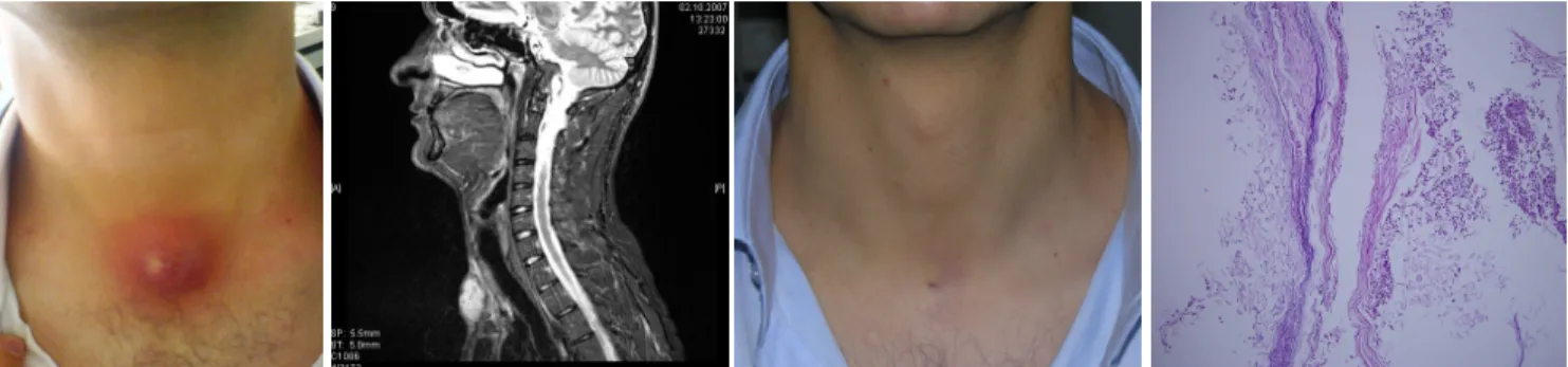
A 29-year-old man was admitted to our otorhinolaryngology clinic with the complaints of midline neck swelling with hyperemia and pain for 4 days. Although this swelling had been present for all his life, he had no complaint until it started to cause pain 4 days prior to his admittance. He was afebrile. Physical examination revealed a 3x3 cm midline neck mass in the infrahyoid region with a smooth surface which was mobile and painful with palpation (Figure 1). No movement with swallowing or protrusion of the tongue could be demonstrated. His blood tests were unremarkable and serum HBs antigen , HIV, HCV,EBV,Toxoplasmosis IgM, IgG antibody results were negative. Magnetic resonance imaging of the neck indicated that the 2x4 cm cervical hypodence mass was lo-cated in front of the anterior cervical muscles and above the incisura of sternum. The mass also presented centrally cystic- necrotic component and ring enhancement by contrast ( Figure 2). he patient underwent abscess drainage and the drainage sample was examined with both microbiologic and pathologic analysis. The patient’s course after drain-age was uneventful. Oral prophylactic antibiotherapy was administered empirically after drainage (Claritromycin 500 mg 2x1, orally). After mi-crobiological culture analysis, Morganella Morganii was found to be the pathogen microorganism and oral antibiothearapy was not changed. After the treatment of 10 days, there was a substantial regression of the neck lesion, without a recurrence after a one month follow up period (Figure 3). Pathologic analysis revealed an inflamed epidermoid kerati-nous cyst with an active inflammation (Figure 4).
Discussion
The differential diagnosis in a patient presenting with a neck mass is broad and extensive. The following conditions can result in midline swelling of the neck: Thyroglossal duct cyst, epidermoid cyst, dermoid cyst, submental lenfadenitis, submental abscess, thyroid gland tumors, ectopic thyroid [1]. Therefore, a thorough history and physical examina-tion make up the critical first step in the evaluaexamina-tion of a neck mass. Information gathered from a detailed history and physical examination alone often narrows the differential diagnosis to a more manageable level [2].
Imaging studies provide useful information in diagnosing the etiology of a neck mass. Computed Tomography (CT) and Magnetic Resonance Imaging (MRI) can differentiate solid, cystic, and vascular masses; lo-calize a mass in relation to the vital structures of the neck; and identify a potential head and neck source for the neck mass. Ultrasonography may be helpful in distinguishing solid from cystic masses, especially in the setting of a suspected thyroid lesion [2]. Fine-needle aspiration and/or surgical biopsy will establish the definitive diagnosis and dictate the final course of treatment [1]. Epidermal cyst have been known for many years as a sebaceous cyst, a misnomer born of a mistaken gross interpretation of the cyst content and perpetuated by uncritical repe-tion [3]. Epidermal cysts are a course of neck masses and historically, two essential types have been identified . The most common type is known as an epidermoid or epidermal inclusion cyst. This represents approximately 90% of keratinous cysts. The second most common type
is termed a pilar or trichilemmal cyst. Cornified epithelium, a very well-demarcated granular layer, and multiple lamellae of keratin without calcification characterize the epidermoid cysts. Additionally, focal cal-cification is common [3].
These lesions can be found in almost any location. They are frequently found on the scalp, ears, face, back, or scrotum.The majority of these lesions in the head and neck (65%) occur in orbital and nasal regions, whereas 24% arise in the oral cavity [4]. Both CT and MRI may be used to evaluate patients with epidermoid cysts. Epidermoid cysts, though lined by squamous epithelium, do not contain the other cutaneous ap-pendages. Epidermoids typically are unilocular cysts, with in a thin wall and contents matching characteristics of water on all MR pulse sequences [4].
On palpation, the cystic mass is firm, globular, movable, and non tender; it seldom causes discomfort unless it has ruptured internally, causing a rapidly enlarging, painful abscess. Keratinous cysts, the most common, are often surmounted with a punctum or pore; their contents are cheesy and often fetid, due to secondary bacterial colonization.
Both bacterial and viral infections can cause inflammatory neck mass-es. Occasionally, the lymph nodes become necrotic, and form abscess. Staphylococcus and Streptococcus species are the organism most com-monly cultured from neck abscess. In many instances, however, the infection is polymicrobial. A neck abscess usually requires antibiotic therapy, and surgical drainage may be necessary [5].
Morganella Morganii is a gram negative, facultative anaerob rod clas-sified within the Enterobacteriaceae family. This bacteria are usually found in the environment and in the intestinal tracts of humans, mam-mals, birds and reptiles as normal flora [6].
Altough nosocomial epidemics with Morganella Morganii have been reported previously in the setting of urinary tract infection, postopera-tive wound infection, or bacteremia [7], we did not find neck abscess infected with Morganella Morganii in literature.
This case report indicates that clinicians should be aware that Morgan-ella Morganii can be one of the causes of neck abscess. Appropriate antibiotic treatment and surgical drainage are required for these infec-tions. Practitioners shouldn’t forget that a wide bacterial spectrum may be a reason for neck abscess and bacterial culture and antibiogram take an important role for the treatment of the neck abscess.
The choice of the proper antibiotic depends on the antimicrobial sus-ceptibility of the etiologic agent that is obtained by culture and sensi-tivity results. Needle aspiration of an acutely infected cyst early in the course of treatment is often helpful.
References
1. Giglio JA, Laskin DM. Estabilshing the differantial diagnosis of neck lesions. Quintessence Int 2000; 31: 609.
2. Anil K. Lalwani. Current Diagnosis & Treatment in Otolaryngologology Head & Neck Surgery In: Anil K. Lalwani, editor. 2nd ed. United States of America: McGraw Hill; 2004 , Neck Masses p. 413-25.
3. Juan Rosai. Rosai and Ackerman’s Surgical Pathology In: 9th ed. London, Mosby; 2004, p. 151.
4. Gujar S, Gandhi D, Mukherji SK. Pediatric Head and Neck Masses. Top Magn Reson Imaging. 2004;15:95
5. Schwetschenau E, Kelley D J. The adult neck mass. Am Fam Physician. 2002 ;66:831.
6. Abdalla J, Saad M, Samnani I, Lee P, Moorman J. Central Nervous System infec-tion caused by
Morganella Morganii. Am J Med Sci 2006; 331: 44.
7. O’Hara CM, Brenner FW, Miller JM. Classification, identification and clinical sig-nificance of
Proteus, Providencia, Morganella. Clin Microbiol Rev 2000; 13: 534.
Figure 4. Keratinous cyst of epidermal type with secondary inflamation.
Figure 1. Inflamed midline neck mass, before the treatment.
Figure 2. Magnetic resonance imagining, sagittal view of lesion.
Figure 3. Neck region, after the treatment.
Journal of Clinical and Analytical Medicine | 453 Epidermal Kist / Epidermal Cyst