The protective effects of propolis and flunixin meglumine on feed
intake, antioxidant status and histological parameters in liver and
kidney tissues against excess copper in rats
Pınar TATLI SEVEN
1, Burcu GÜL BAYKALIR
2, Tuba PARLAK AK
3, İsmail SEVEN
4,
Neşe BAŞAK
5, Mine YAMAN
61University of Fırat, Faculty of Veterinary Medicine, Department of Animal Nutrition & Nutritional Diseases; 2Faculty of Health Sciences, Elazig; 3University of Munzur, Health High School, Department of Nutrition and Dietetic, Tunceli; 4University of Fırat, Vocational School of Sivrice, Department of Plant and Animal Production, Elazig; 5University of İnonü, Faculty of Pharmacy, Department of Pharmaceutical Toxicology, Malatya; 6University of Fırat, Faculty of Veterinary Medicine, Department of Histology
and Embryology, Elazig, Turkey.
Summary: This experiment was conducted to determine the protective effects of flunixin meglumine and propolis on feed
intake, antioxidant status and histological parameters in kidney and liver tissues in rats exposed to excessive copper. In this study individually housed thirty-six male Sprague-Dawley rats were used. Animals were randomly divided into six groups; control, copper sulphate (500 mg/kg BW/day, gavage), flunixin meglumine (2.2 mg/kg BW/day, ip), propolis (100 mg/kg BW/day, gavage), copper sulphate+flunixin meglumine (500 mg/kg BW/day of copper sulphate by gavage and 2.2 mg/kg BW/day of flunixin meglumine, ip) and copper sulphate+propolis (500 mg/kg BW/day of copper sulphate and 100 mg/kg BW/day of propolis by gavage). The study demonstrated that body weight change in the copper sulphate+propolis group significantly ameliorated in comparison with copper group (P<0.01). Propolis and flunixin meglumine administration significantly decreased MDA levels in the kidney and liver tissues, and serum TNF-α levels (P<0.001). Propolis supplementation to rats who were also treated with copper significantly increased the superoxide dismutase, catalase and reduced glutathione activities (P<0.001). Flunixin meglumine and propolis treatments decreased the copper-induced degenerative and necrotic changes with the apoptotic cells in the liver and kidney tissues. In conclusion, propolis appeared to ameliorate the adverse effects on feed intake, liver and kidney tissues seeing in the copper treatment and, apparently caused by the copper toxicity, by scavenging the free radicals and increasing activity of antioxidants.
Keywords: Antioxidant status, copper, flunixin meglumine, histological changes, propolis.
Ratlarda bakır fazlalığına karşı yem tüketimi, antioksidan durum ve karaciğer ile böbrek dokularının
histolojik parametreleri üzerine propolis ve fluniksin meglümin'in koruyucu etkileri
Özet: Bu araştırma, aşırı bakıra maruz bırakılmış sıçanlarda propolis ve fluniksin meglümin’in, yem tüketimi, antioksidan durum
ile karaciğer ve böbrek dokularındaki histolojik parametreler üzerindeki koruyucu etkilerini belirlemek amacıyla yapıldı. Araştırmada bireysel olarak barındırılan 36 adet erkek Sprague-Dawley sıçan kullanıldı. Hayvanlar rastgele; kontrol, bakır sülfat (500 mg/kg BW/gün, gavaj), fluniksin meglümin (2,2 mg/kg BW/gün, ip), propolis (100 mg/kg BW/gün, gavaj), bakır sülfat+fluniksin meglümin (500 mg/kg BW/gün bakır sülfat, gavaj ve 2,2 mg/kg BW/gün fluniksin meglümin, ip) ve bakır sülfat+propolis (500 mg/kg BW/gün bakır sülfat ve 100 mg/kg BW/gün propolis, gavaj) olarak altı gruba ayrıldı. Çalışma, bakır+propolis grubunun vücut ağırlığındaki değişimin bakır grubu ile karşılaştırıldığında önemli ölçüde iyileştiğini göstermiştir (P<0.01). Propolis ve fluniksin meglümin uygulaması, karaciğer ve böbrek dokularındaki MDA düzeylerini ve serum TNF-α düzeylerini önemli ölçüde azalttı (P<0.001). Ayrıca, bakırla muamele edilen ratlara propolis takviyesi, süperoksit dismutaz, katalaz ve indirgenmiş glutatyon aktivitelerini önemli ölçüde arttırdı (P<0.001). Fluniksin meglümin ve propolis tedavileri karaciğer ve böbrek dokularındaki bakır kaynaklı dejeneratif ve nekrotik değişiklikler ile apoptotik hücreleri azalttı. Sonuç olarak, propolisin serbest radikalleri bulunduğu yerden temizleyerek ve antioksidanların aktivitesini arttırarak görünüşte bakır toksisitesinin neden olduğu bakır uygulamasında ortaya çıkan yem tüketimi ve karaciğer ve böbrek dokuları üzerindeki olumsuz etkileri hafiflettiği ortaya çıkmıştır.
Introduction
Copper (Cu) is an important trace element for normal development and growth. It is a complementary part of specialized cuproproteins such as superoxide dismutase (11, 18). It promotes the function of some cellular enzymes. Cu ions can embrance different redox states permitting the metal to play a basic role in cell physiology in the redox chemistry of enzymes, elastin cross-linking and free radical scavenging (37). On the other hand, the accumulation of Cu in amounts that exceed requirements can lead its toxic effects (12). Also, the redox identity of copper, a redox-active metal, contributes to potential toxicity of Cu (1). Cu can induce oxidative stress by increasing production of reactive oxygen species (ROS) (15, 29). The primary affected organ in the copper-induced toxicity is liver. The excessive backlog of Cu might cause renal dysfunction and nephrotoxicity in rats (5).
Generally, nonsteroidal anti-inflammatory drugs (NSAIDs) are used for decreasing fever, pain and inflammation. One of the NSAIDs that was used widely by veterinarians and doctors is flunixin meglumine. It was reported that NSAIDs have the capacity to fight with free radicals by their strong antioxidant effects (29). On the other hand, the use of NSAIDs has considerable adverse effects including mortality and morbidity (27).
Flavonoids with variable phenolic structures are natural compounds found in plants. Flavonoids have health-promoting properties as a dietary component of animal and human, which owe to their high antioxidant capacity (9, 33). Functional hydroxyl groups in flavonoids are responsible for antioxidant effects by chelating metal ions and/or by scavenging free radicals (20, 21). Propolis, collected from the leaves, buds and similar parts of plants like eucalyptus and chestnut by bees, is an adhesive balsam that smells like resin (40). Propolis has been used extensively as a food additive for many purposes such as improving health and preventing diseases like diabetes, inflammation and even cancer (26). Flavonoids in the propolis are one of the compounds responsible for many activities including cancer, inflammatory, anti-microbial and anti-oxidant activities (40). Recently, propolis has extensively begun to attract the attention of scientists. According to results of the studies, propolis may reverse the adverse effects of oxidative stress on the defense system of body (35, 40).
The present study was designed to investigate the effects of flunixin meglumine when it is used as an anti-inflammatory for the treatment of many diseases and of propolis having the flavonoid structure, on the oxidative stress induced by Cu in an animal model using male rats. Histopathological studies were also carried out to assess the effects of flunixin meglumine and propolis on Cu damage to the rats’ liver and kidneys.
Materials and Methods
Drugs: Copper sulphate was purchased from the
SIGMA. Propolis was purchased from Ari Dunyasi Firm, Istanbul, Turkey. Flunixin meglumine was purchased from a commercial firm (50 mg/ml, Vilsan).
Animals, diet, and treatment: In this study, we used,
thirty-six adult male and healthy Sprague Dawley rats (250-300g, 6-8 wks old). The rats were provided from the Experimental Research Centre of the Firat University. They were housed under standard laboratory conditions in stainless steel cages (40-60% humidity, 24±3°C, 12h dark/light cycle). Standard commercial pellet food, containing 2.650 kcal/kg metabolic energy and 23% crude protein (Elazig Food Company, Elazig, Turkey), and fresh water were given as ad libitum. This study was approved by the Animal Experiments Local Ethics Committee (12.31.2014/No:240). The rats were randomly divided into six groups and were housed in individual cages. Over a period of twenty one days, rats in the first group served as control, and those in the second group (Cu) were given copper sulphate 500 mg/kg BW/day by gavage, group 3 (FM) were given flunixin meglumine 2.2 mg/kg BW/day by ip, group 4 (P) were given propolis 100 mg/kg BW/day by gavage, group 5 (Cu+FM) were given 500 mg/kg BW/day of copper sulphate by gavage and 2.2 mg/kg BW/day of flunixin meglumine as ip, group 6 (Cu+P) were given 500 mg/kg BW/day of copper sulphate and 100 mg/kg BW/day of propolis by gavage. Rats were individually weighed at the start of the study and then they were continued to weigh weekly to monitor BW. Additionally, feed intake (FI) and body weight change (BWC) were determined on the 7th, 14th, and 21st days of the experiment. Copper sulphate dose was determined based on previous studies (24, 42). It was given orally with a dosage of 500 mg/kg daily for 21-day and subacute toxicity was formed in the rats given copper sulphate.
Sample collection: 24 hours after the last
administration, rats were decapitated under anesthesia (light inhalation of diethyl ether). From each rat 1.5ml blood sample was collected for identification of serum tumor necrosis factor alpha levels (TNF-α). The liver and kidney tissues were removed for lipid peroxidation (LPO), superoxide dismutase (SOD), catalase (CAT), reduced glutathione (GSH) and histological analysis. The samples were stored at -20°C until analysis.
Extraction procedure of propolis: Chromatographic
analysis was carried out by Prevail C18 reversed-phase column (Grace Davison) (15×4.6mm, 5mm, USA); the mobile phase was methanol/water/acetonitrile (46/46/8, v/v/v), containing 1% acetic acid (43). Quantification was accomplished using by using the integration of the peak external standard methods. Chromatographic procedures were carried out at 25°C.
Analysis of total phenols: The total phenols in
propolis was determined using the method of Velioglu et al. (41). To 0.1ml of each sample were added 0.75ml 0.1 N Folin-Ciocalteau reagent and 0.75 ml Na2CO3 (6%, w/v). This procedure was replicated for three times. Absorbance of the samples after one and a half hours was spectrophotometrically measured at 725 nm. Results were given as mg Gallic Acid Equivalent (GAE)/g fresh weight sample in Table 1.
Analysis of total flavonoids: The total flavonoids
were determined with the colorimetric method (17). The mixture of 1 ml of extract with 0.3 ml of NaNO2 (5%) at t= 0 minute, then 0.3 ml AlCl3(10%) was added at t= 5 minute in the mixture. 2 ml from 1 N NaOH was added in to the solution after 6 minute and mixed. The absorbance was determined at 510 nm against a prepared water blank. Total flavonoid contents were expressed as mg Quercetin Equivalents (QE)/g fresh weight sample in the Table 1.
Analysis of total antioxidant capacity–CUPRAC method: The CUPRAC (cupric reducing antioxidant
capacity) procedure was utilized according to the method of Apak et al. (4). Firstly, in a test tube 1 ml CuCl2 (0.01M), 1 ml neocuproine (0.0075M), and 1 ml NH4Ac buffer (neutral pH) were mixed. Then, 0.1 ml of the sample extract or Trolox was added to this mixture. Lastly, to make the final volume 4.1 ml was added 1 ml from Milli-Q water. The absorbance after 1hour reaction time was measured at 450 nm. The results were given as mg Trolox Equivalent Antioxidant Capacity (TEAC)/g fresh weight sample in the Table 1.
Table 1. The total phenolic content, flavonoid content, and antioxidant capacity values of propolis
Tablo 1. Propolisin toplam fenolik içeriği, flavonoid içeriği ve antioksidan kapasite değerleri
Amounts in 1 g Propolis* Total phenolics 139.1 ± 1.8 mg GAE Total flavonoids 397.6 ± 1.2 mg QE Total antioxidant capacity -
CUPRAC 494.5 ± 1.3 mg TEAC
*Values are given as mean ± standard deviation of the values found for three parallel.
Table 2. Phenolic substances and quantities defined in propolis Tablo 2. Propoliste tanımlanan fenolik maddeler ve miktarlar
Phenolics (mg/g) * Pinobanksin 8.9 ± 0.5 Pinostrobin 5.5 ± 0.3 Pinocembrin 3.4 ± 0.2 Chrysin 2.7 ± 0.1 Galangin 2.2 ± 0.2 Apigenin 1.7 ± 0.1 Kaempferol 0.7 ± 0.0 Luteolin 0.6 ± 0.0
*Values are given as mean ± standard deviation of the values found for three parallel.
Determination of phenolic profile: Filtered extracts
were analyzed using a W600 Waters HPLC system coupled with a Waters 996 photodiode array (PDA) detector (3, 6). The compounds were separated using a C18 column, and applying a gradient from 95 to 25% Milli-Q water and a 5-75% acetonitrile, both in 0.1% trifluoroacetic acid across a fifty-minute period. Phenolics of propolis were detected at 280, 312, and 360 nm. In the quantification, dose-response curves of available were used pure standards (0-500 μg/ml) as reference (Table 2).
LPO: The LPO level of tissues was spectrophotometrically measured at 532 nm according to the concentrations of the Thiobarbituric Acid Reactive Substances (TBARS), and as an index of lipid peroxidation was used the amount of malondialdehyde (MDA) produced (30). 1,1,3,3 Tetraethoxypropane was used as a standard.
GSH: The levels of tissue GSH were measured
spectrophotometrically at 412 nm (10). The protein content of the tissues was measured according to the method of Lowry et al. (25). The levels of GSH were given as nmol/mg protein.
CAT: The CAT activities of kidney and liver were
determined by using the method of Aebi (2) that is based on the determination of the rate constant (k) for the decomposition rate of H2O2 at 240 nm. Results were given as k/g protein.
SOD: Measurements in the tissues were performed
according to the method of Sun et al. (36) that is based on the reduction of xanthine oxidase which produces nitro blue tetrazolium. The product was spectrophotometrically evaluated at 560 nm, and the results were expressed as percent inhibition per mg protein.
Serum TNF-α: Serum TNF-α levels were determined
by enzyme-linked immune-sorbent assay (ELISA, Cat. No, E-EL-R0019) using kits. The plates were read using the automated triturus enzyme immunoassay analyzer (Grifols, Miami, FL) at 450 nm. The cytokine amounts were calculated using linear regression method from standard curves of recombinant cytokines in the samples.
Histopathological and immunohistochemical (IHC) analyses: For histological examinations, the liver and
kidney tissues were fixed in formalin solution (10%), dehydrated in graduated alcohol series, and buried in paraffin. The tissue samples in paraffin were cut into 5 μm thickness, mounted on 1/4 shaven slides and stained with hematoxylin-eosin for histological examination according to the standard procedure (34). The cells which undergo apoptosis in the tissues were evaluated immunohistochemically by using caspase method. For analysis, the sections were mounted on polylysine-coated slides. Following deparaffinization and rehydration of the samples, they were transferred to citrate buffer (pH 7.6) and were heated in a microwave oven for 20 min, were
cooled for 20 min at room temperature, and then were washed with PBS. The sections were immersed 0.3 % H2O2 for 5 min, were washed with PBS, and were incubated with Ultra V block for 5 min at room temperature. Subsequently, they were incubated with a primary rabbit-polyclonal active caspase-3 antibody (Thermo Scientific, CPP32, Ab-4, UK) for 1 h in humidified chamber at 37oC. The sections were then rinsed in PBS before incubation with biotinylated goat anti-polyvalent for 30 min at room temperature and were washed with PBS. Then they were incubated streptavidin peroxidase for 30 min at room temperature and were washed with PBS. Staining was completed after the substrate had been incubated with a 3,3’-diaminobenzidin tetrahidroklorid (DAB) chromogen (Thermo Scientific, Ultra Vision Detection System Anti-Polyvalent, HRP/DAB, UK) for 5-15 min and then the slides were washed distilled water for 5 min. The slides were counterstained with Mayer’s hematoxylin for 30 sec, rinsed in tap water for 5 min, dehydrated and mounted with aqueous mounting medium. The active caspase-3 kit was used according to the manufacturer’s instructions. The routine H&E tissue sections and the active caspase-3 activities were examined in the liver and kidney tissues using a binocular-headed light microscope which have the feature of photomicroscope (Olympus BX- 51, Olympus Optical Co., Ltd., Tokyo, Japan) and photographs were taken. The active caspase-3 positive cells were stained in the tissue sections with a brown color and were evaluated by looking at the staining intensity (-; no, +; slight, ++; moderate, +++; strong) with a semiquantitative scoring system.
Statistical analysis: Results are presented as
mean±standard deviation. Using the one-way analysis of variance and, post-hoc Duncan test, statistically differences of group means were determined (SPSS Statistics 21).
Results
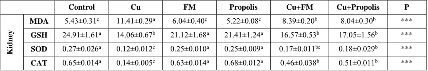
In this study, BW and FI of the rats treated with Cu decreased statistically in comparison with the Control group (P<0.001) (Table 3). The present study demonstrated that BW and BWC numerically increased when flunixin meglumine and propolis supplementation was provided to the rats treated with Cu. Also, BWC of Cu+P group significantly ameliorated in comparison with the Group Cu (P<0.01). Flunixin meglumine administration (Cu+FM) did not significantly attenuate BWC and FI of the rats treated with Cu, while propolis administration significantly increased (P<0.001) (Table 3). Propolis and flunixin meglumine administration significantly decreased MDA levels in the kidney and liver (P<0.001). Significant increase in MDA as a result of excessive Cu indicates oxidative stress in both tissues (Table 4 and 5) (P<0.001). GSH levels, CAT and SOD activities in tissues were significantly lower than those in the control, flunixin meglumine and propolis groups (P<0.001). Serum TNF-α level of the examined rats are given in Table 6. Serum TNF-α levels of the Cu group were significantly higher (P<0.001) than those of other groups. Histopathological and immunohistochemical results in the kidney and liver tissues were showed in the Figure 1-4.
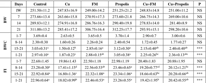
Table 3. Effects of propolis on body weight (BW), body weight change (BWC) and feed intake (FI) of experimental groups (g) Tablo 3. Deney gruplarının vücut ağırlığı (BW), vücut ağırlığı değişimi (BWC) ve yem tüketimi (FI) üzerine propolisin etkileri (g)
Days Control Cu FM Propolis Cu+FM Cu+Propolis P
BW IW 251.50±11.2 247.83±16.9 249.00±14.2 251.25±21.2 248.83±14.8 251.00±11.2 NS 7 273.66±13.4 263.66±15.8 270.91±17.3 273.60±21.8 266.75±14.3 269.00±10.6 NS 14 289.83±12.1 274.91±16.8 286.76±16.3 290.40±19.8 278.83±14.0 281.40±8.9 NS 21 311.00±13.2 285.41±17.2 306.75±16.4 312.25±17.7 293.91±15.1 298.20±10.6 NS BWC 1-7 3.69±0.4 2.63±0.5 3.65±0.5 3.70±1.4 2.90±0.7 3.00±0.6 NS 8-14 2.30±0.30 1.60±0.26 2.26±0.24 2.40±0.54 1.72±0.45 1.77±0.27 NS 15-21 3.03±0.31a 1.50±0.12b 2.85±0.16a 3.12±0.30a 2.15±0.40ab 2.40±0.40ab *** 1-21 2.97±0.10a 1.87±0.23c 2.88±0.15ab 3.05±0.38a 2.25±0.26bc 2.36±0.13abc *** FI 1-7 22.68±1.45 19.86±1.43 22.50±1.18 22.98±1.19 20.40±1.83 20.88±1.95 NS 8-14 23.28±0.30a 17.41±1.15d 22.56±0.53ab 23.46±0.65a 19.20±0.77cd 20.12±1.21bc *** 15-21 22.92±0.84a 16.80±1.36c 22.32±1.08a 23.34±1.06a 18.66±0.63bc 20.28±0.66ab *** 1-21 22.96±0.64a 18.02±0.99b 22.46±0.53a 23.26±0.55a 19.42±1.95b 20.42±0.55ab *** FM: Flunixin meglumine; IW: Initial weight; a,b,c,d: Mean values with different superscripts within a row differ significantly; NS: Non significant; ***: P<0.001
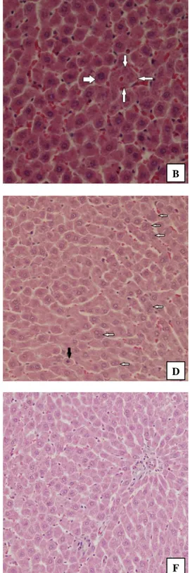
Figure1. Histopathological changes in the liver tissues of the experimental rats. (A) Copper treatment alone; degenerative and necrotic cells (arrows) (H&E staining, Magnification x100), (B) Copper treatment alone; karyomegaly in hepatocyte (thick arrow) and focal
necrosis areas with karyolithic nucleus (thin arrows) (H&E staining, Magnification x200), (C) Copper+flunixin meglumine treatments;
many hepatocytes with double nucleus (arrows) (H&E staining, Magnification x100), (D) Copper+propolis treatments; double nucleus in many hepatocytes (arrows) and hepatocyte with mitotic figure (black arrow) (H&E staining, Magnification x100), (E) Flunixin meglumine treatment alone; normal morphology (H&E staining, Magnification x100), (F) Propolis treatment alone; normal morpohology (H&E staining, Magnification x100).
Şekil 1. Deney ratlarının karaciğer dokularındaki histopatolojik değişiklikler. (A) Sadece bakır tedavisi; dejeneratif ve nekrotik hücreler (oklar) (H&E boyama, Büyütme x100), (B) Sadece bakır tedavisi; hepatositte karyomegali (kalın ok) ve karyolitik nükleuslu fokal nekroz alanları (ince oklar) (H&E boyama, Büyütme x200), (C) Bakır+fluniksin meglümin tedavisi; bir çok çift nükleuslu hepatosit (oklar) (H&E boyama, Büyütme x100), (D) Bakır+propolis tedavisi; bir çok hepatositte çift nükleus (oklar) ve mitotik figürlü hepatosit (siyah ok) (H&E boyama, Büyütme x100), (E) Sadece fluniksin meglümin tedavisi; normal morfoloji (H&E boyama, Büyütme x100), (F) Sadece propolis tedavisi; normal morfoloji (H&E boyama, Büyütme x100).
A B
C D
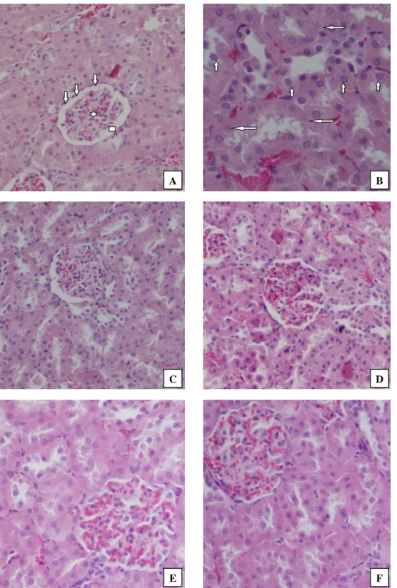
Figure 2. Histopathological changes in the kidney tissues of the experimental rats. (A) Copper treatment alone; shrinkage and congestion in glomeruli (star), thickening in basal membrane of glomeruli (arrows), dilatation and degeneration in Bowman’s capsule (square) (H&E staining, Magnification x100), (B) Copper treatment alone; karyomegaly (small arrows) and karyolysis (big arrows) in epithelial cells of proximal tubules (H&E staining, Magnification x200), (C) Copper+Flunixin meglumine treatments; the decreased degenerative and necrotic changes (H&E staining, Magnification x100), (D) Copper+Propolis treatments; the decreased degenerative and necrotic changes (H&E staining, Magnification x100), (E) Flunixin meglumine treatment alone; normal morphology (H&E staining, Magnification x200), (F) Propolis treatment alone; normal morphology (H&E staining, Magnification x200).
Şekil 2. Deney ratlarının böbrek dokularındaki histopatolojik değişiklikler. (A) Sadece bakır tedavisi; glomerulusta büzülme ve konjesyon (yıldız), glomerulusun bazal membranda kalınlaşma (oklar), Bowman kapsülünde dilatasyon ve dejenerasyon (kare) (H&E boyama, Büyütme x100), (B) Sadece bakır tedavisi; proksimal tübüllerin epitelyal hücrelerinde karyomegali (küçük oklar) ve karyolizis (büyük oklar) (H&E boyama, Büyütme x200), (C) Bakır+fluniksin meglümin tedavisi; azalmış dejeneratif ve nekrotik değişiklikler (H&E boyama, Büyütme x100), (D) Bakır+propolis tedavisi; azalmış dejeneratif ve nekrotik değişiklikler (H&E boyama, Büyütme x100), (E) Sadece Fluniksin meglümin tedavisi; normal morfoloji (H&E boyama, Büyütme x200), (F) Sadece Propolis tedavisi; normal morfoloji (H&E boyama, Büyütme x200).
A B
C D
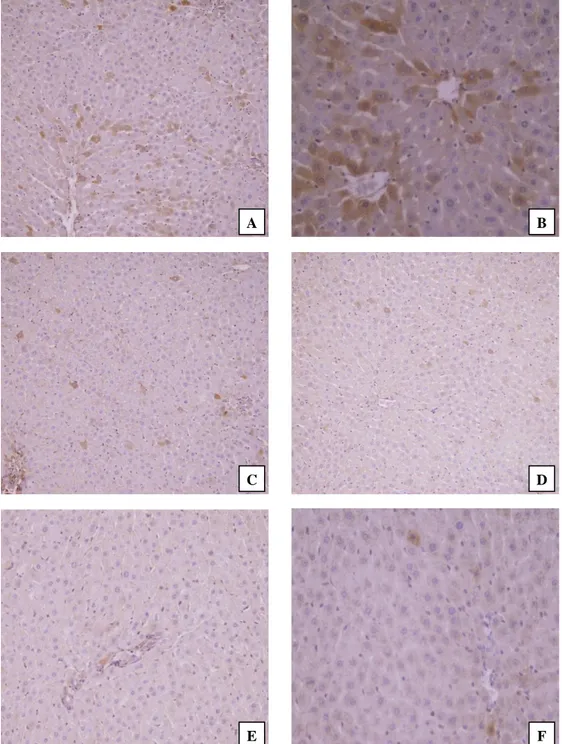
Figure 3. Immunohistochemical findings in the liver tissues of rats. (A) Copper treatment alone; many apoptotic cells showing strong immunoreaction for active caspase-3 with predominantly cytoplasmic and some nuclear locations (IHC, Mayer’s hematoxylin counterstain, Magnification x100), (B) Copper treatment alone; apoptotic cells showing strong immunoreaction for active caspase-3 (IHC, Mayer’s hematoxylin counterstain, Magnification x200), (C) Copper+flunixin meglumine treatments; decreased numbers of strong immunopositive cells for active caspase-3 with the moderate staining intensities (IHC, Mayer’s hematoxylin counterstain, Magnification x100), (D) Copper+propolis treatments; decreased numbers of active caspase-3 immunopositive cells with moderate the staining intensities (IHC, Mayer’s hematoxylin counterstain, Magnification x100), (E) Flunixin meglumine treatment alone; no immunoreactivity for active caspase-3 antibody (IHC, Mayer’s hematoxylin counterstain, Magnification x100), (F) Propolis treatment alone; a few active caspase-3 positive apoptotic cells (IHC, Mayer’s hematoxylin counterstain, Magnification x200).
Şekil 3. Ratların karaciğer dokularındaki immunohistokimyasal bulgular. (A) Sadece bakır tedavisi; baskın olarak sitoplazmik ve bazı nükleer lokasyonlarda aktif kaspaz-3 için güçlü immünoreaksiyon gösteren birçok apoptotik hücre (İHK, Mayer’s hematoksilen zıt boyama, Büyütme x100), (B) Sadece Bakır tedavisi; aktif kaspaz-3 için güçlü immünoreaksiyon gösteren apoptotik hücreler (İHK, Mayer’s hematoksilen zıt boyama, Büyütme x200), (C) Bakır+fluniksin meglümin tedavisi; aktif kaspaz-3 için orta düzeyde boyanma yoğunluğu ile güçlü immünopozitif hücrelerin sayısının azalması (İHK, Mayer’s hematoksilen zıt boyama, Büyütme x100), (D) Bakır+propolis tedavisi; orta derecede boyanma yoğunlukları ile azalmış sayıda aktif kaspaz-3 immünopozitif hücreler (İHK, Mayer’s hematoksilen zıt boyama, Büyütme x100), (E) Sadece Fluniksin meglümin tedavisi; aktif kaspaz-3 antikoru için immünoreaktivite yok (İHK, Mayer’s hematoksilen zıt boyama, Büyütme x100), (F) Sadece Propolis tedavisi; birkaç aktif kaspaz-3 pozitif apoptotik hücre (İHK, Mayer’s hematoksilen zıt boyama, Büyütme x200).
A B
C D
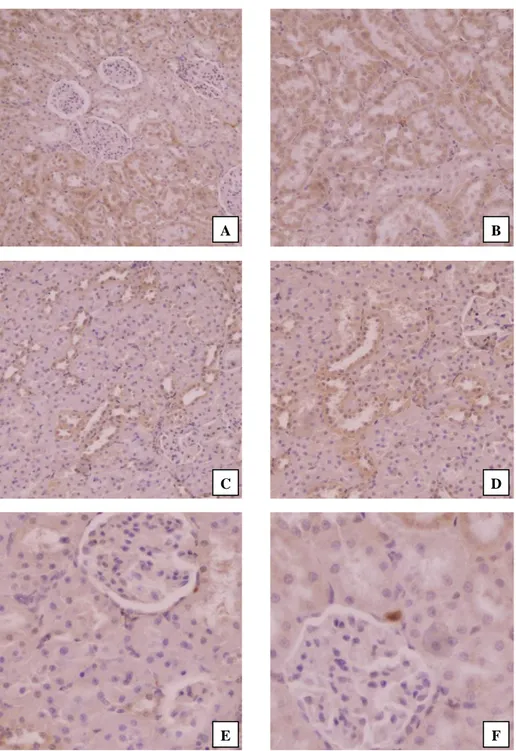
Figure 4. Immunohistochemical findings in the kidney tissues of rats. (A) Copper treatment alone; strong immunoreaction for active caspase-3 in tubular epithelial cells with mostly cytoplasmic and lesser nuclear locations (IHC, Mayer’s hematoxylin counterstain, Magnification x100), (B) Copper treatment alone; tubular cells showing strong immunoreaction for active caspase-3 (IHC, Mayer’s hematoxylin counterstain, Magnification x100), (C) Copper+flunixin meglumine treatments; decreased numbers of cells showing strong positive immunoreaction in mostly proximal and minority distal tubules with moderate the staining intensities (IHC, Mayer’s hematoxylin counterstain, Magnification x100), (D) Copper+propolis treatments; decreased numbers of active caspase-3 immunopositive tubular cells with moderate staining intensities (IHC, Mayer’s hematoxylin counterstain, Magnification x100), (E) Flunixin meglumine treatment alone; no immunoreactivity for caspase-3 antibody (IHC, Mayer’s hematoxylin counterstain, Magnification x200), (F) Propolis treatment alone; no immunoreactivity for caspase-3 antibody (IHC, Mayer’s hematoxylin counterstain, Magnificationx200).
Şekil 4. Ratların böbrek dokularındaki immunohistokimyasal bulgular. (A) Sadece bakır tedavisi; çoğunlukla sitoplazmik ve daha az nükleer lokasyonlara sahip tübüler epitel hücrelerinde aktif kaspaz-3 için güçlü immünoreaksiyon (İHK, Mayer’s hematoksilen zıt boyama, Büyütme x100), (B) Sadece Bakır tedavisi; aktif kaspaz-3 için güçlü immünoreaksiyon gösteren tübüler hücreler (İHK, Mayer’s hematoksilen zıt boyama, Büyütme x100), (C) Bakır+fluniksin meglümin tedavisi; çoğunlukla proksimal ve azınlıkla distal tübüllerde orta derecede boyanma yoğunlukları ile kuvvetli pozitif immünoreaksiyon gösteren azalmış sayıda hücre (İHK, Mayer’s hematoksilen zıt boyama, Büyütme x100), (D) Bakır+propolis tedavisi; orta derecede boyanma yoğunlukları ile aktif kaspaz-3 immünopozitif tübüler hücrelerin sayısındaki azalma (İHK, Mayer’s hematoksilen zıt boyama, Büyütme x100), (E) Sadece Fluniksin meglümin tedavisi; kaspaz-3 antikoru için immünoreaktivite yok (İHK, Mayer’s hematoksilen zıt boyama, Büyütme x200), (F) Sadece propolis tedavisi; kaspaz-3 antikoru için immünoreaktivite yok (İHK, Mayer’s hematoksilen zıt boyama, Büyütme x200).
A B
C D
Table 4. Effects of propolis and flunixin meglumine (FM) on MDA (nmol/ml homogenate), GSH (nmol/mg protein), SOD (% inhibition/ mg protein), and CAT (k/g protein) activities in liver of experimental groups
Tablo 4. Deneme gruplarının karaciğerlerinde propolis ve fluniksin meglüminin (FM) MDA (nmol/ml homojenat), GSH (nmol/mg protein), SOD (% inhibisyon/ mg protein) ve CAT (k/g protein) aktiviteleri üzerine etkileri
Control Cu FM Propolis Cu+FM Cu+Propolis P
Liv er MDA 2.30±0.13c 3.80±0.14a 2.30±0.15c 2.28±0.11c 2.84±0.15b 2.49±0.19bc *** GSH 19.35±0.55a 9.83±0.26c 19.19±2.10a 20.57±1.60a 14.48±0.93b 13.48±0.58b *** SOD 0.23±0.008a 0.15±0.011c 0.22±0.011a 0.23±0.012a 0.18±0.004b 0.18±0.004b *** CAT 0.75±0.014a 0.18±0.009d 0.73±0.017a 0.74±0.017a 0.29±0.008c 0.33±0.009b *** a,b,c,d: Mean values with different superscripts within a row differ significantly; *** P<0.001
Table 5. Effects of propolis and flunixin meglumine (FM) on MDA (nmol/ml homogenate), GSH (nmol/mg protein), SOD (% inhibition/ mg protein), and CAT (k/g protein) activities in kidney of experimental groups
Tablo 5. Deneysel grupların böbreklerinde propolis ve fluniksin meglüminin (FM) MDA (nmol/ml homojenat), GSH (nmol/mg protein), SOD (% inhibisyon/ mg protein) ve CAT (k/g protein) aktiviteleri üzerine etkileri
Control Cu FM Propolis Cu+FM Cu+Propolis P
K id n ey MDA 5.43±0.31c 11.41±0.29a 6.04±0.40c 5.22±0.08c 8.39±0.20b 8.04±0.30b *** GSH 24.91±1.61a 14.06±0.67b 21.12±1.68a 21.41±1.24a 16.57±0.53b 17.05±1.56b *** SOD 0.27±0.026a 0.12±0.012c 0.25±0.010a 0.25±0.009a 0.17±0.011bc 0.18±0.029b *** CAT 0.65±0.014a 0.14±0.005c 0.63±0.014a 0.68±0.012a 0.46±0.038b 0.51±0.011b *** a,b,c: Mean values with different superscripts within a row differ significantly; *** P<0.001
Table 6. Effects of propolis and flunixin meglumine (FM) on serum TNF-α (pg/ml) levels of experimental groups Tablo 6. Deney guruplarının serum TNF-α (pg/ml) düzeyleri üzerine propolis ve fluniksin meglüminin (FM) etkileri
Control Cu FM Propolis Cu+FM Cu+Propolis P
TNF-α 105.42±3.10c 163.26±6.31a 113.87±5.05c 107.59±1.24c 132.33±3.11b 128.51±2.21b *** a,b,c: Mean values with different superscripts within a row differ significantly; *** P<0.001
Discussion and Conclusion
Cu, an essential trace element for life, may cause harmful effects when it is taken for long time and in an excessive amount (28). In the present study, BW and FI of the Cu administered rats decreased significantly in comparison with those of the control group for 21 days (P<0.001) (Table 3). The results of the study demonstrated that Cu intake with dose 500 mg/kg BW might have toxic effects from the 21th day. A previous study (19) determined that feeding with 200 ppm Cu increased growth rate (P<0.01) and FI (P<0.01) during the 5 week trials; 400 ppm Cu depressed growth and FI after week 2 in weanling crossbred pigs (n= 216 and 6.9 kg initially). In another study (7), it was found that the dietary Cu supplements at 400, 500, and 600 ppm significantly lowered FI and egg production in laying hens after 4 weeks (P<0.05). FI of Cu groups in that study (400, 500, and 600 ppm dietary Cu) was found as 109, 107, and 97.8 g/hen/day, respectively (P<0.004). These results are agreed with the findings of the present study. In animals which were steadily exposed to Cu body weight decreased
which was an indicator of poor health. Treatment with antioxidant compounds to rats would partially alleviate the negative effects of oxidative stress induced by Cu (28).
The study demonstrated that BW and BWC numerically increased when flunixin meglumine and propolis supplementations were provided rats treated with Cu. Furthermore, BWC of Cu+P group significantly ameliorated in comparison with that of Cu group (P<0.01). A similar study (35) found that BW of Cyclosporin-A (CsA)-treated animals was lower than that of the control group. The decrease in BW was caused by reduction in FI following CsA administration. These results agree with those of the present study. However, earlier studies have shown protective role of flavonoids against Cu toxicity (13, 28). Flunixin meglumine administration (Cu+FM) did not significantly attenuate BWC and FI of rats treated with Cu, while propolis administration significantly increased (P<0.001) (Table 3). Propolis has delicious substances like vanillin and honey (38). In this study, the reduction in BWC of the group Cu+P could relate to flavonoid content and appetizing characteristic of propolis (Table 1). It could
be also linked to the fact that flavonoids show characteristics of antioxidants, which are also chelating with trace elements or radicals (39).
Excessive Cu in the diet of rats may cause damage of the membrane and LPO. Mladenović et al. (28) reported that increased LPO after incubation of erythrocyte suspension with Cu2+ ions. The Cu+ ion was generated by the reduction of Cu2+ in the presence of O2•- which catalyzed the formation of hydroxyl radicals that readily enter further chemical reactions. In our study, excessive Cu significantly increased MDA which indicates oxidative stress in tissues (Table 4 and 5) (P<0.001). Propolis and flunixin meglumine administration significantly decreased MDA levels in both of tissues (P<0.001). Mladenović et al. (28) noticed that flavonoids can block LPO, which enables them to chelate metal ions or scavenge reactive oxygen species (ROS) as hydrogen- or electron- donating compounds. Thus, they can be curative in conditions that result from oxidative stress or increased metal concentrations (28). GSH levels, CAT and SOD activities in the liver and kidney tissues were found to be significantly lower than those of rats in the control group, flunixin meglumine and propolis groups (P<0.001) (Table 4 and 5). SOD, a cellular antioxidant, is one of the most important antioxidant enzymes. SOD reacts rapidly and as it is removed by CAT it converts O2- into H2O2. It has been reported that excessive Cu accumulation in the liver decreases SOD activity and causes increased MDA levels in serum and liver homogenates (31). Propolis and flunixin meglumine treatments partially ameliorate the Cu-induced imbalance in the oxidant–antioxidant systems of both liver and kidney tissues. Meek et al. (27) reported that flunixin meglumine has antioxidant properties. The cause of its ameliorative effects on the antioxidant status in cases of Cu toxicities can be connected to its antioxidant properties (27). In particular, in rats treated with Cu, propolis supplementation significantly increases SOD, CAT and GSH (P<0.001) (Table 4 and 5). Similarly, another study explained that the first mechanism of this particular effect of propolis may involve in scavenging of free radicals that cause LPO (14). The second mechanism could be its ability to prevent xanthine oxidase activity, which is known to generate free radicals. Propolis contains flavonoid and other phenolic compounds, which seem to be capable of scavenging free radicals and, also avoiding situations in which lipids -and other substances such as vitamin C- could be oxidized or destroyed as a result of oxidative damage (14).
Serum TNF-α level of the examined rats are given in Table 6. Serum TNF-α levels of the Cu group were significantly higher (P<0.001) than those of other groups. However, serum TNF-α levels were significantly lower in the Cu+FM and Cu+P groups (Table 6) (P<0.001). Ciftci et al. (8) suggested that serum TNF-α levels, was given
2,3,7,8-Tetrachlorodibenzo-p-dioxin (TCDD) group, were significantly higher (P<0.05) when compared to other groups on the fifteenth day. TNF-α levels of the Curcumin+TCDD Group (60.81 pg/ml) decreased significantly in comparison to the TCDD group (104.0 pg/ml). These results were similar to our results. Lee et al. (22) reported that TNF-α induces the activation of NF-κB. NF-κB has long been considered as a prototypical pro-inflammatory through TNF-α receptor-dependent signaling pathways. Flavonoids are widely used to inhibit the activation of NF-κB, and they inhibit the expression of pro-inflammatory genes in response to inflammatory mediators such as TNF-α. Propolis administration to rats treated with Cu decreased TNF-α level (Table 6). It may be due to the flavonoid content structure of propolis and anti-inflammatory properties of flunixin meglumine, which are associated with propolis and flunixin (22, 23).
The histopathological changes induced by Cu treatment include confirmed antioxidant status and TNF-α level. Likewise, Abu-Zinadah et al. (1) stated that the hydroxyl radical, which is produced through the biochemical reaction of excessive Cu, may be responsible for devastating cellular damage, including LPO. Cu excess causes damage to liver and kidney tissues. Babaknejad et al. (5) reported that Cu excess toxicity is closely correlated with renal dysfunction. They also stated that tubular necrosis and cellular pleomorphic were reported in rats that had received excessive amounts of Cu.
The livers and kidneys of the control rats and those of the rats treated with both flunixin meglumine alone and propolis alone showed no abnormalities. But the livers from the Cu-supplemented rats showed hepatocellular degeneration and necrosis. Karyolysis and karyomegaly were observed in several hepatocyte cells. Also, it was determined that vasocongestion in most sinusoids, mononuclear inflammatory cell infiltrations in periportal areas, hyperemia in some central veins, and disorganization in some hepatic cord areas were caused by Cu exposure. The flunixin meglumine and propolis treatments decreased the Cu-induced degenerative and necrotic changes. Also, double nucleus in many hepatocytes and a few hepatocytes with mitotic figure were observed (Figure 1). Besides, the kidneys from the Cu-treated rats demonstrated degeneration and necrosis mostly on the proximal and minority distal tubules in the cortex. Swelling, karyomegaly and karyolysis were observed in epithelial cells of the proximal tubules. Shrinkage, congestion, and thickening in the basal membrane of some glomerules located in the cortex were also observed. Dilatation and degeneration were seen in Bowman’s capsules. The flunixin meglumine and propolis treatments decreased tubular degeneration and necrosis and the majority of changes in the cortex had been caused by the Cu supplements and could be reasonably
counteracted with their supplements (Figure 2). When compared to the control, flunixin meglumine alone and propolis alone groups, apoptotic cells which show strong positive reaction in the hepatocytes mostly on the proximal and minority distal tubules was encountered by Cu exposure. But the flunixin meglumine and propolis treatments reduced the Cu-induced apoptotic cells and the staining intensities of these apoptotic cells were moderate (Figure 3 and 4) in the liver and kidney tissues. Rana (32) reported that ROS may be produced by the liver of the experimental animals when administrating Cu. There is some evidence that the liver toxicity may relate to the induction of oxidative damage and the resulting LPO. Lipid peroxidation of mitochondrial membranes of liver cells, rat hepatic mitochondria, membranes of hepatic lysosomes, and MDA in the liver of Cu-loaded animals have been observed in some studies (32). In another a study, apoptosis and necrosis were found in liver and kidney tissues in experimental Cu toxicity in rats which is similar to our study (16). Treatments of propolis and flunixin meglumine modulated the toxic effects of Cu in histopathological and immunohistochemical examinations of the liver and kidney tissues (Figures 1-4). The reason for this may simply be due to the strong antioxidant properties of propolis (40) and flunixin meglumine (27). We determined that propolis which is rich with antioxidants could be used to prevent the adverse effects in liver and kidney tissues resulting the Cu toxicity (40).
In conclusion, propolis that is natural bee product appeared to ameliorate adverse effects on food intake as well as on the liver and kidney tissues caused by the Cu toxicity by increasing antioxidant activities and scavenging the free radicals. Besides, propolis supplementation instead of flunixin meglumine, which is not a natural product, may be used as a good option to diminish the negative effects of Cu on rats. Propolis may be used for treatment of some diseases which cause necrosis in the liver and kidney. However, this subject needs to be further investigated.
Acknowledgement
We would like to thanks for analysis of propolis to Prof. Dr. Dilek BOYACIOGLU and Assoc. Prof. Dr. Esra CAPANOGLU GUVEN.
References
1. Abu-Zinadah OA, Hussein HK, Elshal MF, et al. (2012): Effect of excess dietary copper on proliferation and differentiation of the proerythroblasts and erythrocytes in rats. Afr J Biotechnol, 11, 6187-6191.
2. Aebi H (1984): Catalase in vitro methods in enzymology. In: Willam BJ, editor. Methods in enzymology. New York, USA: Academic Press; 1984. pp. 121-126.
3. Ahn MR, Kumazawa S, Usui Y (2007): Antioxidant activity and constituents of propolis collected in various areas of China. Food Chem, 101, 1383-1392.
4. Apak R, Guclu K, Ozyurek M, et al. (2004): Novel total antioxidantcapacity index for dietary polyphenols and vitamins C and E, using their cupric ion reducing capability in the presence of neocuproine: CUPRAC method. J Agric Food Chem, 52, 7970-7981.
5. Babaknejad N, Moshtaghie AA, Shahanipour K (2015): The toxicity of copper on serum parameters related to renal functions in male wistar rats. Zahedan J Res Med Sci, 15, 29-31.
6. Bino RJ, de Vos CHR, Lieberman M, et al. (2005): The light-hyperresponsive high pigment-2dg mutation of tomato: alterations in the fruit metabolome. New Phytol,
166, 427-438.
7. Chiou PWS, Chen KL, Yu Bi (1998): Effect of dietary organic arsenicals and cupric sulfate on copper toxicity, liver accumulation and residue in eggs and excreta of laying hens. Anim Feed Sci Tech, 73, 161-171.
8. Ciftci O, Tanyildizi S, Godekmerdan A (2010): Protective effect of curcumin on immune system and body weight gain on rats intoxicated with 2,3,7,8-Tetrachlorodibenzo-p-dioxin (TCDD). Immunopharmacol Immunotoxicol, 32, 99-104.
9. Cook NC, Samman S (1996). Flavonoids-chemistry, metabolism, cardioprotective effects, and dietary sources. J Nutr Biochem, 7, 66-76.
10. Ellman GL (1959): Tissue Sulfhydryl groups. Arch Biochem Biophys, 82, 70-77.
11. Ferenci P (2004): Pathophysiology and clinical features of wilson disease. Metab Brain Dis, 19, 229-239.
12. Fuentealba IC, Aburto EM (2003): Animal models of copper-associated liver disease. Comp Hepatol, 2, 5. 13. Gaetke LM, Chow CK (2003): Copper toxicity, oxidative
stress and antioxidant nutrients. Toxicology, 189, 147-163. 14. Gul Baykalir B, Tatli Seven P, Gur S, et al. (2016): The effects of propolis on sperm quality, reproductive organs and testicular antioxidant status of male rats treated with Cyclosporine-A. Anim Reprod, Belo Horizonte, 13, 105-111.
15. Halliwell B, Gutteridge JMC (2007): Oxygen is a toxic gas – an introduction to oxygen toxicity and reactive species. In: Halliwell B, Gutteridge JMC, editors. Free radicals in biology and medicine. New York, USA: Oxford University Press; 2007. pp. 1-29.
16. Kabak YB, Gülbahar MY (2013): Sıçanlarda deneysel bakır zehirlenmesinde karaciğer ve böbrek dokularında apoptozisin belirlenmesi. Ankara Üniv Vet Fak, 60, 39-45. 17. Kim D, Jeong SW, Lee CY (2003): Antioxidant capacity
of phenolic phytochemicals from various cultivars of plums. Food Chem, 81, 321-326.
18. Kodama H, Fujisawa C (2009): Copper metabolism and inherited copper transport disorders: molecular mechanisms, screening and treatment. Metallomics, 1, 42-52.
19. Kornegay ET, van Heugten PHG, Lindemann MD, et al. (1989): Effects of biotin and high copper levels on performance and immune response of weanling pigs. J Anim Sci, 67, 1471-1477.
20. Kumar S, Mishra A, Pandey AK (2013): Antioxidant mediated protective effect of Parthenium hysterophorus against oxidative damage using in vitro models. BMC Complement Altern Med, 13, 120.
21. Kumar S, Pandey AK (2013): Phenolic content, reducing power and membrane protective activities of Solanum xanthocarpum root extracts. Vegetos, 26, 301-307. 22. Lee S, Kim YJ, Kwon S (2009): Inhibitory effects of
flavonoids on TNF-α-induced IL-8 gene expression in HEK 293 cells. BMB reports, 42, 265-270.
23. Lim H, Jin JH, Park H, et al. (2011): New synthetic anti-inflammatory chrysin analog, 5,7-dihydroxy-8-(pyridine-4yl) flavone. Eur J Pharmacol, 670, 617-622.
24. Liu JY, Yang X, Sun XD, et al. (2016): Suppressive effects of copper sulfate accumulation on the spermatogenesis of rats. Biol Trace Elem Res, 174, 356-361.
25. Lowry OH, Rosebrough NJ, Farr AL, et al. (1951): Protein measurement with the folin phenol reagent. J Biol Chem, 193, 265-275.
26. Matsuno T (1995): A new clerodane diterpenoid isolated from propolis. Z Naturforsch, 50C, 93-97.
27. Meek IL, van de Laar MAFJ, Vonkeman HE (2010): Non-steroidal anti-inflammatory drugs: an overview of cardiovascular risks. Pharmaceuticals (Basel), 3, 2146-2162.
28. Mladenović JM, Paunović MG, Matić MM, et al. (2014): Copper-induced changes of lipid peroxidation and hemato-biochemical parameters in rat blood: protective role of flavonoids. Arch Biol Sci Belgrade, 66, 1271-1279. 29. Mouithys-Mickalad AML, Zheng SX, Deby-Dupont GP,
et al. (2000): In vitro study of the antioxidant properties of
non steroidal anti-inflammatory drugs by chemiluminescence and electron spin resonance (ESR). Free Radic Res, 33, 607-621.
30. Ohkawa H, Ohishi N, Yagi K (1979): Assay for lipid peroxides in animal tissues by thiobarbituric acid reaction. Anal Biochem, 95, 351-358.
31. Ozcelik D, Uzun H (2009): Copper intoxication; Antioxidant defenses and oxidative damage in rat brain. Biol Trace Elem Res, 127, 45-52.
32. Rana SVS (2008): Metals and apoptosis: Recent developments. J Trace Elem Med Bio, 22, 262-284. 33. Rice-Evans CA, Miller NJ, Bolwell PG, et al. (1995): The
relative antioxidant activities of plant-derived polyphenolic flavonoids. Free Radic Res, 22, 375-383.
34. Ross MH, Reith EJ, Romrell LJ (1989): Histology: a text and atlas. 2nd ed. Williams and Wilkins: Baltimore.
35. Seven I, Gul Baykalir B, Tatli Seven P, et al. (2014): The ameliorative effects of propolis against cyclosporine A induced hepatotoxicity and nephrotoxicity in rats. Kafkas Univ Vet Fak, 20, 641-648.
36. Sun Y, Oberley LW, Li YA (1988): Simple method for clinical assay of superoxide dismutase. Clin Chem, 34, 497-500.
37. Tapiero H, Townsend DM, Tew KD (2003): Trace elements in human physiology and pathology. Copper. Biomed Pharmacother, 57, 386-398.
38. Tatli Seven P (2008): The effects of dietary Turkish Propolis and vitamin C on performance, digestibility, egg production and egg quality in laying hens under different environmental temperatures. Asian-Aust J Anim Sci, 21, 1164-1170.
39. Tatli Seven P, Seven I, Yilmaz M, et al. (2008): The effects of Turkish Propolis on growth and carcass characteristics in broilers under heat stress. Anim Feed Sci Tech, 146, 137-148.
40. Tatli Seven P, Yilmaz S, Seven I, et al. (2012): Effects of propolis in animals exposed oxidative stress (Chapter 13). In: Lushchak VI, editor. Oxidative stress-environmental induction and dietary antioxidants. Rijeka, Croatia: InTech; 2012. pp. 267-288.
41. Velioglu YS, Mazza G, Gao L, et al. (1998): Antioxidant activity and total phenolics in selected fruits, vegetables, and grain products. J Agric Food Chem, 46, 4113-4117. 42. Zhang SS, Noordin MM, Rahman SO (2000): Effects of
copper overload on hepatic lipid peroxidation and antioxidant defense in rats. Vet Hum Toxicol, 42, 261-264. 43. Zu Y, Li C, Fu Y, et al. (2006): Simultaneous determination of catechin, rutin, quercetin kaempferol and isorhamnetin in the extract of sea buckthorn (Hippophae rhamnoides L.) leaves by RP-HPLC with DAD. J Pharma Biomed Anal, 41, 714-719.
Geliş tarihi: 28.05.2017 / Kabul tarihi: 28.11.2017
Address for correspondence:
Prof. Dr. Pınar TATLI SEVEN
University of Firat, Faculty of Veterinary Medicine, Department of Animal Nutrition & Nutritional Diseases, Elazig, Turkey.