Virulence genes of Shiga toxin-producing Escherichia coli O157:H7
strains isolated from calves and cattle
Yahya KUYUCUOĞLU1, Esra ŞEKER1, Cevdet UĞUZ2, Barış SAREYYÜPOĞLU3,
Selahattin KONAK1
1 Afyon Kocatepe University, Faculty of Veterinary Medicine, Department of Microbiology, 2Department of Medical Biology and Genetics-Afyonkarahisar, 3Ankara University, Faculty of Veterinary Medicine, Department of Microbiology- Ankara, Turkey.
Summary: The aim of this study was to detect E. coli O157:H7 serotype in the faeces samples collected from calves and cattle farms located of Afyonkarahisar province in Turkey and to determine the stx(1) (Shiga toxin 1) and stx(2) (Shiga toxin 2) virulence genes in the strains of E.coli O157:H7 by multiplex PCR (mPCR). In this study, E. coli O157:H7 was isolated from the 3.1% (14 out 457 faeces samples) of the calves and cattle examined, in particular 2.3% of the cattle, 2.6% of non-diarrhoeic calves and 10.6% of diarrhoeic calves were positive for E. coli O157:H7. The stx(1) and stx(2) genes were detected in 6 out of 14 (42.8%) DNA samples extracted from STEC O157:H7 strains. This study demonstrated of E. coli O157:H7 serotype in cattle and calves, which represent an important reservoir for strains that a potential risk for human infections.
Keywords: Calves, cattle, Escherichia coli O157:H7, mPCR, stx(1) and stx(2) genes
İnek ve buzağılardan izole edilen Şiga toksijenik Escherichia coli O157:H7 suşlarının bazı virülens genleri
Özet: Bu çalışmanın amacı, Afyonkarahisar’da bulunan çifliklerdeki inek ve buzağılardan toplanan dışkı örneklerinden E. coli O157:H7 serotiplerini izole etmek, stx(1) (Şiga toksin 1) ve stx(2) (Şiga toksin 2) virülens genlerini multipleks PCR (mPCR) ile belirlemektir. Bu çalışmada, inek ve buzağılardan alınan toplam 457 dışkı örneğinin 14 (%3.1)’ünden E. coli O157:H7 serotipi izole edildi. İzole edilen suşların %2.3’ü ineklerden, %2.6’sı sağlıklı buzağılardan, %10.6’sı ise ishalli buzağılardan elde edildi. Şigatoksijenik E. coli (STEC) O157:H7 suşlarından elde edilen 14 DNA örneğinin 6 (%42.8)’sında stx(1) ve stx(2) geni pozitif bulundu. Bu çalışma, inek ve buzağılardan izole edilen STEC O157:H7 serotiplerinin insan enfeksiyonları için önemli bir risk faktörü olabileceğini göstermektedir.
Anahtar sözcükler: Buzağı, Escherichia coli O157:H7, inek, mPCR, stx(1) ve stx(2) genleri
Introduction
It has been widely reported that Shiga toxin-producing Escherichia coli (STEC) is linked to life threatening human disease such as haemorrhagic colitis (HC) and the haemolytic uremic syndrome (HUS) (20, 27, 34). E. coli O157:H7 was first recognized as a human
pathogen in 1982 (35). As it was associated with consumption of undercooked 'hamburgers', it became known as 'the hamburger bug'. As it has subsequently been found that healthy cattle can harbour the bacterium, ruminants are now regarded as its main reservoir, though STEC O157:H7 has been isolated from other animal species such as sheep, pigs, geese, gulls and pet animals (17). Especially undercooked meat of bovine origin but also unpasteurized milk and other dairy products have been implicated in transmitting STEC O157:H7 to humans. (2, 9, 10, 19, 20, 22). Another route for acquiring the infection is direct transmission from cattle,
especially calves, for instance on 'open farms' where groups of children are welcome to visit. As the bacterium survives well in the environment, drinking water, vegetables irrigated with contaminated water, and public outdoor swimming pools have been mentioned as sources of community outbreaks (17).
E. coli O157:H7 has been shown to have several
virulence factors such as Shiga toxin 1 and 2 (encoded by stx(1) and stx(2) genes), enterohaemolysin (encoded by E-hlyA genes) and intimin (encoded by bacterial eaeA genes) (29). E. coli O157:H7 produced verotoxins are very similar to cytotoxins called stxs. However, stx(1) and stx(2) are encoded by different sets of genes. Therefore, stx(1) and stx(2) are different proteins but their active molecular structure and biological functions are identical to the other Shiga toxins (23). As mentioned above, the family of stxs has been classified into two prominent classes, stx(1) and stx(2). The stx(1) family is
very homogenous while stx(2) has several variants (16). Shiga toxins inhibit protein synthesis by inactivating ribosomal RNA and they induce mortality in host cells. It has been shown that several techniques including immunoassays, verocell assay, PCR, multiplex PCR could be used to detect Shiga toxins (2, 24, 27, 32, 33, 42).
The aim of this study was to detect E. coli O157:H7 serotypes in the faeces samples collected from calves and cattle in dairy cattle enterprises located in small villages and towns in Afyonkarahisar province in Turkey and to determine the stx(1) and stx(2) virulence genes in the strains of E.coli O157:H7 by mPCR.
Materials and Methods
Samples: Faeces samples were collected from 237 calves (38 of them having diarrhea) and 220 cattle with different ages in dairy cattle enterprises in villages, towns and counties of Afyonkarahisar province, Turkey. The sample collection sites and the number of samples are shown in Table 3 and the distribution of samples according to age shown in Table 2. The rectal samples were collected from the rectum and stored in disposable sterile plastic faeces sample container. Separate rectal gloves were used for each animal to avoid cross contamination. Samples were then placed in an ice–pack container and immediately transported to the laboratory. Table 2. Distribution of E. coli O157:H7 strains isolated from faecal samples according to age of animals
Tablo 2. Dışkı örneklerinden izole edilen E. coli O157:H7 suşlarının yaşa göre dağılımı
Age Positivity/n %
1-4 weeks old 5/80 6.2
1-2 months old 3/120 2.5
2-3 months old 1/37 2.7
1 year old and older 5/220 2.3
Total 14/457 3.1
Isolation of E.coli O157:H7: A 10% suspension
was prepared by homogenizing faeces into 10 ml modified tryptone soya broth (mTSB) (Oxoid Basingstoke, Hampshire, UK) containing 20 mg/l novobiocin (Oxoid Basingstoke, Hampshire, UK). Suspension was incubated at 37ºC for 6-12 h. Fifty microliters of the samples from mTSB were transferred into Cefixime-Tellurite Sorbitol MacConkey agar
(CT-SMAC) containing 0.05 mg/l cefixime, 2.5 mg/l tellurite (Oxoid Basingstoke, Hampshire, UK), and 5-bromo-4-chloro-3-indoxyl-β-D-glucuronide (BCIG). The plates were then incubated at 37ºC under aerobic conditions for 18-24 h. Both sorbitol fermentation and β-glucuronidase enzyme activity negative colonies (39) were subjected to agglutination with a latex reagent (Oxoid, Basingstoke, UK) for detecting serogroup O157. Moreover, H7 determination was performed with antisera (Denka Seiken, Tokyo, Japan). Biochemical assays of isolates were completed with triple sugar iron, phenylalanine deaminase, maltose, mannitol, indole, methyl red, Voges-Proskauer, urease, and citrate (18). STEC O157:H7 strain EDL 933 (38) was used as positive control strain and E.
coli ATCC 25922 (Oxoid) as negative control in all tests. Extraction of DNA: DNA purification kit (MBI,
Fermentas, Lithunia) was employed to extract DNA from both control and test strains according to the manufacturer protocols. Briefly, a single bacterial colony grown on TSA (Oxoid Basingstoke, Hampshire, UK) was inoculated into TSB (Oxoid Basingstoke, Hampshire, UK) and incubated at 37ºC for 18h. After incubation, aliquots of one ml was taken from TSB and transferred into sterile DNase and RNase free 1.5 ml eppendorf tubes. Tubes were then centrifuged at 4000 rpm for 2 min. Afterwards, the supernatant was discarded and pellet was re-suspended in 200 μl sterile deionized water. The extraction was completed following the steps as indicated in the kit’s manual.
PCR amplification: The nucleotide sequences and
the product sizes of the primers, purchased from TIB MOLBIOL Syntheselabor (Eresburgstraβe, D-12103 Berlin, Germany), were given in Table 1. mPCR was used for the detection of stx(1) and stx(2) genes (33). The PCR mixture contained 5 μl of 10x PCR buffer, 2.5 mM MgCl2, 0.2 mM from each of dNTPs, 0.25 mM from each primer, 2U Taq DNA polymerase (MBI Fermentas, Lithunai), 2 μl target DNA and the final volume of 50 μl was adjusted by the addition of deionized water. DNA of
E. coli ATCC 25922 dissolved in deionized water kept as
a negative control, while strain carrying virulence genes were employed as positive controls. The PCR amplification conditions for stx(1) and stx(2) genes consisted an initial denaturation step at 95ºC for min, followed by 30 cycles of 95ºC for 30 sec (denaturation), 57ºC for 30 sec (annealing), Table 1. PCR primers used in the present study
Tablo 1. Çalışmada kullanılan PCR primerleri
Gene Primer Oligonucleotide Sequence (5’→ 3’) Size of amplified product (base pairs)
References
stx1 Stx1 F
Stx1 R GCTATTCTGAGTCAACGAAAAATAAC TGTAACTGGAAAGGTGGAGTATACA 210 Otawa et al. 2004
stx2 Stx2 F
Stx2 R
GTTTTTCTTCGGTATCCTATTCC GATGCATCTCTGGTCATTGTATTAC
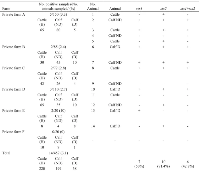
Table 3. The sampling source, origin of animals and distribution of stx genes detected by mPCR Tablo 3. Örnekleme yerleri, hayvanların orijinleri ve mPCR ile saptanan stx genlerinin dağılımı
Farm
No. positive samples/No. animals sampled/ (%) No. Animal Animal stx1 stx2 stx1+stx2 5/150 (3.3) 1 Cattle - + - Cattle (H) Calf (ND) Calf (D) 2 Calf ND + + + 65 80 5 3 Cattle + + + 4 Calf ND - - - Private farm A 5 Cattle - + - 2/85 (2.4) 6 Calf D + + + Cattle (H) Calf (ND) Calf (D) Private farm B 30 45 10 7 Calf ND + + + 2/72 (2.8) 8 Cattle + + + Cattle (H) (ND) Calf Calf (D) Private farm C 42 26 4 9 Calf ND - - - 3/110 (2.7) 10 Calf D + + + Cattle
(H) (ND) Calf Calf (D) 11 Cattle - - -
Private farm D 65 35 10 12 Calf ND - + - 2/20 (10) 13 Calf D + - - Cattle (H) Calf (ND) Calf (D) Private farm E 8 4 8 14 Calf D - + - 0/20 (0) Cattle (H) (ND) Calf Calf (D) - - - - - Private farm F 10 9 1 14/457 (3.1) Cattle (H) (ND) Calf Calf (D) Total 220 199 38 7 (50%) (71.4%) 10 (42.8%) 6 H: Healthy; D: Diarrhoeic; ND: Non-diarrhoeic
Figure 1. PCR amplicons obtained with E.coli O157:H7 strains as tested in mPCR. Lane M, 100 bp DNA marker; Lane +, STEC O157:H7 EDL 933, 1-14 stx1 (210 bp) and stx2 (484 bp) genes were detected in E.coli O157:H7 strains isolated from calves and cattle faeces samples
Şekil 1. E.coli O157:H7 suşlarında mPCR ile saptanan PCR ürünleri. Sütun M, 100 bp DNA marker, Sütun+, STEC O157:H7 EDL 933, 1-14, inek ve buzağı dışkılarından izole edilen E.coli O157:H7 suşlarında saptanan stx1 (210 bp) and stx2 (484 bp) genleri
72 ºC for 30 sec (extension) and a final step at 72ºC for 7 min. All PCR products were analyzed by using 1.5 % agarose gel electrophoresis and visualized by using ethidium bromide under UV light. Product sizes were determined by using DNA size marker (100 bp DNA ladder, Fermentas, Lithuania).
Statistical analysis: Chi-square test (Minitab
version 13.2. Minitab Inc. 2000) was used to detect differences in the prevalence of E.coli O157:H7 for age groups at a significance level of 0.05.
Results
The prevalence of E. coli O157:H7 positive samples: Escherichia coli O157:H7 was detected with a
ratio of 3.1 % (14 out 457 faeces samples) in calves and cattle by the study. Sources of isolation of E.coli O157:H7 strains were those: 4 calves out of 199 non-diarrhoeic calves (2.6%) and 5 out of 38 non-diarrhoeic calves (10.6%) and 5 out 220 cattle (2.3%). The isolation rate of E. coli O157:H7 from calves 1-4 weeks old (6.2%) was higher than other age groups. Distribution of
E. coli O157:H7 strains isolated from faecal samples
according to age are summarized in Table 2.
The distribution of virulence genes: Amplification
of stx(1) and stx(2) genes in E.coli O157:H7 strains by mPCR are shown in Figure 1. The stx(1) and stx(2) genes were detected in 6 out of 14 (42.8%) DNA samples extracted from E. coli O157:H7 strains isolated from faeces samples (Table 3), whereas the stx(1) gene was detected in a total of 7 (50%) strains (Figure 1).
Discussion and Conclusion
Shiga toxin-producing Escherichia coli O157:H7 serotypes are known to be the most important emerged group of pathogens (3, 5, 10, 19, 32). It has been reported that cattle are main reservoir for STEC (2, 20, 26). Investigations carried out in different part of Europe, Asia, North America have showed that 10-80% cattle were infected with STEC (1, 2, 5, 8, 27, 39). The occurrence of E.coli O157:H7 were also detected in different regions of Turkey. For instances, E. coli O157 was found in 14 individuals among 330 cattle slaughtered in five different abattoir in Istanbul (41) and
E. coli O157:H7 were isolated in 4 individuals among
312 cattle in the eastern region of Turkey (14). In another study, the rate of E. coli O157:H7 infection was found to be 13.6% (4). In the present study, E. coli O157:H7 was isolated from the 3.1% (14 out 457 faeces samples) of the calves and cattle examined, in particular 2.3% of the cattle and 3.8% of the calves were positive for E. coli O157:H7. Geographical variations may be the cause of the discrepancy in the isolation rates.
It has been shown that several serotypes of STEC are associated with diarrhea and dysentery in calves (17, 19, 25, 34, 36). An experimental study in which neonatal
calves inoculated with E. coli O157:H7 developed diarrhea and enterocolitis (15). However, this microorganism did not seem to be pathogenic for calves and adults (11, 30). Caprioli et al. (12) reported that the rate of STEC infection in calves younger than 2 months was between 1.8 % and 5%. Similarly, the present study shows that the frequency of E. coli O157:H7 infection is relatively lower in 2 to 3 months old calves than 1 to 4 weeks old calves groups (P<0.05). This study confirms the finding by Wells et al. (39) that calves are more susceptible to E.
coli O157:H7 (Table 2).
The pathogenicity of STEC O157:H7 is associated with a number of virulence factors, including Shiga toxins 1 and 2, as well as intimin and enterohaemolysin (1, 2, 6, 8, 27, 31, 37). Kang et al. (19) reported that stx(1) and stx(2) appear to play a major role in the pathogenesis of haemorrhagic colitis and haemolytic-uremic syndrome and STEC strains predominantly carries either stx(1) or stx(2) or both genes. The distribution and the origin of Shiga toxigenic genes including stx(1), stx(2) and eaeA gene in toxigenic but in non-toxigenic E. coli O157 and O157:H7 isolated from faeces and carcass of cattle could be determined by multiplex PCR (42). The mPCR analysis shows that stx(2) gene is more frequently found than that of stx(1) in STEC strains (33). Similar findings are reported by different researchers by various countries (1, 7, 28, 31, 43). Keen and Elder (21) reported that the frequency of stx(2) genes could be 93.1 %, whereas the frequency of stx(1) 0 % in any isolated strains. In another study conducted by Chapman et al. (13), stx(2) and intimin gene were found in 71.3 % of isolated E.coli O157:H7 strains. In this study, mPCR amplified genes isolated from E. coli 0157:H7 are shown in Figure 1 showing that stx(2) (71.4%) is found in higher frequency than that of stx(1) gene alone (42.8%) or stx(1) gene in combination (50%) (Table 3).
Blanco et al. (10) demonstrated that the strains of STEC can easily be isolated from healthy animals in comparison to animals having diarrhea since this strain present in the normal flora of intestine. According to Blanco et al. (10) stx(1), pathogenic strains could only be isolated from animals having diarrhea, while both stx(1) and stx(2), could be isolated from healthy calves. For instance, stx(1) gene was found significantly higher in diarrheic calves than healthy calves (40). Leomil et al. (24) reported that the diarrheic calves carry the higher frequency of stx was than that of non-diarrhoeic animals. Aslantas et al. (4) showed that E. coli 157:H7 containing either stx(2) or both stx(1) and stx(2), could be isolated predominantly in 1- to 3- year old cattle group studied in Turkey. In the present study, stx(1) was more prevalent in diarrhoeic calves in comparison to other studies (Tablo 3).
In conclusion, E. coli O157:H7 virulence genes such as stx(1) and stx(2) were detected in faeces samples collected from calves and cattle using mPCR. Therefore, we believe that the E. coli O157:H7 serotypes isolated and analyzed in Turkey will be beneficial to the many research and researcher in this field. This study demonstrated of E. coli O157:H7 serotype in cattle and calves, which represent an important reservoir for strains that a potential risk for human infections.
Acknowledgments
This study was supported by Afyon Kocatepe University Scientific Research Projects Council, Project No: 06.VF.18
References
1. Abu-Ali GS, Oulette LM, Henderson ST, Whittam TS, Manning SD (2010). Differences in adherence and
virulence gene expression between two outbreak strains of enterohemorrhagic Escherichia coli O157:H7. Microbiol,
156, 408-419.
2. Aidar Ugrinovich L, Blanco J, Blanco M, Blanco JE, Leomil L, Dahbi G, Mora A, Onuma DL, Siveira WD, Pestane de Castro AF (2007). Serotypes, virulence genes,
and intimin types of Shiga toxin-producing Escherichia coli (STEC) and enteropathogenic E.coli (EPEC) isolated from calves in Sao Paulo, Brazil. Int J Food Microbiol,
115, 297-306.
3. Armstrong G, Hollingsworth J, Morris JG (1996).
Emerging foodborne pathogens: Escherichia coli O157:H7 as a model of entry of a new pathogen into the food supply of developed world. Epidemiol Rev, 18, 29-51.
4. Aslantas Ö, Erdogan S, Cantekin Z, Gülacti I, Evrendilek GA (2006). Isolation and characterization of
verocytotoxin-producing Escherichia coli O157 from Turkish cattle. Int J Food Microbiol, 106, 338-342.
5. Baines D, Lee B, McAllister T (2008). Heterogeneity in
enterohemorrhagic Escherichia coli O157:H7 fecal shedding in cattle is related to Escherichia coli O157:H7 colonization of the small and large intestine. Can J
Microbiol, 54, 984-95.
6. Barret TJ, Kaper JB, Jarse AE, Wachsmuth IK (1992).
Virulence factors in Shiga-like toxin-producing Escherichia coli isolated from humans and cattle. J Infect
Dis, 165, 979-980.
7. Beutin L (1999). Escherichia coli O157 and Other Types
of Verocytotoxigenic E.coli (VTEC) Isolated from Humans, Animals and Food in Germany. 121-145. In: Stewart, C.S.,
and H.J. Flint (eds), Escherichia coli O157 in Farm Animals. Wallingford, UK
8. Beutin L, Geiger D, Steinruck H, Zimmermann S, Scheutz F (1993). Prevalence and some properties of
verotoxin (Shiga-like toxin)-producing Escherichia coli in seven different species of healthy domestic animals. J Clin
Microbiol, 31, 2483-2488.
9. Bielaszewska M, Janda J, Blahova K, Minarikova H, Jikova E, Karmali MA, Laubova J, Sikulova J, Preston MA, Khakhria R, Karch H, Klazorova H, Nye O (1997). Human Escherichia coli O157:H7 infection
associated with the consumption of unpasteurized goat’s milk. Epidemiol Infec, 119, 299-305.
10. Blanco J, Blanco M, Blanco JE, Mora A, Alonso MP, Gonzalez EA, Bernardez MI (2001). Verocytotoxigenic
Escherichia coli. 113-148. In: G. Duffy, P. Garvey, and D.
A. McDo- well (eds), Epidemiology of Verocytotoxigenic Escherichia coli (VTEC) in Ruminants, Food & Nutrition Press Inc., Trumbull, CT, USA.
11. Brown CA, Harmon BG, Zhao T, Doyle MP (1997).
Experimental Escherichia coli O157:H7 carriage in calves. Appl Environ Microbiol, 63, 27-32.
12. Caprioli A, Morabito S, Brugere H, Oswald E (2005).
Enterohaemorrhagic Escherichia coli: emerging issues on virulence and modes of transmission. Vet Res, 36,
289-311.
13. Chapman PA, Siddons CA, Wright DJ, Norma P, Fox J, Crick E (1993). Cattle as a possible source of
verocytotoxin-producing Escherichia coli O157 infections in man. Epidemiol Infect, 111, 439-447.
14. Cabalar M, Boynukara B, Gülhan T, Ekin IH (2001).
Van yöresinde sağlıklı görülen süt sığırcılığı işletmelerinde Rotavirus, E. coli K99 and O157:H7’nin varlığı üzerine araştırmalar. Turk J Vet Anim Sci, 25, 191-196.
15. Dean-Nystrom EA, Bosworth BT, Cray WC, Moon HW (1997). Pathogenicity of Escherichia coli O157:H7 in the
intestines of neonatal calves. Infect Immun, 65,
1842-1848.
16. Gioffre′ A, Meichtri L, Miliwebsky E, Baschkier A, Chillemi G, Romano MI, Sosa Estani S, Cataldi A, Rodrı′guez R, Rivas M (2002). Detection of Shiga
toxin-producing Escherichia coli by PCR in cattle in Argentina Evaluation of two procedures. Vet Microbiol, 87, 301-313.
17. Gyles CL (2007). Shiga toxin-producing Escherichia coli:
an overview. J Anim Sci, 85, 45–62.
18. Holth JG, Krieg, NR, Sneath, PHA, Staley JT, Williams ST (2000). Bergey’s manual of determinative bacteriology. Lippincott Williams & Wilkins, Philadelphia
19. Kang SJ, Ryu SJ, Chae JS, Eo SK, Woo GJ, Lee LH (2004). Occurrence and characteristics of enterohemorrhagic
Escherichia coli O157 in calves associated with diarrhoea.
Vet Microbiol, 98, 323-328.
20. Karnali MA, Gannon V, Sargeant JM (2009).
Verocytotoxin-producing Escherichia coli (VTEC). Vet
Microbiol, 140(3-4), 360-70.
21. Keen EJ, Elder RO (2002). Isolation of Shiga-toxigenic
Escherichia coli O157 from hide surface and oral cavity of finished beef feedlot cattle. J Am Vet Med Assoc, 220,
756-763.
22. Keene W, Hedberg K, Herriot D, Hancock D, Mckay R, Barret T, Fleing D (1997). A prolonged outbreak of
Escherichia coli O157:H7 infection caused by commercially distributed raw milk. J Infect Dis, 176,
815-818.
23. Law D (2000). Virulence factors of Escherichia coli O157
and other Shiga toxin–producing E. coli. J Appl Microbiol,
88, 729-745.
24. Leomil L, Aidar-Ugrinovich L, Guth BEC, Irino K, Vettorato MP, Onuma DL, de Castro AFP (2003).
Frequency of Shiga toxin-producing Escherichia coli (STEC) isolates among diarrheic and non-diarrheic calves in Brazil. Vet Microbiol, 97, 103-109.
25. Mainil JG, Duchesnes CJ, Whipp SC, Marques LRM, O’brien AD, Casey TA, Moon, HW (1987). Shiga-like
toxin production and attaching effacing activity of Escherichia coli associated with calf diarrhea. Am J Vet.
Res, 48, 743-748.
26. Masana MO, Leotta GA,Del Castillo LL, D’Astek BA, Palladino PM, Galli L, Vilacoba E, Carbonari C, Rodriguez HR, Rivas M (2010). Prevalence,
characterization, and genotypic analysis of Escherichia coli O157:H7/NM from selected beef exporting abattoirs of Argentina. J Food Protect, 73, 649-656.
27. Mainil JG, Jacquemin ER. Kaeckenbeeck AE (1993).
Association between the effacing (eae) gene and shiga-like toxin-encoding genes in Escherichia coli isolates from cattle. Am J Vet. Res, 54, 1064-1068.
28. Miyao Y, Kataoka T, Nomoto T, Kai A, Itoh T (1998).
Prevalence of verotoxin-producing Escherichia coli harbored in the intestine of cattle in Japan. Vet Microbiol,
61, 137-143.
29. Nataro JP, Kaper JB (1998). Diarrheagenic Escherichia coli. Clin Microbiol Rev, 11, 142-201.
30. Ohya T, Ito H (1999): Experimental infection of calves
with Escherichia coli O157:H7. J Vet Med Sci, 61,
1187-1189.
31. Oporto B, Esteban JI, Aduriz G, Juste RA, Hurtado A (2008). Escherichia coli O157:H7 and non-O157 Shiga
toxin-producing E.coli in healthy cattle, sheep and swine herds in Northern Spain. Zoonoses and Puplic Health, 55,
73-81.
32. Osek J (2003). Development of a multiplex PCR approach
for the identification of shiga toxin-producing Escherichia coli strains and their major virulence factor genes. J Appl
Microbiol, 95, 1217-1225.
33. Otawa K, Sato M, Sasaki T, Nonaka, J., Ito, K., Kuroki, T. and Nakai, Y (2004): Genetic analysis of
shiga-toxigenic Escherichia coli isolates from cattle in a limited region. J Anim Sci, 75, 261-269.
34. Paton JC, Paton AW (1998). Pathogenesis and diagnosis
of shiga toxin producing Escherichia coli infections. Clin
Microbiol Rev, 11, 450-479.
35. Riley LW, Remis RS, Helgerson SD, McGee HB, Wells JG, Davis BR, Hebert RJ, Olcott ES, Johnson LM, Hargrett NT (1983). Hemorrhagic colitis associated with
a rare Escherichia coli serotype. N Engl J Med , 308, 681– 685.
36. Sandhu KS, Gyles CL (2002). Pathogenic Shiga
toxin-producing Escherichia coli in the intestine of calves. Can J
Vet Res, 66, 65-72.
37. Schmidt H, Beutin L, Karch H (1995). Molecular
analysis of the plasmid-encoded hemolysin of Escherichia coli O157:H7 strain EDL 933. Infect Immun, 63,
1055-1061.
38. Wells JG, Davis BR, Wachsmuth IK, Riley LW, Remis RS, Sokolow R, Morris GK (1983). Laboratory
investigation of hemorrhagic colitis outbreaks associated with a rare Escherichia coli serotype. J Clin Microbiol, 18,
512-520.
39. Wells JG, Shipman L.D, Greene KD, Sowers EG, Green JH, Cameron DN, Downes FP, Martin ML, Grıffın PM, Ostroff SM, Potter ME, Tauxe RV, Wachsmuth KI (1991). Isolation of Escherichia coli
serotype O157:H7 and other Shiga-like toxin-producing E.coli from dairy cattle. J Clin Microbiol, 29, 985-989.
40. Wieler LH, Vieler E, Erpenstein C, Schlapp T, Steinrück H, Bauerfeind R, Byomi A, Baljer G (1996).
Shiga toxin-producing Escherichia coli strains from bovines: Association of adhesion with carriage of eae and other genes. J Clin Microbiol, 34, 2980-2984.
41. Yilmaz A, Gun H, Yilmaz H (2002). Frequency of
Escherichia coli O157:H7 in Turkish cattle. J Food
Protect, 10, 1637-1640.
42. Yilmaz A, Gun H, Ugur M, Turan N, Yilmaz H (2006).
Detection and frequency of VT1, VT2 and eaeA genes in Escherichia coli O157 and O157:H7 strains isolated from cattle, cattle carcasses and abattoir environment in Istanbul. Int J Food Microbiol, 106, 213-217.
43. Zhao T, Doyle MP, Shere J, Garber L (1995).
Prevalence of enterohemorrhagic Escherichia coli O157:H7 in a survey of dairy herds. Appl Environ
Microbiol, 61, 1290-1293.
Geliş tarihi: 14.09.2010 / Kabul tarihi: 03.03.2011
Address for Correspondence
Doç.Dr. Yahya KUYUCUOĞLU
Afyon Kocatepe Üniversitesi, Veteriner Fakültesi, Mikrobiyoloji Anabilim Dalı,
A.N. Sezer Kampüsü, 03200, Afyonkarahisar,
Phone: +90 272 228 13 12/148, Fax:+90 272 228 13 49 , E-mail: [email protected]