ORIGINAL ARTICLE
The influence of selenium on expression levels of the rbcL gene
in Chlorella vulgaris
Gulru Ozakman1 · Sinem Gamze Yayman1 · Cigdem Sezer Zhmurov2 · Emel Serdaroglu Kasikci1 · Tunc Catal1,3
Received: 19 August 2017 / Accepted: 13 March 2018 / Published online: 20 March 2018 © Springer-Verlag GmbH Germany, part of Springer Nature 2018
Abstract
In this study, the effects of selenium on the microalgae Chlorella vulgaris were examined. Four groups of C. vulgaris were cultivated using Bristol medium: group I (control), no sodium selenite (Se); group II, 1 µM Se; group III, 10 µM Se; and group IV, 100 µM Se. Algal biomass samples were collected for biochemical evaluation and gene expression studies on the 21st day of cultivation. The following parameters were investigated: chlorophyll a (Cla), chlorophyll b (Clb) and total
caro-tene content, total protein, and total glutathione (GSH) and malondialdehyde (MDA) levels. Gene expression levels of large subunits of Rubisco (rbcL) were analyzed using real-time quantitative polymerase chain reaction. Total Cla and total carotene in C. vulgaris decreased in high concentrations of Se (100 µM) (around 23 and 42%, respectively) when compared to con-trols while, Clb content increased by about 10%. 10 µM of Se led to increased GSH levels (3.04 ± 0.02 µg GSH/mg protein)
and decreased MDA levels (2.02 ± 0.1 µmol MDA/mg protein) when compared to control groups (1.18 ± 0.04 µg GSH/mg protein and 0.94 ± 0.23 µmol MDA/mg protein), while a significant decrease in GSH and an increase in MDA levels in the presence of 100 µM Se showed the opposite effect. rbcL gene expression increased 1.76 ± 1.37-fold and 0.86 ± 1.33-fold in 10 and 100 µM selenium experiments when compared to control groups. Our results suggest both pro-oxidant and antioxidant activities of Se on C. vulgaris and upregulation of the rbcL gene for the first time. Treatment with low concentrations of Se improves the antioxidant features of the microalgae, C. vulgaris.
Keywords Biotechnology · Chlorella vulgaris · Malondialdehyde · rbcL gene expression · Selenium
Introduction
Selenium is an important trace element whose health ben-efits include the prevention of various diseases such as cor-onary heart disease and cancer by activating the immune system (Reich and Hondal 2016; Gunes et al. 2016; Chen et al. 2013). Selenium and selenium compounds prevent DNA damage by scavenging free radicals (Tinggi 2008). In fighting against viral infections, selenium was shown to slow the growth of HIV. Its protective effects against stom-ach and colon cancer were demonstrated (Kieliszek and
Błażejak 2016). Selenium is found in poultry, sea food and meat, in onions, garlic and red peppers, and recently sele-nium enriched foods have drawn great attention because of enhanced health benefits (Yuan et al. 2016). Selenium interacts with various vitamins and aminoacids such as glu-tathione, which play a role in the body as an antioxidant. Selenium-rich foods have become very popular in recent years because of its improved health benefits and microalgae have been suggested as a food source and supplement.
Trace elements are used in very small amounts as constit-uents of all organisms and are largely acquired from plants. Trace elements such as selenium plays a vital role not only in microalgae but also in plants such as promoting growth, decreasing injury caused by reactive oxygen species and inducing chlorophyll amounts under light stress (Araie and Shiraiwa 2009). The main emphasis is on selenium, which is by far the most widely studied among the essential toxic metals/metalloids. On the other hand, excess selenium has a negative effect on the growth and development of micro-algae, although it remains unclear what concentrations of * Tunc Catal
1 Department of Molecular Biology and Genetics, Uskudar
University, Uskudar, 34662 Istanbul, Turkey
2 Department of Genetics and Bioengineering, Istanbul Bilgi
University, Eyup, 34060 Istanbul, Turkey
3 Istanbul Protein Research and Innovation Center, Uskudar
Se are toxic to microalgae. We investigated the effects of Se at different concentrations on oxidative stress in micro-algae to provide a better understanding of its toxic effects and how selenium-rich microalgae might act as a possible food supplement.
Chlorella vulgaris microalgae are 2–8 µm long,
single-celled photosynthetic green microalgae. They have an important nutritional value since they produce more carbo-hydrate and fat as they age. Chlorella vulgaris provides an important source of vitamins, proteins, fatty acids, enzymes and carotenoids with its 50–60% protein content and high amounts of chlorophyll (Panahi et al. 2016). It is a photo-lithoautotrophic organism and growth media strongly affect its metabolic products (Panahi et al. 2016). Besides these characteristics, it acts as an antioxidant (Panahi et al. 2016).
Chlorella vulgaris can also be used for other purposes such
as bioremediation (Zeraatkar et al. 2016). Previously, the potential use of C. vulgaris has been suggested for food pur-poses and the metabolic activity of the microalgae has been studied. However, the effect of selenium on photosystem-related activities including ribulose-1,5-bisphosphate car-boxylase/oxygenase has not been reported, and this is the first study highlighting the effect of selenium on the rbcL gene in the photosystem of C. vulgaris.
Ribulose-1,5-bisphosphate carboxylase/oxygenase (Rubisco) has an important role in carbon fixation by which atmospheric carbon dioxide is converted into other carbon compounds, such as glucose, by photosynthetic organisms (Dhingra et al. 2004). The large subunit rbcL (Rubisco, large subunit gene) (ribulose-1,5-biphosphate carboxylase) is encoded by chloroplast DNA. It is enzymatically active and forms dimers, in which aminoacids conduce to binding sites (Cooper 2000). During carbon fixation, the substrate mol-ecules for Rubisco are ribulose-1,5-biphosphate and carbon dioxide or molecular oxygen (Feller et al. 2008). Rubisco activation is directly related to photosynthetic performance in photosynthetic organisms [Crafts–Brandner and Salvucci
2000).
In this study, C. vulgaris microalgae were used to analyze the effects of various concentrations of selenium. Total chlo-rophyll (Cla and Clb), total carotenoids, total protein, total
GSH, and total malondialdehyde (MDA) levels in C.
vul-garis were measured. For the first time, expression levels of
the rbcL gene in C. vulgaris were compared in the presence of different selenium concentrations using real-time PCR.
Materials and methods
Culture conditions of C. vulgaris
Chlorella vulgaris was obtained from UTEX (UTEX No.
26, Texas, USA) and grown using Bristol agar medium. The
following stock solutions were prepared: NaNO3 solution
(25 g L−1), CaCl
2·2H2O solution (2.5 g L−1), MgSO4·7H2O
solution (7.5 g L−1), K
2HPO4 solution (7.5 g L−1), KH2PO4
solution (17.5 g L−1) and NaCl solution (2.5 g L−1). 10 mL
of each stock solution was added to 940 mL of distilled water and then 1 g L−1 of peptone was added. For agar plates, 1.5%
of agar was added to the solution for solidification. The Bris-tol medium was autoclaved at 121 °C for 15 min. Seed cul-ture of C. vulgaris was inoculated using loop on agar plates and cultures remained at 25 °C under led-light illumination. Active cultures of C. vulgaris were transferred into shake-flasks (100 mL) containing 60 mL of Bristol medium for enrichment before Se experiments. Sodium selenite (VWR, USA) was added to the Bristol medium and four experimen-tal groups were prepared as follows: 0, 1, 10, and 100 µM of sodium selenite (n = 3). The same amount of microalgae was inoculated in shake-flasks (100 mL) containing 60-mL medium. Then, Chlorella vulgaris was grown continuously for 21 days in a rotary shaker (Thermo Scientific, Q4000-04, USA) at 185 rpm at 25 °C under led-light illumination. The aseptic technique was followed during all inoculations in the experiments. C. vulgaris samples were collected on the 21st day of cultivation for biochemical and real-time PCR analysis.
Biochemical analysis
1 mL of each experimental group was sampled and the cells were centrifuged at 4000 rpm for 5 min for total chloro-phyll content. The supernatant was discarded, and 2 mL of methanol was added over the pellets. The mixture was homogenized at 10,000 rpm for 1 min using an homogenizer (MagnaLyser/Roche). Cells were re-suspended in 2 mL of methanol and homogenized at 10,000 rpm for 1 min. The supernatant was placed in absorbance tubes. Methanol was used as a blank and absorbance values at 666, 653 and 470 nm were recorded. The pigments were calculated using the following formula (Dere et al. 1998):
where Cx+c corresponds to total carotenes.
Total protein concentrations were determined accord-ing to the Lowry method usaccord-ing Folin’s phenol reagent from Sigma (F-5292) and bovine serum albumin as the stand-ard (Lowry et al. 1951). Lipid peroxidation levels were measured spectrophotometrically according to a previous report (Ledwozyw et al. 1986). Levels of malondialde-hyde, a lipid peroxidation product, were measured using Cla= 15.65A666− 7.340A653
Clb= 27.05A653− 11.21A666
C
1,1,3,3-tetraethoxypropane as the standard. Total GSH in
C. vulgaris was measured using Elman’s indicator (Beutler
1975).
Total RNA isolation and reverse transcription
Total RNA was isolated using a commercial kit for gene expression study according to the manufacturer’s protocol (Zymo Research, Quick-RNA MiniPrep, CA, USA). Con-centrations of RNA were measured using a spectrophotom-eter (Thermo, USA) and calculated based on the ratio of absorbance of 260/280 nm (Qian et al. 2008). RNA extracts were used to synthesize cDNA. cDNA mastermix (Roche) contained 8 µL of buffer, 2 µL of reverse transcriptase and 10 µL of DNase-/RNase-free-water. The mastermix was split into two halves, one treated and the other untreated. The fol-lowing program was set up in a thermal cycler (BIO-RAD, T100 Thermal Cycler): 25 °C for 10 min (primer annealing), 42 °C for 15 min (reverse transcription), 85 °C for 5 min (inactivation), and 4 °C hold. The following mixture was prepared for total RNA (10 µL): 5× TransAmp buffer (4 µL), reverse transcriptase (1 µL) and DNase-/RNase-free-water (5 µL). Reaction products were stored at − 80 °C until real-time PCR analysis.
Real‑time PCR analysis
Reverse transcription (RT) was carried out using a RT–PCR (Roche Light Cycler, USA). To facilitate the real-time poly-merase chain reaction (PCR) analysis of the selected genes under the same reaction conditions, primers were obtained from Medsantek (Istanbul, Turkey). The following primers were used in the study: for 18S rRNA (forward) 5′-TTG ACG GAA GGG CACCA-3′, (reverse) 5′-CAC CAC CCA TAG AAT CAA GAA AGA G-3′. For rbcL (forward) 5′-CTT GGA CGA CTG TAT GGA CTG-3′, (reverse) 5′-ATA CCG TGA GGA GGA CCT TG-3′. Their gene bank accession numbers are X13688 and AF499684 (Qian et al. 2008). The 18S rRNA transcript was used to normalize the results by eliminating variations in the quantity and quality of mRNA and cDNA. Each mRNA level was expressed as the ratio of itself to 18S rRNA. Mastermix Rbcl-(6×) including 6 µL SYBR, 12 µL buffer without Mg, 9.6 µL MgCl2, 2.4 µL DNTP, 51 µL H2O, 3µ Taq, 3 µL R-Primer and 3 µL Primer-R, 15 µL was split as 5 µL cDNA. The strips were positioned and RT–PCR was started. The cycle parameters consisted of one cycle of 10 s at 95 °C and then 40 cycles of 5 s at 95 °C followed by 31 s at 60 °C. Data were collected at the end of each extension step. The relative quantification of gene expressions among the treatment groups was analyzed by the 2−ΔΔCT method
(Livak and Schmittgen 2011).
Statistical analysis
All experiments were done in triplicate. Data were expressed as mean ± SD in all graphs. All the result groups were evalu-ated statistically via one-way ANOVA followed by the inde-pendent sample’s t test for comparisons of the groups using the IBM Statistical Package for the Social Sciences (SPSS) for Windows (version 24). The results were considered sig-nificant at the level of P < 0.05.
Results and discussion
Total chlorophyll and carotene content changes
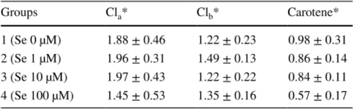
Table 1 shows the effects of selenium on chlorophyll a (Cla), chlorophyll b (Clb) and total carotene amount in C.
vulgaris. Total Cla and total carotene production was gradu-ally decreased together with a gradual increase in selenium concentration (P > 0.05) (Table 1). Cla and total carotene
production was decreased in group IV (1.45 ± 0.53 and 0.57 ± 017 µg gfw−1, respectively) at around 23 and 42%,
respectively, while their concentration was similar in group 1 and group 2 experiments (Table 1). Interestingly, the Clb
amount increased (1.35 ± 0.16 µg gfw−1) in a higher
con-centration of selenium (100 µM) when compared to those of the control (1.22 ± 0.23 µg gfw−1) and other groups treated
with lower selenium concentrations (P > 0.05). These results indicate that selenium negatively affects total carotene and total Cla production. However, Clb production is improved in the presence of selenium.
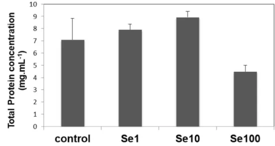
Total protein content changes
Total protein levels were gradually increased in groups treated with 1 and 10 µM concentrations of selenium (7.9 ± 0.45 and 8.9 ± 0.44 mg mL−1, respectively). 100 µM
sele-nium resulted in a decrease in total protein amount (4.5 ± 0.5 mg mL−1) when compared to the 10 µM group (8.9 ±
1.7 mg mL−1) (P < 0.0001 between group 3 and group 4)
(Fig. 1) (PANOVA 0.05 between groups). Our results indicate Table 1 Effect of different concentrations of selenium (1, 10, 100 µM) on Cla, Clb and carotenoids amount (µg gfw−1) in C.
vul-garis *Mean ± SD Groups Cla* Clb* Carotene* 1 (Se 0 µM) 1.88 ± 0.46 1.22 ± 0.23 0.98 ± 0.31 2 (Se 1 µM) 1.96 ± 0.31 1.49 ± 0.13 0.86 ± 0.14 3 (Se 10 µM) 1.97 ± 0.43 1.22 ± 0.22 0.84 ± 0.11 4 (Se 100 µM) 1.45 ± 0.53 1.35 ± 0.16 0.57 ± 0.17
that in concentrations of selenium up to 10 µM, total pro-tein production is promoted in biomass of C. vulgaris while 100 µM selenium negatively affected the production of proteins.
Total malondialdehyde levels
Figure 2 shows malondialdehyde (MDA) levels affected by selenium treatment (PANOVA 0.0001 between groups). 4.34 ±
0.49 µmol MDA/mg protein of MDA levels was detected in group four biomass samples while MDA levels were similar in 1 µM selenium-treated samples (1.02 ± 0.06 µmol MDA/ mg protein) and the control group (0.94 ± 0.23 µmol MDA/ mg protein) (P < 0.0001 group 1 vs. group 4; P < 0.0001 group 2 vs. group 4; P < 0.001 group 3 vs. group 4). MDA levels were lower up to 10 µM selenium treatment. These results suggest that the increased concentrations of selenium (100 µM) lead to oxidative stress in microalgae while similar
MDA levels were observed caused by low concentrations of selenium.
Total glutathione levels
Total glutathione (GSH) levels were increased in the group III (3.04 ± 0.02 µg GSH/mg protein) experiment compared to lower GSH levels found in control biomass samples (1.18 ± 0.04 µg GSH/mg protein) (Fig. 3) (PANOVA 0.0001
between groups) (P < 0.05 group 1 vs. group 3). On the other hand, it seems that total GSH is consumed in 100 µM selenium experiments (P < 0.0001 group 1 vs. group 4). Our GSH results highlight the fact that up to 10 µM selenium treatment improves the antioxidant capacity of C. vulgaris. Schiavon et al. (2017) reported that microalgae enriched with selenium might improve antioxidant capacity by act-ing as an anticarcinogenic compound.
Fig. 1 Total protein content changes in C. vulgaris in the presence of selenium. Control, Se1, Se10 and Se100 corre-sponds to the following con-centrations; 0, 1, 10, 100 µM, respectively
Fig. 2 Total malondialdehyde (MDA) levels in control (no selenium) and selenium-treated C. vulgaris. 100 µM concen-tration of selenium increased MDA levels while lower MDA was observed in 1 and 10 µM treatment with selenium
Chlorella vulgaris is resistant against high concentrations
of selenium levels demonstrating that the microalgae are good candidates for selenium bio-accumulation (Neumann et al. 2003). However, it was shown that in high concentra-tions, selenium leads to toxicity in C. vulgaris. Selenopro-teins contain enzymes such as glutathione peroxidase and have obscure functions (Kryukov et al. 2003). Many stud-ies have shown that selenium is required for protein and lipid synthesis and to increase cellular division (Furness and Rainbow 1990). Several studies showed that the effects of selenium could be attributed to its antioxidative function as demonstrated by excessive contents of chlorophyll a, and reduced LPO and ROS (Sun et al. 2014; Vítová et al. 2011). In a previous report, C. vulgaris cultures were exposed to various selenite concentrations for 144 h to investigate the effects of different concentrations of selenite on algal growth (Sun et al. 2014). It was reported that low selenite concentra-tions (≤ 75 mg L−1) increased the growth of C. vulgaris by
decreasing lipid peroxidation levels and intracellular reactive oxygen species whereas 100 mg L−1 of selenium resulted
in toxicity. Similarly, 100 mg L−1 concentrations of Se led
to an increase in MDA in our research. In our study, glu-tathione peroxidase activities were not measured, but levels of GSH, which is an antioxidant amino acid, significantly decreased at 100 µM concentrations indicating that Se pro-motes antioxidation up to concentration levels of 10 µM. It can be suggested that under stress conditions some free-radical species are generated inside algal cells. To prevent oxidative cell damage, protective mechanisms are induced. Because of stress caused by triggering agents including trace and heavy metals, algal cells increase the activity of some antioxidant enzymes and the synthesis of compounds such as carotenoids and GSH (Li et al. 2006). Glutathione is the other important antioxidative molecule that protects
cells against damage caused by free radicals. It has been shown that GSH utilization was increased in the microalgae cells Scenedesmus bijugatus under copper stress (Nagalak-shmi and Prasad 2001). Similar results were observed in our study, and in addition, it was shown that selenium also affected the photosystem of C. vulgaris.
Relative gene expression levels of rbcL
Following RNA isolation and cDNA synthesis, quantitative PCR analysis was performed to determine the gene expres-sion levels of the large subunit of Rubisco (rbcL) to assess selenium effects on transcript abundance and related pro-teins involved in oxidative stress responses. Figure 4 shows relative gene expression levels of rbcL. Although there was no statistically significant difference, the expression level of
rbcL increased about 1.76 ± 1.37-fold (P = 0.09, t = 2.26, df
= 4) and 0.86 ± 1.33-fold (P = 0.325, t = 1.120, df = 4) in 10 µM and 100 µM selenium experiments when compared to control groups. On the other hand, about 0.36 ± 0.67 down-regulation (P = 0.408, t = − 0.924, df = 4) in lower selenium treatment was observed. rbcL gene expression results sug-gest that selenium treatment may lead to the upregulation of the Rubisco gene. In a previous study, a strong relationship between rbcL gene and chlorophyll content was shown while the expression of the rbcL gene is upregulated together with increased chlorophyll content, indicating that the transcrip-tion of rbcL is coordinated with chlorophyll accumulatranscrip-tion (Ohmiya et al. 2014).
Rubisco is found in the chloroplasts of bundle-sheath cells in higher plants performing C4-type photosynthesis (Leegood 2008). Catalytic activity of Rubisco is decreased and cysteine residues are oxidized leading to the dena-turation of the enzyme when chloroplasts are subject to Fig. 3 Total GSH levels in
con-trol and Se-treated C. vulgaris. GSH levels increased in 10 µM selenium treatment
oxidizing conditions (Marin-Navarro and Moreno 2003). Enzyme activities such as superoxide dismutase (SOD) and glutathione peroxidase are enhanced by selenium treatment, which activates defense mechanisms against the deleterious effects of reactive oxygen species. Moreover, Se induces lipid peroxidation at higher concentrations causing an inhi-bition in gene expression triggering cell death. Increased GSH levels and upregulation of the rbcL gene in C.
vul-garis seems to be the result of the antioxidant effect of Se at
10 µM concentrations while at 100 µM concentration tox-icity of Se was shown with enhanced LPO together with insignificant upregulation of the rbcL gene (below onefold change). It seems that the rbcL gene, which is responsi-ble for CO2 fixation, is upregulated significantly referring
to enhanced photosynthetic capacity although chlorophyll production decreased. These results indicate that toxic lev-els of Se significantly affect rbcL gene expression blocking synthesis of chlorophyll production. In our study, compared to the control, 1 μM of Se led to increased GSH levels but decreased LPO levels. For better-value food supplementa-tion, C. vulgaris Rubisco-related genes should be focused on to enhance the quality of possible food supplements with Se-enriched ingredients. Previously, Kouba et al. (2014) showed that the bioactivity of Se-enriched C. vulgaris would be more effective than that of inorganic forms of Se (Kouba et al. 2014). Similar pro-oxidant and antioxidant features of selenium were shown in fungi. Microalgae also regulate their metabolic conditions at different concentration levels of selenium (Catal et al. 2008).
Qian et al. (2008) showed that supplementation of C.
vulgaris with free-radical scavengers led to the
upregu-lation of photosynthesis genes such as rbcL. Similarly,
at 10 µM concentrations, Se showed antioxidant features including increase in GSH levels and upregulation in
rbcL genes. These results are also supported by enhanced
Cla content indicating that the photosynthetic activity is enhanced by low concentrations of Se.
Conclusions
In conclusion, dual effects of selenium on C. vulgaris were shown indicating that lower concentrations of Se (10 µM) increase GSH levels, while GSH levels decrease because of oxidative stress caused by high concentrations of Se (100 µM). Increased GSH levels and upregulation of the
rbcL gene in C. vulgaris show the antioxidant features of
Se and treatment with up to 10 µM Se improve the anti-oxidant features of C. vulgaris. Therefore, treatment of C.
vulgaris with 10 µM concentrations of Se may increase
the antioxidant features of the microalgae with a pos-sible Se-rich and enhanced chlorophyll content through Rubisco-related genes. In future, other Rubisco-related genes should be analyzed using different trace elements to understand the exact mechanisms to enhance the food benefits of the microalgae, C. vulgaris.
Acknowledgements This study was funded by the Republic of Tur-key, Istanbul Development Agency (ISTKA) (Grant No. TR10/16/ YNY/0167).
Compliance with ethical standards
Conflict of interest No conflict of interest declared. Fig. 4 Relative gene expression
levels of rbcL in C. vulgaris. Group 1, 2, 3 and 4 corresponds 0, 1, 10, 100 µM treatment with selenium, respectively
References
Araie H, Shiraiwa Y (2009) Selenium utilization strategy by microal-gae. Molecules 14:4880–4891
Beutler E (1975) Glutathione in red cell metabolism a manual of bio-chemical methods, 2nd edn. Grune and Stratton, New York Catal T, Liu H, Bermek H (2008) Selenium induces
manganese-dependent peroxidase production by the white-rot fungus Bjer-kandera adusta (Willdenow) P. Karsten. Biol Trace Elem Res 123:211–217
Chen YC, Prabhu KS, Mastro AM (2013) Is selenium a potential treat-ment for cancer metastasis?. Nutrients 5:1149–1168
Cooper GM (2000) The chloroplast genome. The Cell: a molecu-lar approach, 2nd edn. ASM Press, Washington, DC. ISBN 0-87893-106-6
Crafts-Brandner SJ, Salvucci ME (2000) Rubisco activase constrains the photosynthetic potential of leaves at high temperature and CO2. Proc Natl Acad Sci 97:13430–13435
Dere S, Gunes T, Sivaci R (1998) Spectrophotometric determination of chlorophyll—a, b and total carotenoid contents of some algae species using different solvents. Turk J Bot 22:13–17
Dhingra A, Portis AR, Daniell H (2004) Enhanced translation of a chloroplast-expressed RbcS gene restores small subunit levels and photosynthesis in nuclear RbcS antisense plants. Proc Natl Acad Sci 101:6315–6320
Feller U, Anders I, Mae T (2008) Rubiscolytics: fate of Rubisco after its enzymatic function in a cell is terminated. J Exp Bot 59:1615–1624
Furness RW, Rainbow S (1990) Heavy metals in the marine environ-ment. CRC Press Inc., Boca Raton
Gunes S, Sahinturk V, Karasati P, Sahin IK, Ayhanci A (2016) Car-dioprotective effect of selenium against cyclophosphamide-induced cardiotoxicity in rats. Biol Trace Elem Res. https ://doi. org/10.1007/s1201 1-016-0858-1
Kieliszek M, Błażejak S (2016) Current knowledge on the importance of selenium in food for living organisms: a review. Molecules 21(5):609
Kouba A, Velíšek J, Stará A, Masojídek J, Kozák P (2014) Supple-mentation with sodium selenite and selenium-enriched microalgae biomass show varying effects on blood enzymes activities, anti-oxidant response, and accumulation in common barbel (Barbus barbus). Biomed Res Int. https ://doi.org/10.1155/2014/40827 0
Kryukov GV, Castellano S, Novoselov SV, Lobanov AV, Zehtab O et al (2003) Characterization of mammalian selenoproteoms. Science 300:1439–1443
Ledwozyw A, Michalak J, Stepien A, Kadziolka A (1986) The relation-ship between plasma tryglicerides, cholesterol, total lipids and lipid peroxidation products during human atherosclerosis. Clin Chim Acta 155:275–284
Leegood RC (2008) Roles of the bundle sheath cells in leaves of C3 plants. J Exp Bot 59:1663–1673
Li M, Hu C, Zhu Q, Chen L, Kong Z, Liu Z (2006) Copper and zinc induction of lipid peroxidation and effects on antioxidant enzyme
activities in the microalga Pavlova viridis (Prymnesiophyceae). Chemosphere 62:565–572
Li M, Zhu Q, Hu C, Chen L, Liu Z, Kong Z (2007) Cobalt and manga-nese stress in the microalga Pavlova viridis (Prymnesiophyceae): effects on lipid peroxidation and antioxidant enzymes. J Environ Sci 19:1330–1335
Livak KJ, Schmittgen TD (2011) Analysis of relative gene expression data using real-time quantitative PCR and the 2(− Delta Delta C(T)) method. Methods 25:402–408
Lowry OH, Rosebrough NJ, Farr AL, Randall RJ (1951) Protein meas-urement with the Folin phenol reagent. J Biol Chem 193:265–275 Marin-Navarro J, Moreno J (2003) Modification of the proteolytic
fragmentation pattern upon oxidation of cysteines from ribu-lose 1,5-bisphosphate carboxylase/oxygenase. Biochemistry 42:14930–14938
Nagalakshmi N, Prasad MNV (2001) Responses of glutathione cycle enzymes and glutathione metabolism to copper stress in Scened-esmus bijugatus. Plant Sci 160:291–299
Neumann PM, De Souza MP, Pickering IJ, Terry N (2003) Rapid microalgal metabolism of selenate to volatile dimethylselenide. Plant Cell Environ 26:897–905
Ohmiya A, Hirashima M, Yagi M, Tanase K, Yamamizo C (2014) Identification of genes associated with chlorophyll accumulation in flower petals. PLoS ONE 9(12):e113738
Panahi Y, Darvishi B, Jowzi N, Beiraghdar F, Sahebkar A (2016) Chlo-rella vulgaris: a multifunctional dietary supplement with diverse medicinal properties. Curr Pharm Des 22:164–173
Qian H, Daniel Sheng G, Liu W, Lu Y, Liu Z, Fu Z (2008) Inhibitory effects of atrazine on Chlorella vulgaris as assessed by real-time polymerase chain reaction. Environ Toxicol Chem 27:182–187 Reich HJ, Hondal RJ (2016) Why nature chose selenium. ACS Chem
Biol 11:821–841
Schiavon M, Ertani A, Parrasia S, Vecchia FD (2017) Selenium accu-mulation and metabolism in algae. Aquat Toxicol 189:1–8 Sun X, Zhong Y, Huang Z, Yang Y (2014) Selenium accumulation in
unicellular green alga Chlorella vulgaris and its effects on anti-oxidant enzymes and content of photosynthetic pigments. PLoS ONE 9(11):e112270
Tinggi U (2008) Selenium: its role as antioxidant in human health. Environ Health Prev Med 13:102–108
Vítová M, Bišová K, Hlavová M, Zachleder V, Rucki M, Cížková M (2011) Glutathione peroxidase activity in the selenium-treated alga Scenedesmus quadricauda. Aquat Toxicol 102:87–94 Yuan H, Wang W, Chen D, Zhu X, Meng L (2016) Effects of a
treat-ment with Se-rich rice flour high in resistant starch on enteric dysbiosis and chronic inflammation in diabetic ICR mice. J Sci Food Agric. https ://doi.org/10.1002/jsfa.8011
Zeraatkar AK, Ahmadzadeh H, Talebi AF, Moheimani NR, McHenry MP (2016) Potential use of algae for heavy metal bioremediation, a critical review. J Environ Manage 181:817–831