ORIGINAL INVESTIGATION ÖZGÜN ARAŞTIRMA
1Department of Pathology, Faculty of Medicine, İstanbul Bilim University, İstanbul, Turkey 2Department of Gynecology
and Obstetrics, Faculty of Medicine, İstanbul Bilim University, İstanbul, Turkey 3Department of Pathology,
Faculty of Medicine, Yeni Yüzyıl University, İstanbul, Turkey
Available Online Date Çevrimiçi Yayın Tarihi
15.02.2013 Submitted/Geliş Tarihi 26.04.2012 Accepted/Kabul Tarihi 21.09.2012 Correspondance/Yazışma
Dr. İlknur Çetinaslan Türkmen, Department of Pathology, Faculty of Medicine, İstanbul Bilim University, 34780 İstanbul, Turkey Phone: +90 212 288 44 56 e.mail: ilknurcetinaslan@hotmail.com
This study was presented at the 36th European Congress of Cytology, Istanbul, Turkey, September 22-25, 2011. Presentation Number: P42, P68
Bu çalışma, 36. Avrupa Sitoloji Kongresi’nde (22-25 Eylül 2011 İstanbul, Türkiye) sunulmuştur. Sunum Numarası: P42, P68
©Copyright 2013 by Erciyes University School of Medicine - Available on-line at www.erciyesmedicaljournal.com
©Telif Hakkı 2013 Erciyes Üniversitesi Tıp Fakültesi Makale metnine www.erciyesmedicaljournal.com web sayfasından ulaşılabilir.
Interobserver Variability in Cervical Smears
from Patients with a History of Abnormal
Cytology: Comparison of Conventional Pap Smears
and Liquid-Based Cytology
Anormal Sitoloji Hikayesi Olan Hastalarda Servikal Sitoloji
Değerlendirmesinde Gözlemciler Arası Değişkenlik: Geleneksel Pap
Smear ve Sıvı Bazlı Sitoloji Karşılaştırması
İlknur Çetinaslan Türkmen1, Nuray Başsüllü1, Banu Bingöl2, Gülen Bülbül Doğusoy1, Sema Arıcı3
ABSTRACT ÖZET
Introduction
Cervical cytology is one of the best cancer screening programs, resulting in a dramatic decrease in the incidence of cervical cancer in many developed countries since conventional Pap smears (CPS) began to be used for cervical cancer screening in the 1960s (1). However, it must be kept in mind that many factors influence the success of this screening program, and the disparities in diagnostic assessment of cervical cytology and the inaccuracy of cytologi-cal diagnosis have emerged as being very important (2).
Liquid-based cytology (LBC) is an alternative technique for transferring the cellular material collected from the transformation zone of the uterine cervix. In contrast to CPS, the cells are not directly spread on a slide, but rather into a vial containing fixative liquid (3). The commonly used automated LBC techniques are ThinPrepTM (Cytyc
Corporation, Boxborough, MA, USA), SurePathTM (TriPath Imaging, Burlington, NC, USA), PapSpinTM
(ThermoElec-tron, Pittsburgh, PA, USA), DNACITOLIQ (Digene Brazil, Sao Paulo, Brazil), and Liqui-PrepTM (LGMInternational,
Fort Lauderdale, FL, USA) (4, 5).
The reproducibility of cervical cytological interpretations are low to moderate, and there are many studies with conflicting results regarding the superiority of LBC to CPS in this context (1, 3-8).
This paper focuses on the reproducibility of diagnostic classification between the use of CPS and LBC among three pathologists.
Objective: One of the problems encountered when assessing
cervical precancerous lesions is intra- and interobserver vari-ability. The aim of this study was to determine the degree of interobserver variation in conventional PAP smears (CPS) and liquid-based cytology (LBC).
Materials and Methods: The diagnostic variability among three
pathologists was assessed using 120 smears (67 conventional CPS and 53 LBC). The cases were selected retrospectively from the archives of the Pathology Department among the patients with follow-up, such as biopsy confirmation and/or persistent/ resolving disease in the follow-up smear. The observers exam-ined the slides in a blinded fashion.
Results: Diagnostic agreement on the presence or absence of
intraepithelial lesions was found in 30 of 51 slides (58.82%) of LBC (kappa=0.42) and in 44 of 67 slides (65.67%) of con-ventional cytology (kappa=0.50). The agreement was slightly higher in conventional smears. The highest agreement was in the LSIL category with a kappa value of 0.50 in LBC and 0.62 in conventional cytology, while ASCUS was the least reproduc-ible diagnosis.
Conclusion: Our results are in agreement with the literature in
that the reproducibility of cervical cytology shows low to mod-erate consistency. The study showed no significant difference between LBC and CPS in the reproducibility of the diagnosis.
Key words: Cytological technique, intraepithelial neoplasia,
cervical, cervical smear
Amaç: Servikal prekanseröz lezyonlar sözkonusu olduğunda
tanının tekrarlanabilirliği problemlerden biri olarak karşımıza çıkmaktadır. Çalışmamızda geleneksel pap smear ve sıvı bazlı sitoloji de gözlemciler arasındaki farlılık derecesinin belirlen-mesi amaçlanmıştır.
Gereç ve Yöntemler: Yüz yirmi smear (67 konvansiyonel, 53
sıvı bazlı sitoloji) incelenerek, 3 patolog arasındaki tanısal değişkenlik araştırılmıştır. Olgular patoloji departmanı arşivin-den, biyopsi ile kanıtlı, gerilemiş veya ısrarcı hastalık şeklinde takipli hastalar arasından seçilmiştir. İnceleme kör olarak ya-pılmıştır.
Bulgular: İntraepitelyal lezyon varlığı veya yokluğu
konusunda-ki tanısal uzlaşma, sıvı bazlı sitolojilerde %58,82 (kappa=0,42) geleneksel yaymalarda %65,57 (kappa=0,50) olarak bulundu. Sonuçlar geleneksel yaymalarda biraz daha yüksek olup en yüksek tutarlılık LSIL kategorisinde (geleneksel yaymada kappa 0,62, sıvı bazlı sitolojide 0,50) izlenirken ASCUS uyumun en düşük olduğu tanı olarak belirlendi.
Sonuç: Sonuçlarımız literatür bilgilerine paralel olup, servikal
sitolojide tekrarlanabilirlik düşük-orta derecededir. Bu çalış-mada sitoloji yöntemleri arasında belirgin fark gözlenmemiştir.
Anahtar kelimeler: Sitolojik teknik, intraepitelyal neoplazi,
Materials and Methods
Case Selection and Evaluation
In this retrospective study, cervical cytology specimens were re-evaluated in 120 smears (67 CPS and 53 LBC) from 90 patients. Biopsy confirmation was present in 61 of them. The cases were selected retrospectively from the archives of the Pathology Depart-ment among the patients with follow-up, as biopsy confirmation and/or persistent/resolving disease in the follow-up smear.
The observers were three pathologists (two of with 7 years of expe-rience as pathology specialists (first and second pathologists) and one professor of pathology (third pathologist) with 15 years of ex-perience and a gynecopathology subspeciality) and the slides were examined blindly. Two slides were not examined by the second pathologist, so the total number of LBC in the statistical evaluation was 51 in this group.
PapSpinTM Procedure
The specimens were collected using a cervix brush. The brush was first smeared on a glass side for a CPS, then the head of the brush was removed and placed in the PapSpinTM preservation fluid and
submitted to the laboratory. Some of the patients had only CPS, while a group had only LBS.
The vial containing the head of the brush was vigorously shaken using a vortex for 5-10 seconds. For specimens containing blood or mucus, 0.2-2.0 mL of cleaning solution were added. Then the con-tents of each vial were transferred to a megafunnel, centrifuged and transferred onto glass slides in a 21x14 mm rectangle. The slide was then fixed in alcohol for 10 minutes and Pap stained.
Data and Statistics
All pathologists independently examined the slides, blinded to the diagnosis made on the CPS or the previous reported diagnosis. The cytological interpretation was classified into eight categories: nega-tive for epithelial abnormality (NEA); atypical squamous epithelial cells of undetermined significance (ASCUS); ASCUS having a few cells suspicious of low grade (LSIL) squamous intraepithelial lesion (ASCUS-L); atypical squamous epithelial cells with a high grade squamous intraepithelial lesion (HSIL) that cannot be excluded (ASC-H); LSIL; LSIL with a few cells suspicious of HSIL (LSIL-H); HSIL; and suboptimal smear (SUBOP).
Interobserver variability was tested using weighted kappa statistics and Fleiss’ kappa statistics. Specifically, the weights were 0-0.19: very low accordance, 0.20-0.39: low accordance, 0.40-0.59: mod-erate accordance, 0.60-0.79: good accordance, 0.80-1.00: excel-lent accordance. The percentage of cases with diagnostic agree-ment between pathologists was reported.
Results
Among the 90 selected cases, 37 had CPS, 23 had LBC, and 30 had both LBC and CPS preparations.
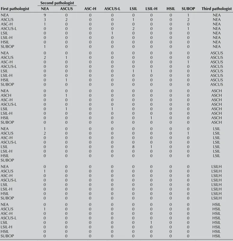
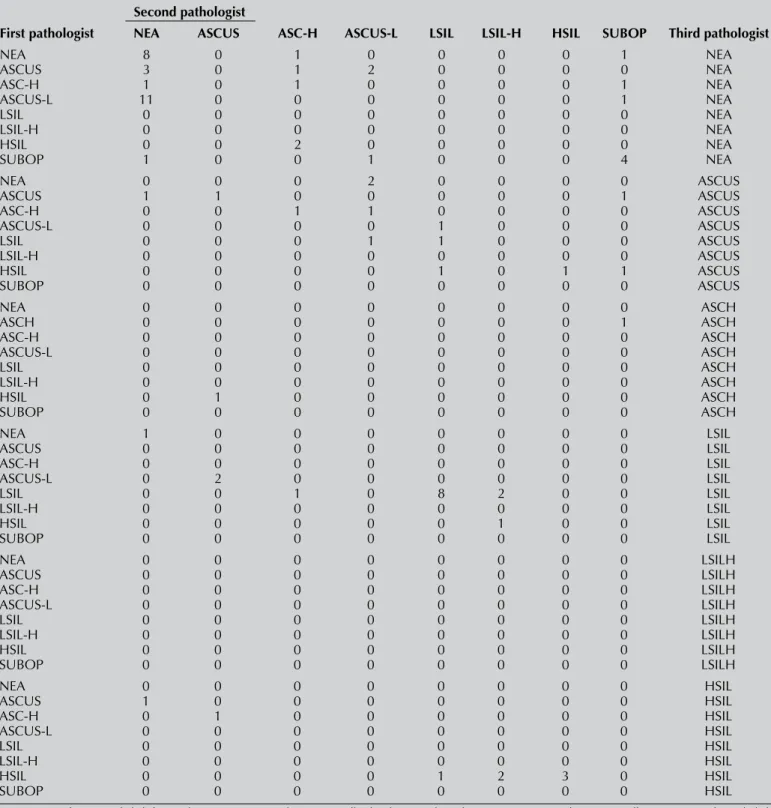
The findings for LBC and CPS are summarized in Table 1 and Table 2, respectively. There was a slightly better agreement in the CPS preparations. The triple agreement percentage was 58.82% (30/51) in LBC with a kappa value of 0.427 and 65.67% (44/67) in CPS with a kappa value of 0.505. The highest agreement between di-agnosis was in the LSIL group (kappa=0.625), while the lowest one was in the ASCUS group (kappa=0.045) (Table 3 and Table 4).
Table 1. The distribution of 51 cases diagnosed as negative or positive for intraepithelial abnormalities and epithelial abnormalities in liquid-based cytology preparations
Third pathologist Second pathologist Total
Negative Positive
Negative First pathologist Negative 10 2 12
Positive 7 4 11
Total 17 6 23
Positive First pathologist Negative 1 0 1
Positive 7 20 27
Total 8 20 28
Table 2. The distribution of 67 cases diagnosed as negative or positive for intraepithelial abnormalities and epithelial abnormalities in conventional Pap smear preparations
Third pathologist Second pathologist Total
Negative Positive
Negative First pathologist Negative 13 3 16
Positive 7 6 13
Total 20 9 29
Positive First pathologist Negative 1 2 3
Positive 4 31 35
The kappa values of interobserver variability varied between 0.366-0.567 with a moderate degree of agreement. The results are presented in Table 5.
Discussion
In order to evaluate the interobserver reproducibility of cervical smears prepared by conventional Pap smear or by the PapSpinTM
method, a set of 120 cervical smears from 90 patients were evalu-ated by three pathologists. The interobserver variability showed a moderate degree of agreement with a slightly higher percentage in CPS. There have been several studies investigating interobserver variability in the diagnosis of cervical epithelial cell abnormalities including a large group by comparing CPS and LBC. In their study of a group of 20,000 patients, Yobs et al. (9) found 82-96.8%
agree-Table 3. The results of liquid-based cytology preparations according to three pathologists Second pathologist
First pathologist NEA ASCUS ASC-H ASCUS-L LSIL LSIL-H HSIL SUBOP Third pathologist
NEA 9 0 0 0 0 0 0 1 NEA ASCUS 3 2 0 0 1 0 0 2 NEA ASC-H 1 0 0 0 0 0 0 0 NEA ASCUS-L 0 0 0 0 2 0 0 1 NEA LSIL 0 0 0 1 0 0 0 0 NEA LSIL-H 0 0 0 0 0 0 0 0 NEA HSIL 0 0 0 0 0 0 0 0 NEA SUBOP 1 0 0 0 0 0 0 0 NEA NEA 0 0 0 0 0 0 0 0 ASCUS ASCUS 2 1 0 0 0 0 0 0 ASCUS ASC-H 0 0 0 0 0 0 0 1 ASCUS ASCUS-L 0 0 0 0 0 0 0 0 ASCUS LSIL 0 0 0 0 1 1 0 0 ASCUS LSIL-H 0 0 0 0 0 0 0 0 ASCUS HSIL 0 1 0 0 0 0 0 0 ASCUS SUBOP 0 0 0 0 0 0 0 0 ASCUS NEA 0 0 0 0 0 0 0 0 ASCH ASCH 0 1 0 0 0 0 0 0 ASCH ASC-H 0 0 0 0 0 0 0 0 ASCH ASCUS-L 0 0 0 0 0 0 0 0 ASCH LSIL 0 1 0 1 0 0 0 0 ASCH LSIL-H 0 0 0 0 0 0 0 0 ASCH HSIL 0 0 0 0 0 1 0 0 ASCH SUBOP 0 0 0 0 0 0 0 0 ASCH NEA 1 0 0 0 0 0 0 0 LSIL ASCUS 2 0 0 0 0 0 0 1 LSIL ASC-H 0 0 0 0 0 0 0 0 LSIL ASCUS-L 0 0 0 0 0 0 0 0 LSIL LSIL 0 0 0 0 8 1 0 0 LSIL LSIL-H 0 0 0 0 0 0 0 0 LSIL HSIL 0 0 0 0 0 0 0 0 LSIL SUBOP NEA 0 0 0 0 0 0 0 0 LSILH ASCUS 1 0 0 0 0 0 0 0 LSILH ASC-H 0 0 0 0 0 0 0 0 LSILH ASCUS-L 0 0 0 0 0 0 0 0 LSILH LSIL 0 0 0 0 0 0 0 0 LSILH LSIL-H 0 0 0 0 0 0 0 0 LSILH HSIL 0 0 0 0 0 0 0 0 LSILH SUBOP 0 0 0 0 0 0 0 0 LSILH NEA 0 0 0 0 0 0 0 0 HSIL ASCUS 1 0 0 0 0 0 0 0 HSIL ASC-H 0 0 0 0 0 0 0 0 HSIL ASCUS-L 0 0 0 0 0 0 0 0 HSIL LSIL 0 0 0 0 0 1 0 0 HSIL LSIL-H 0 0 0 0 0 0 0 0 HSIL HSIL 0 0 0 0 0 0 0 0 HSIL SUBOP 0 0 0 0 0 0 0 0 HSIL
NEA: Negative for intraepithelial abnormalities. ASCUS: Atypical squamous cells of undetermined significance. ASCUS-L: ASCUS having few cells suspicious of a low grade squamous intraepithelial lesion. ASC-H: Atypical squamous cells, HGSIL cannot be excluded. HSIL: High grade squamous intraepithelial lesion. LSIL: Low grade squamous intraepithelial lesion. SUBOP: suboptimal smear
ment, with the lowest percentage in moderate dysplasia. Duca et al. (2) studied 120 CPS slides assessed by three cytotechnicians and found interobserver agreement with kappa values in the range of 0.418–0.575. Similarly, Klinkhamer et al. (10) noted 83.3% consis-tency with no more than one grade of disagreement. Confortini et al. (11), in their study reviewing a set of 100 slides assessed by 16 cytopathologists, found a moderate to good degree of agreement
with kappa values varying from 0.35-0.57; the best agreement was seen in severe dysplasia. The results were similar in the case of CPS reproducibility. However, when compared with LBC, there are conflicting results. While some studies state a high degree of repro-ducibility with LBC, there are also studies showing no difference or less reproducibility (4-8, 12, 13).
Table 4. The results of conventional Pap smear preparations according to three pathologists Second pathologist
First pathologist NEA ASCUS ASC-H ASCUS-L LSIL LSIL-H HSIL SUBOP Third pathologist
NEA 8 0 1 0 0 0 0 1 NEA ASCUS 3 0 1 2 0 0 0 0 NEA ASC-H 1 0 1 0 0 0 0 1 NEA ASCUS-L 11 0 0 0 0 0 0 1 NEA LSIL 0 0 0 0 0 0 0 0 NEA LSIL-H 0 0 0 0 0 0 0 0 NEA HSIL 0 0 2 0 0 0 0 0 NEA SUBOP 1 0 0 1 0 0 0 4 NEA NEA 0 0 0 2 0 0 0 0 ASCUS ASCUS 1 1 0 0 0 0 0 1 ASCUS ASC-H 0 0 1 1 0 0 0 0 ASCUS ASCUS-L 0 0 0 0 1 0 0 0 ASCUS LSIL 0 0 0 1 1 0 0 0 ASCUS LSIL-H 0 0 0 0 0 0 0 0 ASCUS HSIL 0 0 0 0 1 0 1 1 ASCUS SUBOP 0 0 0 0 0 0 0 0 ASCUS NEA 0 0 0 0 0 0 0 0 ASCH ASCH 0 0 0 0 0 0 0 1 ASCH ASC-H 0 0 0 0 0 0 0 0 ASCH ASCUS-L 0 0 0 0 0 0 0 0 ASCH LSIL 0 0 0 0 0 0 0 0 ASCH LSIL-H 0 0 0 0 0 0 0 0 ASCH HSIL 0 1 0 0 0 0 0 0 ASCH SUBOP 0 0 0 0 0 0 0 0 ASCH NEA 1 0 0 0 0 0 0 0 LSIL ASCUS 0 0 0 0 0 0 0 0 LSIL ASC-H 0 0 0 0 0 0 0 0 LSIL ASCUS-L 0 2 0 0 0 0 0 0 LSIL LSIL 0 0 1 0 8 2 0 0 LSIL LSIL-H 0 0 0 0 0 0 0 0 LSIL HSIL 0 0 0 0 0 1 0 0 LSIL SUBOP 0 0 0 0 0 0 0 0 LSIL NEA 0 0 0 0 0 0 0 0 LSILH ASCUS 0 0 0 0 0 0 0 0 LSILH ASC-H 0 0 0 0 0 0 0 0 LSILH ASCUS-L 0 0 0 0 0 0 0 0 LSILH LSIL 0 0 0 0 0 0 0 0 LSILH LSIL-H 0 0 0 0 0 0 0 0 LSILH HSIL 0 0 0 0 0 0 0 0 LSILH SUBOP 0 0 0 0 0 0 0 0 LSILH NEA 0 0 0 0 0 0 0 0 HSIL ASCUS 1 0 0 0 0 0 0 0 HSIL ASC-H 0 1 0 0 0 0 0 0 HSIL ASCUS-L 0 0 0 0 0 0 0 0 HSIL LSIL 0 0 0 0 0 0 0 0 HSIL LSIL-H 0 0 0 0 0 0 0 0 HSIL HSIL 0 0 0 0 1 2 3 0 HSIL SUBOP 0 0 0 0 0 0 0 0 HSIL
NEA: Negative for intraepithelial abnormalities. ASCUS: Atypical squamous cells of undetermined significance, ASC-H: Atypical squamous cells, HGSIL cannot be excluded, HSIL: High grade squamous intraepithelial lesion, LSIL: Low grade squamous intraepithelial lesion, ASCUS-L: Atypical squamous cells of undetermined significance, LSIL cannot be excluded, LSIL-H: Low grade squamous intraepithelial lesion, HGSIL cannot be excluded, SUBOP: Suboptimal smear
In our study, we found a slight better agreement in the CPS prepa-rations. The triple agreement percentage was 58.82% (30/51) in LBC with a kappa value of 0.427 and 65.67% (44/67) in CPS with a kappa value of 0.505. The highest agreement between diagnosis was in the LSIL group (kappa=0.625), while the lowest value was in the ASCUS group (kappa=0.045). These differences may be due to the LBC method. In a study using PapSpinTM as the LBC method (4),
the results showed no great differences. However, the consensus of opinion is that the highest agreement is achieved in HSIL carci-noma, while the lowest agreement is in the ASCUS group (2, 5-9, 11, 12). Our results agree with this consensus opinion.
Conclusion
The interobserver reproducibility of cervical cytology is moderately independent from the method used. LBC is more comfortable for the pathologist but has a higher cost. The selection of technique will be made according to the socio-economic status of the patient and the country. However, screening of the population should be as broad as possible, regardless of which method is used.
Acknowledgments
We would like to thank Assoc. Prof. Ferhan Elmalı for his great sup-port and intensive labor in the statistical analysis of the study and Pınar Korkmaz, Reyhan Yaşar, Gülcin Civan, Tülay Akgün, Arzu Özturk, Ozan Aydoğumus for their dedicated efforts in the comple-tion of this study.
Conflict of interest
No conflicts of interest were declared by the authors.
Authors’ contributions: Conceived and designed the experiments
or case: İÇT, NB, SA. Performed the experiments or case: İÇT, NB, SA. Analyzed the data: GBD. Wrote the paper: İÇT. All authors read and approved the final manuscript.
References
1. Koss LG. Melamet MR. Koss’ Diagnostic Cytology And Its Histopatho-logic Bases. 5th edition. Lipingot Williams and Wilkins. 2010.
2. Duca P, Braga M, Chiappa L, Piffer R, Turolla E. Intralaboratory rep-roducibility of interpretation of Pap smears: results of an experiment. Tumori 1988; 74(6): 737-44.
3. Arbyn M, Bergeron C, Klinkhamer P, Martin-Hirsch P, Siebers AG, Bul-ten J. Liquid compared with conventional cervical cytology: a systema-tic review and meta-analysis. Obstet Gynecol 2008; 111(1): 167-77.
[CrossRef]
4. Rosenthal DL, Geddes S, Trimble CL, Carson KA, Alli PM. The PapS-pin: a reasonable alternative to other, more expensive liquid-based Papanicolaou tests. Cancer 2006; 108(3): 137-43. [CrossRef]
5. Settakorn J, Rangdaeng S, Preechapornkul N, Nateewatana S, Pongsira-lai K, Srisomboon J, et al. Interobserver reproducibility with LiquiPrep liquid-based cervical cytology screening in a developing country. Asi-an Pac J CAsi-ancer Prev 2008; 9(1): 92-6.
6. Coste J, Cochand-Priollet B, de Cremoux P, Le Galès C, Cartier I, Molinié V, et al. Cross sectional study of conventional cervical smear, monolayer cytology, and human papillomavirus DNA testing for cervi-cal cancer screening. BMJ 2003; 326(7392): 733. [CrossRef]
7. Duggan MA, Khalil M, Brasher PM, Nation JG. Comparative study of the ThinPrep Pap test and conventional cytology results in a Canadian cohort. Cytopathology 2006; 17(2): 73-81. [CrossRef]
8. Celik C, Gezginç K, Toy H, Findik S, Yilmaz O. A comparison of liqu-id-based cytology with conventional cytology. Int J Gynaecol Obstet 2008; 100(2): 163-6. [CrossRef]
9. Yobs AR, Plott AE, Hicklin MD, Coleman SA, Johnston WW, Ashton PR, et al. Retrospective evaluation of gynecologic cytodiagnosis. II. Interlaboratory reproducibility as shown in rescreening large consecu-tive samples of reported cases. Acta Cytol 1987; 31(6): 900-10. 10. Klinkhamer PJ, Vooijs GP, de Haan AF. Intraobserver and interobserver
variability in the diagnosis of epithelial abnormalities in cervical sme-ars. Acta Cytol 1988; 32(6): 794-800.
11. Confortini M, Biggeri A, Cariaggi MP, Carozzi FM, Minuti PA, Russo A, et al. Intralaboratory reproducibility in cervical cytology. Results of the application of a 100-slide set. Acta Cytol 1993; 37(1): 49-54. 12. Stoler MH, Schiffman M; Atypical Squamous Cells of Undetermined
Significance-Low-grade Squamous Intraepithelial Lesion Triage Study (ALTS) Group. Interobserver reproducibility of cervical cytologic and histologic interpretations: realistic estimates from the ASCUS-LSIL Tri-age Study. JAMA 2001; 285(11): 1500-5. [CrossRef]
13. Biro C, Hyne S, Roberts J, Thurloe J, Bowditch R. Liquid-based ver-sus conventional cervical cytology. Lancet 2006; 367(9521): 1481-2.
[CrossRef] Table 5. Percentage of agreement and weighted kappa scores between pathologists
Method Agreement Kappa
First pathologist - Second pathologist Liquid-based cytology 68.6% 0.366 Second pathologist - Third pathologist Liquid-based cytology 72.5% 0.450 First pathologist - Third pathologist Liquid-based cytology 73.5% 0.456 First pathologist - Second pathologist Conventional Pap smear 76.0% 0.463 Second pathologist - Third pathologist Conventional Pap smear 79.0% 0.567 First pathologist - Third pathologist Conventional Pap smear 80.5% 0.493


