Serum eosinophilic cationic protein is correlated with
food impaction and endoscopic severity in eosinophilic
esophagitis
Cem Cengiz
Department of Gastroenterology, TOBB University of Economics and Technology School of Medicine, Ankara, Turkey
ABSTRACT
Background/Aims: The aim of the present study was to analyze the diagnostic accuracy of serum eosinophilic cationic protein (ECP) for eosinophilic esophagitis (EoE) and the correlation of ECP with clinical, histopathological, laboratory, and endoscopic features of EoE. Materials and Methods: Fifteen patients with EoE and 14 healthy controls were included in the study. Demographic parameters were recorded. EoE Endoscopic Reference Score (EREFS) was calculated according to endoscopic features, and esophageal biopsies were obtained by a single experienced endoscopist in a patient group. Serum ECP levels (μg/mL), absolute eosinophil count (U/mm3), and maximum peak of eosinophils/high-power field in esophageal biopsies were analyzed.
Results: The median age of all participants was 33.0 (min-max: 18-46) years. There were 27 (93.1%) male patients. Serum ECP level was significantly higher in patients with EoE than in healthy volunteers (20.4 vs. 8.8, p<0.0001). According to the receiver operating characteristic (ROC) curve analysis, ECP had 80% sensitivity and 92.8% specificity to diagnose EoE with a cut-off value of 13.9 μg/mL (area under the ROC curve 0.895; p<0.0001; 95% CI: 0.725-0.978). EREFS (p<0.0001) and the presence of food impaction (p=0.04) were significantly correlated with ECP.
Conclusion: Serum ECP is an accurate non-invasive biomarker for EoE with high specificity and sensitivity. In addition, ECP is strongly correlated with EREFS and the symptom of food impaction.
Keywords: Eosinophilic esophagitis, eosinophilic cationic protein, diagnosis
INTRODUCTION
Eosinophilic esophagitis (EoE) is a chronic esophageal inflammatory disease that is mediated by immunogen-ic/antigenic (primarily food allergen) mechanisms. It is characterized by esophageal symptoms (dysphagia, heartburn, and chest pain) caused by esophageal dys-function and eosinophilic esophageal inflammation (>15 eosinophilic granulocytes/high-power field (hpf)) (1,2). The prevalence of EoE in children and adults has been increasing steadily over the past decade (3-5). However, it is not clear if this is a true increase in prevalence or an artificial result due to improved understanding of the dis-ease by physicians, enabling a higher rate of diagnosis (6). Eosinophilic esophagitis Endoscopic Reference Score (EREFS) has been recently described to guide physicians for recognizing and reporting the main endoscopic find-ings of EoE (Table 1) (8). It has been documented that EREFS has a good inter- or intra-observer agreement. It is a simple scoring system with an accurate diagnostic util-ity that shows the severutil-ity of the disease and probabilutil-ity of response to treatment (8,9).
In addition to endoscopic/histopathological findings, some non-invasive serological markers have been studied for the diagnosis of EoE. Increased serum total IgE levels have been shown in active EoE disease (10). The diagnos-tic role of peripheral blood eosinophil count and eosino-phil-derived proteins (eosinoeosino-phil-derived neurotoxin and major basic protein) has been documented in previous studies. Another eosinophil-derived protein, eosinophilic cationic protein (ECP), has been studied and shown to be accurate in the clinical monitoring of asthma and allergic rhinoconjunctivitis (11). However, data regarding the di-agnostic accuracy of ECP for EoE are extremely rare. In the present study, we aimed to analyze serum ECP levels in patients with EoE and controls to predict the diagnostic accura-cy of ECP for EoE and to determine the correlation of ECP with clinical symptoms, eosinophilia (histological/peripheral blood), and endoscopic reference score of EoE in a patient group. MATERIAL AND METHODS
Fifteen adult patients with EoE were included consecu-tively according to the current diagnostic criteria (12) as Cite this article as: Cengiz C. Serum eosinophilic cationic protein is correlated with food impaction and endoscopic severity in eosinophilic esophagitis. Turk J Gastroenterol 2019; 30(4): 345-9.
Corresponding Author: Cem Cengiz; [email protected]
Received: June 29, 2018 Accepted: September 11, 2018 Available online date: January 21, 2019
© Copyright 2019 by The Turkish Society of Gastroenterology • Available online at www.turkjgastroenterol.org DOI: 10.5152/tjg.2019.18529
≥15 eosinophils/hpf (eo/hpf) in esophagus biopsies be-fore and after treatment with proton pump inhibitors for 8 weeks. The control group included 14 healthy partici-pants to provide blood sample only for ECP.
Endoscopic procedures were performed by an experi-enced gastroenterologist (CC) using a flexible 9.9 mm gastroscope with a 2.8 mm work channel (GIF-H180J; Olympus. Hamburg, Germany). Biopsies from the prox-imal esophagus were obtained using conventional for-ceps when there is an endoscopic stigmata of EoE. No complications were observed during the endoscopic procedure.
Biopsy specimens were sent to the pathology depart-ment of the TOBB University of Economics and Tech-nology Hospital. A diagnosis of “histologically compatible with EoE” was made with the presence of ≥15 eo/hpf clustered in microabscesses.
Venous blood was collected from the participants. Abso-lute eosinophil count (AEC) was analyzed via an automat-ed hematology analyzer at the central laboratory of the TOBB Economy and Technology University Hospital. ECP assay was performed in an affiliated commercial labora-tory specialized for this type of tests.
ECP assay procedure
1. All reagents, samples, and standards were prepared according to the manufacturer’s instructions.
2. One hundred µL of sample was added into each well and incubated for 2 h at 37°C.
3. The liquid of each well was removed, and 100 µL of biotin antibody was added into each well and incu-bated for 1 h at 37°C.
4. Each well was aspirated and washed three times. 5. One hundred µL of horseradish peroxidase-avidin
was added into each well and incubated for 1 h at 37°C.
6. Each well was aspirated and washed five times. 7. Ninety µL of 3,3′,5,5′-tetramethylbenzidine
sub-strate was added into each well and incubated for 15-30 min at 37°C.
8. Fifty µL of stop solution was added into each well, and the assay was read at 450 nm within 5 min. Statistical analysis
The Statistical Package for the Social Sciences (SPSS) version 16 software (SPSS Inc.; Chicago, IL, USA) was used for statistical analysis. Kolmogorov-Smirnov test was used to determine the normality of the distribu-tion of data. Comparisons between the groups were performed by the Mann-Whitney U test for non-para-metric variables. The group characteristics were com-pared using the Fisher’s exact test and the chi-square test. A receiver operating characteristic (ROC) curve was used to determine the diagnostic ability of ECP for EoE. Pearson’s test was used for correlation analyses. A p-value of <0.05 was considered statistically signif-icant.
RESULTS
Study population
Our study included 29 participants (15 consecutive pa-tients with EoE and 14 age-sex-matched control group). The median age of all participants was 33.0 (min-max: 18-46) years. Twenty-seven (93.1%) participants were male. Table 2 shows the demographic characteristics of the participants.
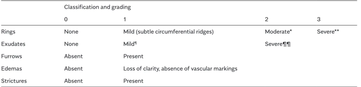
Table 1. Eosinophilic Esophagitis Endoscopic Reference Score (EREFS) Classification and grading
0 1 2 3
Rings None Mild (subtle circumferential ridges) Moderate* Severe**
Exudates None Mild¶ Severe¶¶
Furrows Absent Present
Edemas Absent Loss of clarity, absence of vascular markings Strictures Absent Present
*Distinct rings that do not impair passage of a standard diagnostic adult endoscope of outer diameter 8-9.5 mm **Distinct rings that do not permit passage of a diagnostic endoscope
¶Lesions involving <10% of the esophageal surface ¶¶Lesions involving >10% of the esophageal surface
Clinical characteristics, laboratory findings, and EREFS of patients with EoE
Eleven (73.3%) patients with EoE had at least one allergic disease. Eight out of the 11 patients had food allergy, three had allergic rhinitis, and four (26.7%) had bronchial asthma. The most common symptom was dysphagia (9/15, 60.0%) among patients with EoE. The median eo/hpf count in esophageal biopsy was 40 (min-max: 15-100) while periph-eral blood eosinophilia was observed in 4 (26.7%) patients with EoE. The median EREFS was found to be 3.0 (min-max: 1.0-6.0) in patients with EoE. Table 2 shows the clinical characteristics and laboratory findings of patients with EoE. Predictive value of ECP for EoE
Eosinophilic cationic protein level was significantly high-er in patients with EoE than in healthy voluntehigh-ers (20.4 vs. 8.8, p<0.0001) (Figure 1). An ROC curve was used to
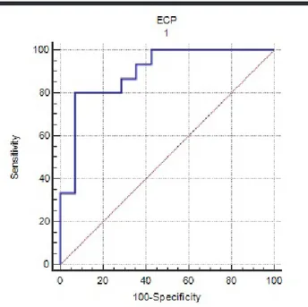
determine the predictive value of ECP for EoE. Accord-ing to this ROC curve analysis, the cut-off value of ECP was found to be 13.9 µg/mL (sensitivity: 80% (95% CI: 51.9-95.7), specificity: 92.8% (95% CI: 66.1-99.8), posi-tive predicposi-tive value: 92.3 (95% CI: 64.0-99.8), and neg-ative predictive value: 81.2 (95% CI: 54.4-96.0), with an area under the ROC curve (AUC) 0.895; p<0.0001; 95% CI: 0.725-0.978) (Fig. 2).
Table 2. Demographic, clinical characteristics, and laboratory findings of the study groups
EoE (+),
n=15 EoE (-), n=14 p Age (median, min-max) 33.0 (18-44) 33.0 (18-46) 0.7 Male gender (n, %) 14 (93.3) 13 (92.9) 1.0 Atopic disease Asthma (n, %) 4 (26.7) -Allergic rhinitis (n, %) 3 (20.0) Other AD (n, %) 1 (6.7) Symptoms Dysphagia (n, %) 9 (60.0) Heartburn (n, %) 6 (40.0) Food impaction 2 (13.3) Laboratory/histopathological finding
Peripheral blood eosinophilia
(n, %) 4 (26.7)
EC/hpf (esophageal biopsy)
(median, min-max) 40 (15-100) AEC (median, min-max) 285
(205-450) EREFS (median, min-max) 3.0 (1.0-6.0)
EoE: eosinophilic esophagitis; AD: allergic disease; EC: eosinophil count; hpf: high-power field; AEC: absolute eosinophil count; EREFS: Eosinophilic Esophagitis Endoscopic Reference Score
Figure 1. Comparison of the median ECP between patients with EoE and control group
Figure 2. A receiver operating characteristic curve analysis regarding the diagnostic ability of ECP for EoE
Correlation of ECP with symptoms, laboratory findings, and EREFS
When correlation of ECP with AEC, esophageal tissue eo-sinophil count, EREFS, and EoE symptoms was analyzed, only EREFS (p<0.0001) and the presence of food impac-tion (p=0.04) were significantly correlated with ECP (Ta-ble 3).
DISCUSSION
Eosinophilic esophagitis is predominantly seen in male patients between 30 and 40 years old. Although it is as-sumed that the prevalence of EoE is increasing due to improvement in the understanding of the disease, it is still apparent that EoE is an underdiagnosed entity with a delay of diagnosis (6).
Histological examination of the esophageal mucosa is required for the exact diagnosis of EoE. Non-invasive di-agnostic markers have been analyzed in previous studies to assist in the diagnosis and monitoring of EoE. With the establishment of these non-invasive markers, it is aimed to prevent multiple endoscopies that are costly, invasive, and associated with risks (13). Several prospective stud-ies have assessed the potential role of various biomark-ers, including peripheral eosinophil count, cytokines, and chemokines involved in the pathogenesis of EoE (14-17). Contradictory results have been documented in studies analyzing the diagnostic role of several non-invasive bio-markers for EoE. In the study by Dellon et al., 14 different serum biomarkers showed no significant correlation or difference between subjects with EoE and controls (18). Eosinophils have cytoplasmic granules that degranulate on stimulation and release toxic mediators, causing tissue damage and inflammation (19). Several studies have re-ported that in addition to the aforementioned non-inva-sive markers, the presence of these degranulation prod-ucts in the esophageal tissue can help to diagnose EoE (13,17). In a recent study, Min et al. showed that serum ECP is significantly higher in patients with EoE than in controls (13). Since elevation of ECP may also be due to bronchial inflammation, it is suggested that ECP will be
beneficial only in patients without asthma (6). However in the study by Min et al., higher serum ECP level was an independent predictor of EoE when age, sex, and atopic disease were evaluated in multivariate analysis. The au-thors concluded that although ECP can be elevated in other atopic diseases, such as asthma or allergic rhinitis, it was noteworthy to report since it has remained signifi-cantly associated with EoE by the multivariate analysis (13). In our study, we determined that ECP levels were significantly higher in subjects with EoE than in controls. In addition, we documented that ECP has very accurate sensitivity and specificity values (AUC 0.895; p<0.0001) to diagnose EoE.
There are contradictory results in previous studies ana-lyzing the correlation of ECP with AEC and esophageal tissue eosinophil count. In the study by Chehade et al., ECP has been found to be significantly higher in pa-tients with EoE, and a positive correlation was obtained between ECP and peripheral eosinophilia, whereas this correlation could not be observed between ECP and eo-sinophil count in esophagus biopsy (20). The findings of the study by Rodriguez-Sanchez were similar with the aforementioned study (21). However, Min et al. showed a significant correlation between ECP and esophageal tis-sue eosinophil count (13). In our study, differently from previous studies, ECP was correlated neither with AEC nor with esophageal tissue eosinophil count.
Endoscopic reference score is a recent classification sys-tem that is used to define the endoscopic features (ede-mas, rings, exudates, furrows, and strictures) of EoE. This grading system has an accurate intra- and inter-observer agreement that has been independently validated by sev-eral investigators from North America and Europe (7,22). Shoepfer et al. (23) showed that EREFS is associated with symptom severity due to a validated patient-report-ed outcome instrument for EoE.
Although correlation of non-invasive biomarkers of EoE and laboratory/histopathological parameters has been studied in several studies, to the best of our knowledge, Table 3. Correlation of ECP with symptoms, laboratory findings, and EREFS
Laboratory Symptoms Endoscopy
AEC HPEC Heartburn Dysphagia FI EREFS
r -0.077 0.072 -0.201 0.398 0.525 0.900
p 0.7 0.7 0.4 0.1 0.04 <0.0001
ECP: eosinophilic cationic protein; EREFS: Eosinophilic Esophagitis Endoscopic Reference Score; AEC: absolute eosinophil count; HPEC: histopathological eosinophil count; FI: food impaction
there is no study analyzing the correlation of non-inva-sive biomarkers with endoscopic features. In the current study, we analyzed the correlation of ECP (a non-invasive biomarker) with endoscopic grade of EoE (EREFS), AEC, esophageal tissue eosinophil count, and EoE symptoms. Importantly, we found that EREFS and the presence of food impaction symptom were strongly correlated with ECP.
In conclusion, we have shown that a non-invasive bio-marker ECP is an accurate diagnostic tool with high specificity and sensitivity. The most striking finding of the present study is the significant correlation between ECP and EREFS that has not been studied in the literature to date. However, a small sample size is a limitation of the present study, and further prospective studies with larger sample size are needed to confirm the role of biomarkers in assessing the diagnosis and symptomatic/endoscopic severity of EoE.
Ethics Committee Approval: Ethics committee approval was
re-ceived for this study from the Ethics Committee of TOBB University of Economics and Technology (Decision No: MED/2171/2011; Deci-sion Date: August 8, 2011).
Informed Consent: Written informed consent was obtained from
the patients who participated in this study.
Peer-review: Externally peer-reviewed.
Conflict of Interest: The author has no conflict of interest to
de-clare.
Financial Disclosure: The author declared that this study has
re-ceived no financial support.
REFERENCES
1. Furuta GT, Liacouras CA, Collins MH, et al. First International Gas-trointestinal Eosinophil Research Symposium (FIGERS) Subcommit-tees. Eosinophilic esophagitis in children and adults: a systematic review and consensus recommendations for diagnosis and treat-ment. Gastroenterology 2007; 133: 1342-63. [CrossRef]
2. Liacouras CA, Furuta GT, Hirano I, et al. Eosinophilic esophagitis: updated consensus recommendations for children and adults. J Al-lergy Clin Immunol 2011; 128: 3-20, e6; quiz 21-2.
3. Simon D, Wardlaw A, Rothenberg ME. Organ-specific eosinophilic-disorders of the skin, lung, and gastrointestinal tract. J Allergy Clin Immunol 2010; 126: 3-13. [CrossRef]
4. Roy-Ghanta S, Larosa DF, Katzka DA. Atopic characteristics ofadult patients with eosinophilic esophagitis. Clin Gastroenterol Hepatol 2008; 6: 531-5. [CrossRef]
5. Atkins D, Furuta GT. Mucosal immunology, eosinophilic esophagi-tis, and other intestinal inflammatory diseases. J Allergy Clin Immu-nol 2010; 125: S255-61. [CrossRef]
6. Castro Jiménez A, Gómez Torrijos E, García Rodríguez R, et al. De-mographic, clinical and allergological characteristics of Eosinophil-ic Esophagitis in a Spanish central region. Allergol Immunopathol (Madr) 2014; 42: 407-14. [CrossRef]
7. Hirano I, Moy N, Heckman MG, Thomas CS, Gonsalves N, Achem SR. Endoscopic assessment of the oesophageal features of eosino-philic oesophagitis: validation of a novel classification and grading system. Gut 2013; 62: 489-95. [CrossRef]
8. Dellon ES, Cotton CC, Gebhart JH, et al. Accuracy of the Eosino-philic Esophagitis Endoscopic Reference Score in Diagnosis and De-termining Response to Treatment. Clin Gastroenterol Hepatol 2016; 14: 31-9. [CrossRef]
9. van Rhijn BD, Warners MJ, Curvers WL, et al Evaluating the en-doscopic reference score for eosinophilic esophagitis: moderate to substantial intra- and interobserver reliability. Endoscopy 2014; 46: 1049-55. [CrossRef]
10. Gupta SK. Noninvasive markers of eosinophilic esophagitis. Gas-trointest Endosc Clin N Am 2008; 18: 157-67. [CrossRef]
11. Cheng KJ, Xu YY, Liu HY, Wang SQ. Serum eosinophil cationic protein level in Chinese subjects with nonallergic and local allergic rhinitis and its relation to the severity of disease. Am J Rhinol Allergy 2013; 27: 8-12. [CrossRef]
12. Liacouras CA, Furuta GT, Hirano I, et al. Eosinophilic esophagitis: Updated consensus recommendations for children and adults. J Al-lergy Clin Immunol 2011; 128: 3-20. [CrossRef]
13. Min SB, Nylund CM, Baker Tpet al. Longitudinal Evaluation of Noninvasive Biomarkers for Eosinophilic Esophagitis. J Clin Gastro-enterol 2017; 51: 127-35. [CrossRef]
14. Schlag C, Miehlke S, Heiseke A, et al. Peripheral blood eosino-phils and other non-invasive biomarkers can monitor treatment re-sponse in eosinophilic oesophagitis. Aliment Pharmacol Ther 2015; 42: 1122-30. [CrossRef]
15. Furuta GT, Kagalwalla AF, Lee JJ, et al. The oesophageal string test: a novel, minimally invasive method measures mucosal in-flammation in eosinophilic oesophagitis. Gut 2013; 62: 1395-405. [CrossRef]
16. Konikoff MR, Blanchard C, Kirby C, et al. Potential of blood eo-sinophils, eosinophil-derived neurotoxin, and eotaxin-3 as biomark-ers of eosinophilic esophagitis. Clin Gastroenterol Hepatol 2006; 4: 1328-36. [CrossRef]
17. Schlag C, Pfefferkorn S, Brockow K, et al. Serum eosinophil cat-ionic protein is superior to mast cell tryptase as marker for response to topical corticosteroid therapy in eosinophilic esophagitis. J Clin Gastroenterol 2014; 48: 600-606. [CrossRef]
18. Dellon ES, Rusin S, Gebhart JH, et al. Utility of noninvasive se-rum biomarker panel for diagnosis and monitoring of eosinophilic esophagitis: a prospective study. Am J Gastroenterol 2015; 110: 821-7. [CrossRef]
19. Rothenberg ME. Biology and treatment of eosinophilic esophagi-tis. Gastroenterology 2009; 137: 1238-49. [CrossRef]
20. Chehade M, Yershov O, Sampson HA. Serum eosinophil cationic protein and eosinophil derived neurotoxin are potential non-inva-sive biomarkers for eosinophilic esophagitis. Gastroenterology 2007; 132(4 Supl. 2): A-6.
21. Rodríguez-Sánchez J, Gómez-Torrijos E, de-la-Santa-Belda E, et al. Effectiveness of serological markers of eosinophil activity in monitoring eosinophilic esophagitis. Rev Esp Enferm Dig 2013; 105: 462-7. [CrossRef]
22. Peterson KA, Thomas KL, Hilden K, et al. Comparison of esome-prazole to aerosolized, swallowed fluticasone for eosinophilic esoph-agitis. Dig Dis Sci 2010; 55: 1313-9. [CrossRef]
23. Schoepfer AM, Straumann A, Panczak R, et al. Development and validation of a symptom-based activity index for adults with eosino-philic esophagitis. Gastroenterology 2014; 147: 1255-66. [CrossRef]