https://doi.org/10.1177/2515841419861856 https://doi.org/10.1177/2515841419861856 Ther Adv Ophthalmol
2019, Vol. 11: 1–7 DOI: 10.1177/ 2515841419861856 © The Author(s), 2019. Article reuse guidelines: sagepub.com/journals-permissions
journals.sagepub.com/home/oed 1
Introduction
Intravitreal injection (IVI) is commonly used to treat ocular pathologies that require effective drug supply to the back of the eye. Although IVI has been used for years for intraocular delivery of antibiotics to treat endophthalmitis,1 nowadays
they are used to inject steroids for intraocular inflammation2 and antivascular endothelial
growth factor agents for macular edema in dia-betic retinopathy,3 retinal vein occlusion (RVO),4
and neovascularization in age-related macular degeneration.5
IVIs may be the most performed procedure in ophthalmology. Numerous studies have reported the rate of pain related to IVI.6,7 Some of them
evaluated the pain related to a single injection,
whereas others evaluated the pain associated with repeated injections. Therefore, it requires anes-thesia like other ophthalmic procedures. Usually, more than one injection is required in most patients, and it may cause anxiety and discom-fort, which may also increase the risk of complica-tions. This worrisome condition decreases the treatment compliance of patients who require more than one injection, as in diabetic macular edema and age-related macular degeneration. Ozurdex (Allergan Inc., Irvine, CA, USA) is a dexamethasone drug delivery system. It is an intravitreal device of 6 mm in length and 0.46 mm in diameter that contains 0.7 mg of dexametha-sone and is inserted into the vitreous cavity with a 22-gauge needle. It is used to treat macular edema
Evaluation effectiveness of 0.1% nepafenac
on injection-related pain in patients
undergoing intravitreal Ozurdex injection
Tevfik Ogurel , Reyhan Ogurel, Fatma Ozkal, Yas¸ar Ölmez, Nurgül Örnekand Zafer Onaran Abstract
Purpose: To evaluate the analgesic effect of topical 0.1% nepafenac solution during intravitreal
Ozurdex injection.
Methods: This prospective, randomized, double-blind placebo-controlled study included 59
patients who were diagnosed with retinal vein occlusion or pseudophakic cystoid macular edema and were selected to receive intravitreal Ozurdex injection. The patients were divided into two groups. Group 1, consisting of 31 eyes of 31 patients, received topical 0.1% nepafenac with topical anesthesia (0.5% proparacaine HCl, Alcaine; Alcon, TX, USA), and group 2,
consisting of 28 eyes of 28 patients, received placebo with topical anesthesia.
Results: There were 14 (45.2%) men and 17 (54.8%) women in group 1 and 16 (57.1%) men and
12 (42.9%) women in group 2. The mean age of the subjects was 64.42 ± 5.51 years in group 1 and 62.32 ± 7.54 years in group 2. The median visual analog scale pain score was 2 (1–3) in group 1 and 4 (1–6) in group 2. The visual analog scale pain score was significantly lower in group 1 than in group 2 (p < 0.001).
Conclusion: Topical 0.1% nepafenac has an additive analgesic effect when combined with
topical anesthesia for intravitreal Ozurdex injection.
Keywords: intravitreal injection, nepafenac, Ozurdex, pain
Received: 13 November 2018; revised manuscript accepted: 1 June 2019.
Correspondence to: Tevfik Ogurel Department of Ophthalmology, Faculty of Medicine, Kırıkkale University, Kırıkkale University Campus, Ankara Road 6. Km, Yahsihan 71100, Kırıkkale, Turkey. [email protected] Tevfik Ogurel Fatma Ozkal Nurgül Örnek Zafer Onaran Department of Ophthalmology, Faculty of Medicine, Kırıkkale University, Yahsihan, Turkey Reyhan Ogurel Reyhan Ogurel Eye Clinic, Kırıkkale, Turkey Yas¸ar Ölmez Adıyaman Besni State Hospital, Besni, Turkey
secondary to RVO as well as diabetic retinopa-thy, noninfectious uveitis, and pseudophakic cystoid macular edema (PCME; Irvine–Gass syndrome).8–10
Nepafenac ophthalmic suspension is a topical ocular nonsteroidal anti-inflammatory drug (NSAID). Although it is approved to treat inflam-mation and pain after cataract surgery, it has also been used to treat exudative age-related macular degeneration, prevent cystoid macular edema, and reduce diabetic macular edema.11–13 It is
metabolized into its active form, amfenac, because it is a prodrug. Studies have shown that nepafenac had greater inhibition of prostaglandin synthesis, longer duration, and greater corneal penetration than diclofenac.14 Nepafenac is a more potent at
inhibiting COX-2 than bromfenac and ketorolac.15
Today, there is still controversy regarding the most effective procedure of anesthesia for lessening dis-ruption and pain during IVI. Previously, several local anesthetic techniques for IVIs have been com-pared, including topical eye drops, gel, peribulbar injection, and subconjunctival injection.7,16–18
In this study, we aimed to evaluate the analgesic effect of topical 0.1% nepafenac in patients undergoing intravitreal Ozurdex injection.
Materials and methods
This prospective, randomized, double-blind pla-cebo-controlled study included 59 patients who were diagnosed with RVO or PCME and were selected to have intravitreal Ozurdex injections. The research was confirmed by Institutional Review Board and all concerned patients had provided informed consent in keeping with the Helsinki Declaration.
Patients with a major psychiatric disorder, demen-tia, or other neurological diseases affecting mem-ory and cognitive function; diabetic patients with known peripheral neuropathy; or a previously
known allergic reaction to the agents to be used were excluded.
Patients were randomized by Y. Ölmez using the block randomization method. The patients were distributed into two groups. Group 1, consisting of 31 eyes of 31 patients, received topical 0.1% nepafenac with topical anesthesia (0.5% propa-racaine HCl, Alcaine; Alcon, TX, USA), and group 2, consisting of 28 eyes of 28 patients, received placebo (sterile saline solution) with top-ical anesthesia. All injections were performed by the same specialist (T. Ogurel).
Topical 0.1% nepafenac was used half an hour and again 5 min just before the injection in group 1, and placebo was used in group 2. Both topical agents were given in camouflaged bottles with trial-specific tags to hide the identity of the test agent and were placed in a tamper-evident box. One of two nurses who had been accustomed to the technique was administered the agents. Two drops of proparacaine, 0.1% nepafenac, and pla-cebo were applied each time.
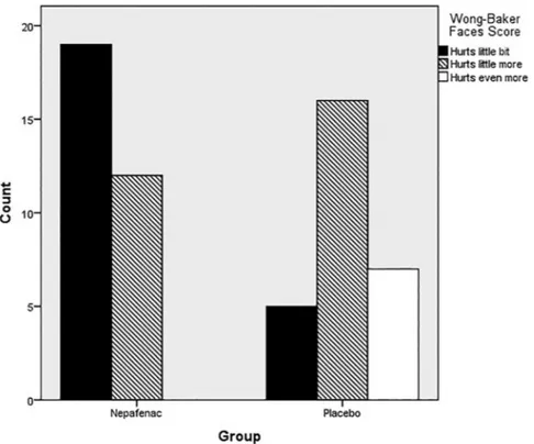
One drop of 10% povidone-iodine was supplied to each patient before the IVI. Injections were per-formed at 4.0 mm site from the limbus for phakic patients and at 3.5 mm site from the limbus for pseudophakic patients in the superotemporal quadrant of each eye. Immediately following the injection, the visual analog scale (VAS) was explained to the patients for pain, and they were tested to categorize their pain from 0 to 10, with 0 = no pain/no distress and 10 = worst possible pain/unbearable distress (Figure 1). Also, the Wong-Baker Faces Pain Rating Scale was evalu-ated as an observer scale (0 = no hurt, 1–2 = hurts a little bit, 3–4 = hurts a little more, 5–6 = hurts even more, 7–8 = hurts a whole lot, and 9–10 = hurts the worst) (Figure 2). All patients were injected with Ozurdex for the first time. Ofloxacin, a third-generation fluoroquinolone, was prescribed to all patients for 3–5 days as a postinjection antibiotic.
All data analyses were performed using a statisti-cal package (version 20.0, IBM), and software was used for the power analyses. The determined effect size was 1.13; considering a type I error (α) of 0.05 and accepting a power of 95%, a mini-mum sample size was calculated. The power cal-culation analysis revealed that the minimum required sample size was 19 patients for each group estimation based on the VAS pain score according to the data from a previous study.19
The Kolmogorov–Smirnov test was used to examine whether the data fit a normal distribu-tion. The independent samples t test and Mann– Whitney U test were used to compare variables. Comparisons of nominal data were evaluated with chi-square test. Spearman’s Rho correlation test was used to check the correlation between quantitative variables. In all analyses, a value of p < 0.05 was considered statistically significant.
Results
There were 14 (45.2%) men and 17 (54.8%) women in group 1 and 16 (57.1%) men and 12 (42.9%) women in group 2. The mean age of the patients was 64.42 ± 5.51 years in group 1 and 62.32 ± 7.54 years in group 2. There was no sig-nificant difference between the two groups in terms of sex and age (p = 0.411 and p = 0.284). Table 1 shows the demographic data of groups. The number of subjects with RVO and PCME was 25 and 6, respectively, in group 1 and 20 and 8, respectively, in group 2.
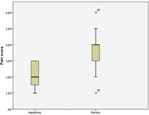
The median VAS pain score was 2 (1–3) in group 1 and 4 (1–6) in group 2. The VAS pain score was significantly lower in group 1 compared with group 2 (p < 0.001) (Figure 3). The Wong-Baker Faces scores were statistically reduced in group 1 (p < 0.001). Figure 4 shows the number of Figure 2. Wong-Baker Faces scores.
Table 1. Demographic characteristics and VAS score of two groups.
Variable Nepafenac Placebo p value
Age (mean ± SD) 64.42 ± 5.51 62.32 ± 7.54 0.411a Sex 0.284b Male, n (%) 14 (45.2) 16 (59.3) Female, n (%) 17 (54.8) 11 (40.7) Ocular disease 0.553b RVO, n (%) 25 (80.6) 20 (74.1) PCME, n (%) 6 (19.4) 7 (24.9)
VAS score (median/mean–max) 2 (1–3) 4 (1–6) <0.001c
PCME, pseudophakic cystoid macular edema; RVO, retinal vein occlusion; VAS, Visual Analog Scale.
aIndependent t test. bChi-square test. cMann–Whitney U test.
patients according to their Wong-Baker Faces scores in both groups.
There was no correlation between VAS pain score and sex, age, or underlying disease (p > 0.05; Table 2)
No eyes experienced complications during the injections.
Discussion
This study found that the analgesic effect of 0.1% nepafenac reduces the pain of patients during intravitreal Ozurdex injection.
IVI is an effective procedure to deliver the desired concentration of drugs or pharmacologic agents into the eye. As most of the topical and periocular drugs used in ophthalmology have lower pene-trance and, therefore, reduced effectiveness in the back of the eye, they are not as effective as IVIs for targeted treatment. Today, there are several new treatment agents for retinal diseases, espe-cially for diabetic retinopathy and age-related macular degeneration. Moreover, numerous intravitreal pharmacologic agents have become available with continued delivery systems. Many
patients may necessitate as frequently as once-a-month injections, possibly for years.
Like other procedures in ophthalmology, IVIs cause pain at the site of injection and raise the concerns of patients. This pain can be associated with age, sex, anxiety, or number of injec-tions.6,20,21 The views on this matter are,
how-ever, controversial. While some authors reported that women and older patients perceived more pain,6 other authors claimed opposite views.19
This situation causes distress for patients and reduces treatment compliance, especially in patients who need multiple injections to attain and sustain a treatment effect. Therefore, per-forming the procedure as painlessly as possible will make the patient more comfortable and agreeable.
Ozurdex is an intravitreal implant in an applica-tor. The applicator is a disposable injection device that contains a rod-shaped implant which is not visible and has a relatively large 22-gauge needle compared with other needles. The implant is approximately 0.46 mm in diameter and 6 mm in length. Therefore, patients may experience more pain than other procedures with a smaller size needle (27.5–32 gauges).22
Figure 3. Nonparametric box plots for the distribution of VAS pain scores are shown (horizontal lines are medians and quartiles, and circles indicate outliers and extreme values, respectively, with more than two to three times deviation of the interquartile range from the upper quartile).
Standard application of an IVI is to perform it under topical anesthesia. There are some studies which compared the effectiveness of various anes-thetic methods or agents for IVIs.23–25 Still, no
anesthesia technique for this procedure has been proven to eliminate pain completely.
Rifkin and Schaal23 compared the analgesic efficacy
of Tetravisc, proparacaine, and tetracaine and reported that patients receiving tetracaine had lower pain score than patients receiving propa-racaine or Tetravisc. Blaha and colleagues24 showed
that topical anesthesia is a more effective method for reducing pain related to IVI than lidocaine-applied pledget. In another study, Cintra and col-leagues18 compared three different anesthetic
methods for IVI of bevacizumab, including topical, subconjunctival, and peribulbar anesthesia, and found that peribulbar anesthesia was more effective
in limiting injection-related pain but was associated with minimum effectiveness in reducing entire pro-cedure pain. Also, there is a study that evaluated anesthetic methods for intravitreal Ozurdex injec-tion. Karabaş and colleagues25 compared topical
proparacaine drops and lidocaine-applied pledget with subconjunctival lidocaine injection and found that there was no difference in pain scores.
Topical NSAIDs have been used in ophthalmol-ogy to control postoperative inflammation, pre-vent PCME,25 maintain intraoperative mydriasis,26
and, in the last years, reduce pain after ocular sur-gery.27 In this study, 0.1% nepafenac combined
with topical anesthesia was used to control injec-tion-related pain.
Modi and colleagues28 found that nepafenac
0.3% used once-daily was as effective as three Figure 4. The number of patients according to the Wong-Baker Faces scores in both groups.
Table 2. Correlation with VAS score and age, sex, and ocular disease.
VAS score Age Sex Ocular disease
r 0.145 −0.228 0.055
p 0.273 0.08 0.677
significant analgesic effect during intravitreal Ozurdex injection. We think that these results may enhance patient comfort and improve compliance with treatment because pain is the most important factor affecting the patients’ adherence to treat-ment.29 Makri and colleagues30 reported that a
sin-gle drop of nepafenac before IVI was effective in reducing IVI-related pain immediately and up to 6 h after the injection. In another study, Georgakopoulos and colleagues19 compared nepafenac 0.1% and
0.3% with placebo in patients undergoing IVI, and they found that immediately after IVI, the VAS pain scores were statistically significantly lower in patients treated with nepafenac 0.1% and 0.3% compared with placebo. These studies support the findings of the present study because intravitreal Ozurdex injection is one type of IVI.
In the present study, VAS scores were relatively higher than in previous studies.7,31 We think that
this is due to the patient group in our study that received Ozurdex injections for the first time. The limitation of this study is the absence of an objective test to assess the pain sensitivity of patients before injection. Unfortunately, there is currently no test that can assess the pain sensitiv-ity of subjects.
In this study, the analgesic effectiveness and patient satisfaction with 0.1% nepafenac were evaluated with the VAS and Wong-Baker Faces scores. The VAS used in this study has been shown to be a reproducible and dependable method for evaluating a patient’s pain level.32,33
Previous studies have demonstrated the effective-ness of nepafenac in pain reduction after cataract surgery and IVIs. To our knowledge, this is the first study evaluating the analgesic effect of nepafenac on injection-related pain during intra-vitreal Ozurdex injection. Topical anesthesia is used as a standard anesthetic technique for IVIs, but it may not always provide complete analgesia. The present study has shown that 0.1% nepafenac combined with topical anesthesia may improve
in some patients.
Funding
The authors received no financial support for the research, authorship, and/or publication of this article.
Conflict of interest statement
The authors declared no potential conflicts of interest with respect to the research, authorship, and/or publication of this article.
ORCID iD
Tevfik Ogurel https://orcid.org/0000-0003- 0658-2286
References
1. Daily MJ, Peyman GA and Fishman G. Intravitreal injection of methicillin for treatment of endophthalmitis. Am J Ophthalmol 1973; 76: 343–350.
2. Kok H, Lau C, Maycock N, et al. Outcome of intravitreal triamcinolone in uveitis. Ophthalmology 2005; 112: 1916.e1–e7.
3. Crawford TN, Alfaro DV, Kerrison JB, et al. Diabetic retinopathy and angiogenesis. Curr
Diabetes Rev 2009; 5: 8–13.
4. Stahl A, Agostini H, Hansen LL, et al.
Bevacizumab in retinal vein occlusion: results of a prospective case series. Graefes Arch Clin Exp
Ophthalmol 2007; 245: 1429–1436.
5. Penfold PL, Gyory JF, Hunyor AB, et al. Exudative macular degeneration and intravitreal triamcinolone. A pilot study. Aust N Z J
Ophthalmol 1995; 23: 293–298.
6. Rifkin L and Schaal S. Shortening ocular pain duration following intravitreal injections. Eur J
Ophthalmol 2012; 22: 1008–1012.
7. Moisseiev E, Regenbogen M, Rabinovitch T,
et al. Evaluation of pain during intravitreal
Ozurdex injections vs. intravitreal bevacizumab injections. Eye (Lond) 2014; 28: 980–985.
9. Donati S, Gandolfi C, Caprani SM, et al. Evaluation of the effectiveness of treatment with dexamethasone intravitreal implant in cystoid macular edema secondary to retinal vein occlusion. Biomed Res Int 2018; 2018: 3095961. 10. Klamann A, Böttcher K, Ackermann P, et al.
Intravitreal dexamethasone implant for the treatment of postoperative macular edema.
Ophthalmologica 2016; 236: 181–185.
11. Hariprasad SM, Akduman L, Clever JA, et al. of cystoid macular edema with the new-generation NSAID nepafenac 0.1%. Clin Ophthalmol 2009; 3: 147–154.
12. Libondi T and Jonas JB. Topical nepafenac for treatment of exudative age-related macular degeneration. Acta Ophthalmol 2010; 88: e32–e33. 13. Callanan D and Williams P. Topical nepafenac
in the treatment of diabetic macular edema. Clin
Ophthalmol 2008; 2: 689–692.
14. Ke T-L, Graff G, Spellman JM, et al. Nepafenac, a unique nonsteroidal prodrug with potential utility in the treatment of trauma-induced ocular inflammation: II. Inflammation 2000; 24: 371–384.
15. Walters T, Raizman M, Ernest P, et al. In vivo pharmacokinetics and in vitro pharmacodynamics of nepafenac, amfenac, ketorolac, and bromfenac.
J Cataract Refract Surg 2007; 33: 1539–1545.
16. Kozak I, Cheng L and Freeman WR. Lidocaine gel anesthesia for intravitreal drug administration.
Retina 2005; 25: 994–998.
17. Kaderli B and Avci R. Comparison of topical and subconjunctival anesthesia in intravitreal injection administrations. Eur J Ophthalmol 2006; 16: 718–721.
18. Cintra LP, Lucena LR, Da Silva JA, et al. Comparative study of analgesic effectiveness using three different anesthetic techniques for intravitreal injection of bevacizumab. Ophthalmic
Surg Lasers Imaging 2009; 40: 13–18.
19. Georgakopoulos CD, Plotas P, Kagkelaris K, et al. Analgesic effect of a single drop of nepafenac 0.3% on pain associated with intravitreal injections: a randomized clinical trial.
J Ocul Pharmacol Ther 2019; 35: 168–173.
20. Haas P, Falkner-Radler C, Wimpissinger B,
et al. Needle size in intravitreal injections—pain
evaluation of a randomized clinical trial. Acta
Ophthalmol 2016; 94: 198–202.
22. Rodrigues E, Grumann A Jr, Penha FM, et al. Effect of needle type and injection technique on pain level and vitreal reflux in intravitreal injection.
J Ocul Pharmacol Ther 2011; 27: 197–203.
23. Rifkin L and Schaal S. Factors affecting patients’ pain intensity during in office intravitreal injection procedure. Retina 2012; 32: 696–700. 24. Blaha GR, Tilton EP, Barouch FC, et al.
Randomized trial of anesthetic methods for intravitreal injections. Retina 2011; 31: 535–539. 25. Karabaş VL, Ozkan B, Kocer CA, et al.
Comparison of two anesthetic methods for intravitreal Ozurdex injection. J Ophthalmol 2015; 2015: 861535.
26. Zanetti FR, Fulco EA, Chaves FR, et al. Effect of preoperative use of topical prednisolone acetate, ketorolac tromethamine, nepafenac, and placebo, on the maintenance of intraoperative mydriasis during cataract surgery: a randomized trial.
Indian J Ophthalmol 2012; 60: 277–281.
27. Rajpal RK, Ross B, Rajpal SD, et al. Bromfenac ophthalmic solution for the treatment of postoperative ocular pain and inflammation: safety, efficacy, and patient adherence. Patient
Prefer Adherence 2014; 8: 925–931.
28. Modi SS, Lehmann RP, Walters TR, et al. Once-daily nepafenac ophthalmic suspension 0.3% to prevent and treat ocular inflammation and pain after cataract surgery: phase 3 study. J Cataract
Refract Surg 2014; 40: 203–211.
29. So J. Improving patient compliance with biopharmaceuticals by reducing injection-associated pain. J Mucopolysacch Rare Dis 2015; 1: 15–18.
30. Makri OE, Tsapardoni FN, Plotas P, et al. Analgesic effect of topical nepafenac 0.1% on pain related to intravitreal injections: a randomized crossover study. Curr Eye Res 2018; 43: 1061. 31. Kessel L, Tendal B, Jørgensen KJ, et al.
Post-cataract prevention of inflammation and macular edema by steroid and nonsteroidal anti-inflammatory eye drops a systematic review.
Ophthalmology 2014; 121: 1915–1924.
32. Huskisson EC. Measurement of pain. Lancet 1974; 304: 1127–1131.
33. Price DP, McGrath PA, Rafii A, et al. The validation of visual analog scales as ratio scale for measures for chronic and experimental pain. Pain 1983; 17: 45–56.
Visit SAGE journals online journals.sagepub.com/ home/oed