556 Current Medical Science 40(3):2020
#Corresponding author, E-mail: [email protected] *This study was supported by Ege University School of Medicine-Research Funds, Izmir, Turkey (No. 2011-TIP-090).
The Role of Ankaferd Blood Stopper and Oxytocin as Potential
Therapeutic Agents in Endometriosis: A Rat Model
*Ismet Hortu1, 2#, Gokay Ozceltik1, Elif Karadadas3, Oytun Erbas4, Gurkan Yigitturk5, Murat Ulukus1
1Department of Obstetrics and Gynecology, School of Medicine, Ege University, Izmir 35100, Turkey
2Department of Stem Cell, Institute of Health Sciences, Ege University, Izmir 35100, Turkey
3Department of Biochemistry, School of Medicine, Ege University, Izmir 35100, Turkey
4Department of Physiology, School of Medicine, Istanbul Bilim University, Istanbul 34394, Turkey
5Department of Histology and Embryology, School of Medicine, Mugla Sitki Kocman University, Mugla 48000, Turkey
Huazhong University of Science and Technology 2020
Summary: To evaluate the potential effect of Ankaferd Blood Stopper (ABS) and oxytocin (OT) in an experimental endometriosis model, 18 female Sprague Dawley rats were used in this study. The animals were divided randomly into three groups after surgical induction of endometriosis: group 1: control group (isotonic NaCl, 1 mL/kg/day, intramuscular, n=6); group 2: OT group (OT, 80 U/kg/day, intramuscular, n=6); group 3: ABS group (ABS, 1.5 mL/kg/day, intraperitoneal, n=6). Each group was treated for four weeks (two times per week). Volumes of endometriotic explants were measured in biopsy samples for histopathological analysis. Vascular endothelial growth factor (VEGF), monocyte chemotactic protein-1 (MCP-1), and tumour necrosis factor (TNF-α) levels were measured in plasma and peritoneal fluid. Endometriotic explant volumes were significantly decreased after OT administration (P<0.0001). The epithelial score was significantly decreased in both treatment groups compared to the control group (P<0.05). TUNEL immunohistochemistry showed more apoptotic changes in the endometriosis foci (gland epithelium and surrounding tissue) in the OT group than in the control group (P<0.05). The levels of VEGF, MCP-1, and TNF-α were significantly reduced in the OT group (P<0.05), whereas no significant changes in protein levels were found in the ABS-applied group. The results indicate that OT has greater potential as a therapeutic agent in experimentally induced peritoneal endometriosis, where ABS, which is a VEGF modulator, appears to act through different mechanisms to show its palliative effects on a rat model of peritoneal endometriosis.
Key words: endometriosis; Ankaferd Blood Stopper; oxytocin; cytokines
Endometriosis is an enigmatic, chronic gynaeco-logical disease in which endometrial glandular and stromal structures are found outside the uterine cavity. Although endometriosis affects about 10% of women of childbearing age, the reported prevalence in women with pelvic pain and infertility ranges from 35% to 50%[1]. Bleeding from peritoneal foci
causes an increase in the number of cytokines within the peritoneal region, which is associated with inflammation, infertility, painful intercourse, and chronic pelvic pain. Although different theories about the pathogenesis of endometriosis are still debated, the most accepted theory is Sampson’s theory of retrograde
menstruation[2]. However, the etiopathogenesis of the
disease is not completely explained in all aspects, and the modalities of treatment are prone to changes and improvements.
Cytokines play a crucial role in the pathophysiology of the disease, where tumour necrosis factor-α (TNF-α) concentration was shown to be increased in the peritoneal fluid (PF) of women with endometriosis compared to that of women without the disease[3].
Furthermore, it has been reported that activated macrophages secrete TNF-α, which enhances both fibroblast proliferation favouring adhesion formation and angiogenesis by generating further cascades of inflammatory cytokines[4]. Vascular endothelial growth
factor (VEGF) is a growth factor specific to endothelial cells. It has been shown that the level of VEGF in the PF is increased in women with endometriosis[5]. Monocyte
chemotactic protein-1 (MCP-1) is responsible for DOI https://doi.org/10.1007/s11596-020-2213-1 40(3):556-562,2020
localisation of monocytes in response to injury or inflammation. It is a stimulatory factor for endometrial fibroblast cell proliferation, where its level is increased in the PF of women with endometriosis[6].
Oxytocin (OT) is a neuromodulator peptidic molecule. Peritoneal smooth muscle cells express oxytocin receptors (OTRs)[7]. OT binds to membrane
receptors of endometrial cells, increases Ca+2 influx
through the cells, and activates prostaglandin E2 (PGE2). In addition, it has been shown that OT functions as an anti-inflammatory molecule[8].
Ankaferd Blood Stopper (ABS) is a haemostatic agent composed of a combination of folkloric plant extracts. ABS comprises a standardized mixture of the plants Thymus vulgaris (dried leaf), Glycyrrhiza glabra (dried leaf), Vitis vinifera (dried leaf), Alpinia officinarum (dried leaf), and Urtica dioica (dried root); each has known effects on the endothelium, blood cells, angiogenesis, cell proliferation, vascular dynamics, and/or cell mediators[9, 10]. Extracts in ABS have been
shown to be effective with inflammatory, anti-angiogenic, and antioxidant properties in different studies[11, 12].
In this study, we aimed to evaluate OT and ABS as potential therapeutic agents in endometriosis to alleviate inflammation and, specifically for ABS, to modulate haemostatic regulation. The effects of the candidate therapeutics were assessed based on macroscopic, histological, and cytokine-level changes (VEGF, TNF-α, and MCP-1) with each treatment option in an endometriosis-induced rat model.
1 MATERIALS AND METHODS 1.1 Animals
Twenty-four mature female Sprague Dawley rats (weighing 180–260 g) were housed for eight weeks in groups of three rats per cage and fed ad libitum. The procedures used in this study were approved by the Institutional Animal Care and Ethical Committee of Ege University, Izmir, Turkey (2011/090). The animals were kept at 22±2°C with a 12-h light/dark cycle. The oestrous stage of each rat was determined by taking a vaginal smear at an interval of 6 to 12 h. Cell types in the smear were subsequently examined under a microscope according to the staining procedure of Papanicolaou. Twenty-four rats that had confirmed smear (oestrous stage) were included in the experiments.
1.2 Experimental Protocol
Rats were anesthetised by ketamine hydrochloride i.p. (40 mg/kg, Ketalar; Eczacibasi, Turkey) and xylazine hydrochloride i.p. (7 mg/kg, Alfazyne; Alfasan International BV, Holland). Initial endometriosis was induced under sterile conditions as described previously[13]. The first surgical procedure was started
with a longitudinal abdominal incision of 5 cm where
the peritoneal area was to be explored after abdominal shaving. After incision, penicillin (i.m.) was used against the possible risk of surgical infections (1000 U/kg, Iecilline; I.E. Ulugay, Turkey). The right uterine horn was ligated at the utero-tubal junction, and the cervical end was subsequently removed. The segmental horn was immersed in a sterile saline solution, the endometrium was exposed by bisecting along its antimesenteric axis, and 5 mm × 5 mm × 1 mm- sections were prepared. Explants were sutured to the anterolateral peritoneal surface by 5/0 vicryl polyglactin 910 suture (Ethicon, New Jersey, USA). The peritoneal area was closed, and after the implantation period for four weeks, the peritoneal area was incised to measure the endometrial explant volumes (V in mm3). The
spherical volume of each focus was calculated using the conventional prolate ellipsoid formula: V (mm3) =
0.524×W×L×T, in which W = width, L = length, T = thickness (all in millimetres)[14]. Since the presence of
peritoneal endometriotic foci was not confirmed in six rats, they were excluded from the study. The remaining 18 rats were randomly divided into three groups with six rats each (group 1: control; group 2: OT; group 3: ABS). All the tissues were photographed with a digital camera, and measurements were recorded.
After induction of peritoneal endometriosis, established experimental groups were treated with NaCl, OT, or ABS two times per week for a total of four weeks. Group 1 was administered with isotonic NaCl i.m. (1 mL/kg/day), group 2 was administered with OT i.m. (80 IU/kg, Pituisan, Ege Vet, Turkey), and group 3 was administered with ABS (Immun Ilac Kozmetik, Istanbul, Turkey) i.p. (1.5 mL/kg)[15, 16].
1.3 Histological Evaluation
After the given period, the peritoneal area was explored (third laparotomy) to measure endometrial foci volumes and to take PF samples. Peritoneal lavage was done with 1-mL NaCl isotonic solution. All the animals were sacrificed after necessary sampling procedures. Peritoneal explant volumes were again measured with the same method by the same investigator, who was blinded to group assignment. The tissues were sectioned as 5-μm-thick samples via microtome (Leica RM 2145, German). The tissue samples in 10% formaldehyde (Merck, German) were dehydrated and embedded in paraffin blocks. Histopathological scoring of implants was performed based on epithelial cell persistence, as previously described under a light microscope[17]. Scoring was
based on the following criteria: 0, no epithelium; 1, poorly preserved epithelium; 2, moderately preserved epithelium with leukocyte infiltration; and 3, well-preserved epithelial layer. All the sections were photographed with an Olympus C-5050 digital camera mounted on the Olympus BX51 microscope (Olympus Corp., Japan). Apoptotic changes were evaluated with
the Apoptag Peroxidase In Situ Apoptosis Detection Kit (TUNEL; terminal deoxynucleotidyl transferase dUTP nick-end labelling, Chemicon-Millipore), as previously described[18]. To calculate the apoptotic
index, TUNEL-positive cells were counted under 40 magnification in random sections. The numbers of positive and negative cells were noted for each group, and the ratio of TUNEL-positive cells was calculated[18]. All immunohistochemical measurements
were performed by the same histologist, who was blinded to the treatment groups.
1.4 Biochemical Analysis
Plasma samples were collected into heparin-containing tubes by cardiac puncture with a 1-mL syringe. Plasma and PF samples were centrifuged at 3000 rpm for 10 min and kept at –20°C until the day of measurements. VEGF and MCP-1 were measured with an enzyme-linked immunosorbent assay (ELISA) kit (Ray Biotech, Inc., USA), and TNF-α levels were evaluated with an ELISA kit (Invitrogen, USA). Samples from each rat were measured in duplicate according to the manufacturer’s guidelines. The detection limit for each ELISA kit was 2 pg/mL, <15 pg/mL, and <4 pg/mL, respectively.
1.5 Statistical Analysis
GraphPad Prism 8.1.1 software (GraphPad Software, USA) was used for all statistical analyses. Parametric measurements were performed with one-way ANOVA tests, where non-parametric tests were performed with the Mann–Whitney-U test. P values < 0.05 were considered statistically significant. Results were expressed as the mean ± standard error of the
mean (SEM).
2 RESULTS
2.1 Macroscopic Findings
The changes in experimental ectopic implant volumes for all treatment groups are shown in fig. 1. Mean explant volume was increased in the control group with placebo injection of isotonic NaCl until the end of the treatment period (28.6 ± 9.1 mm3 and 43.2
± 10.4 mm3; P<0.01). Explant volumes were decreased
significantly in the group treated with OT, whereas the changes in explant volumes were not statistically significant in the group treated with ABS (29.8 ± 7.2 mm3 and 8.1 ± 3.3 mm3, 30.7 ± 6.9 mm3 and 24.6 ± 5.8
mm3; P<0.0001, P=0.125, respectively).
2.2 Histopathological Evaluation and Scoring of Epithelium
Histopathological evaluation of endometriotic foci was performed on TUNEL-stained tissue samples based on epithelial cell persistence. Apoptotic changes were highly increased on endometriosis foci (gland epithelium and surrounding tissue) in the OT group in comparison to the control group by TUNEL-stained sections (P<0.05). Similarly, apoptotic changes in the endometriotic foci were also increased in the ABS group compared to the control group; however, they were not statistically significant. Representative microscopic views of the evaluated samples of the corresponding experimental group are given in fig. 2. Immunoreactivity percentages of all groups are listed in table 1. Epithelial and glandular structures are
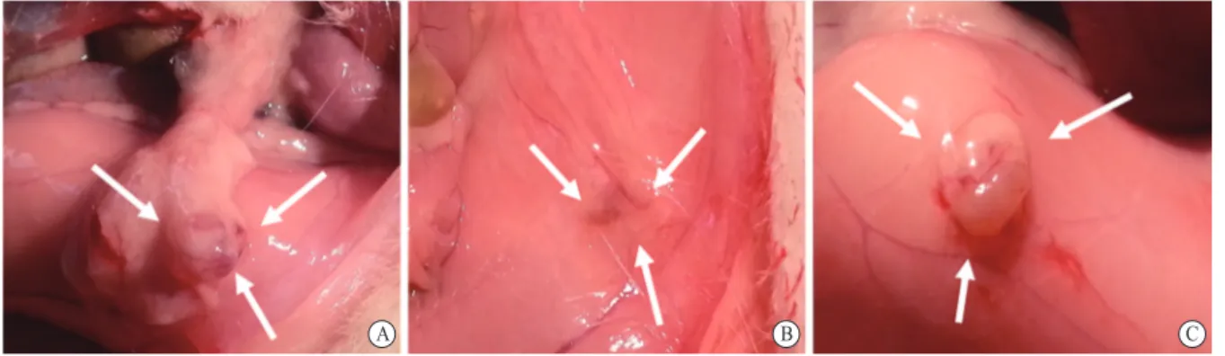
Fig. 1 Macroscopic evaluation and quantitative measurements of mean endometrial implant volumes
The figure shows the mean endometrial implant volumes measured before and after the treatment protocol in control, oxytocin-applied, and ABS-applied groups. While the implant volume was increased significantly in the control group (P<0.01), the mean implant volume was significantly decreased in the oxytocin-applied group only (P<0.0001). The white arrows indicate endometriotic foci located in the anterolateral peritoneum in the control group (A), oxytocin-applied group (B), and ABS-applied group (C).
B
A C
Table 1 TUNEL immunoreactivity percentage of three groups
Control group (n=6) Oxytocin group (n=6) ABS group (n=6) TUNEL immunoreactivity percentage (3.02±1.6)% (13.5±2.08)%* (5.7±1.1)%**
Numerical variables are expressed as mean±standard error of the mean; *P<0.05, oxytocin group compared with the control group; **: TUNEL immunoreactivity percentages were not significantly different between the ABS-applied group and the control group. ABS: Ankaferd Blood Stopper
moderately preserved and infiltration of leukocytes is visible with an epithelial score of 2.7 ± 0.3 in the control group. Vascular structures around endometriotic foci were more obvious in the histopathology of control group samples. However, related structural architecture was not observed in the OT and ABS groups, marking a decreased vascularisation surrounding the endometriotic tissue. In the OT-applied group, the integrity of epithelial tissue was not maintained, with a loosened cellular architecture. The epithelial score was found to be decreased in the OT group compared to the control group (1.1 ± 0.2; P<0 .0001). In the ABS-applied group, an epithelial structure was preserved in a moderately higher level than in the OT-applied group, where the epithelial scoring was lower than in the control group (1.8 ± 0.5; P<0 .0015). Semi-quantitative measurements of epithelial scoring are shown in fig. 3.
2.3 Cytokine Levels in Plasma and Peritoneal Fluid
In the control group, levels of VEGF in plasma and PF were 175.2 ± 16.5 pg/mL and 49.6 ± 8.2 pg/mL, respectively. In the OT-treated group, VEGF levels
were significantly lower than those of the control group in both plasma and PF (plasma: 56.5 ± 6.6 pg/ mL; PF: 22.4 ± 5.3 pg/mL; P<0.0001). In the ABS-treated group, VEGF levels in both plasma and PF were similar to the control group (plasma: 168.4 ± 21.5 pg/mL; PF: 45.2 ± 9.3 pg/mL).
Levels of MCP-1 in plasma and PF were found at 105.8 ± 7.4 ng/mL and 17.2 ± 3.1 ng/mL, respectively, in the control group. Compared to the control group, plasma and PF levels of MCP-1 were significantly lower in the OT-treated group (plasma: 45.2 ± 7.5 ng/ mL; PF: 8.6 ± 1.2 ng/mL; P<0.05), whereas in the ABS-treated group, MCP-1 levels were similar to that of the control group (plasma: 97.1 ± 7.5 ng/mL; PF: 16.3 ± 4.4 ng/mL).
Finally, levels of TNF-α in plasma and PF were 49.6 ± 2.3 pg/mL and 26.5 ± 4.8 pg/mL, respectively. Compared to the control group, plasma and PF levels of TNF-α were significantly lower in the OT-treated group (plasma: 38.1 ± 3.5 pg/mL; PF: 14.3 ± 1.6 pg/ mL; P<0.05). In the ABS-treated group, TNF-α levels were similar to that of the control group (plasma: 45.4 ± 1.8 pg/mL; PF: 18.9 ± 6.1 pg/mL).
Cytokine levels in plasma and PF are summarised in table 2.
3 DISCUSSION
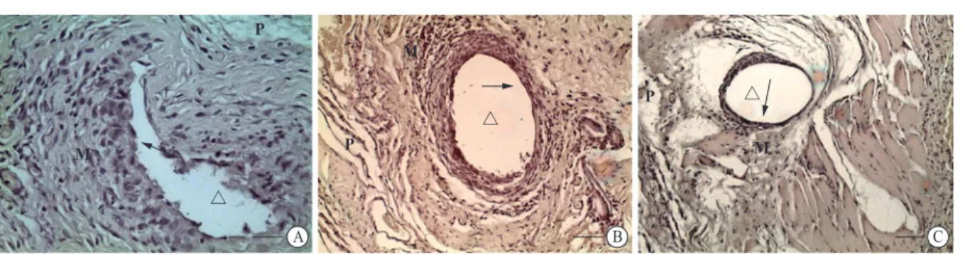
In the current study, we established a rat experimental model of peritoneal endometriosis, and we investigated the effects of OT and ABS on explant size, histopathological scores of the epithelium, and plasma and PF levels of VEGF, TNF-α, and MCP-1. One of the most important outcomes of our study is that endometriotic explant volume was significantly decreased in the OT-treated group compared to that of the pre-treatment level. Additionally, the histopathological scores of the epithelium in the OT-treated and ABS-treated groups were significantly lower than those in the control group. This decline was also confirmed by TUNEL staining and immunoreactivity percentage Fig. 2 Histopathological evaluation of endometriotic foci stained by TUNEL (magnification ×40).
Integrity and structure of epithelium were poorly preserved in the OT group (B) in comparison to the control group (A), shown by the arrow. The epithelial structure is conserved at a better level in the ABS-applied group (C) compared to the OT group; nevertheless, the preservation is not as good as observed in the control group. Scale bars represent 250 µm. P: peritoneum; M: myometrium; △: gland B A C P P P M M M ŗ ŗ ŗ
Fig. 3 Semi-quantitative measurements of epithelium scoring Scoring of the epithelium was based on the persistence of the epithelium. A scale of scoring is between 0 and 3, where a score of 0 represents no epithelium and 3 is a well-conserved epithelial cell layer. ***P<0 .0001, OT vs. control group; **P<0.0015, ABS vs. control group
3 2 Epithelial score 1 Control group OT group ABS group *** **
Control groupOT groupABS group
in the OT group. Moreover, the present study clearly showed that the levels of VEGF, MCP-1, and TNF-α in plasma and PF were significantly lower in the OT-treated group than in the control group. On the other hand, the aforementioned cytokine levels were decreased in the ABS-treated group; however, they did not reach a significant level.
Pelvic pain and/or infertility are the nonspecific but most common symptoms of this enigmatic disease and can negatively affect the woman’s quality of life[19].
The pathogenesis of endometriosis is still poorly understood despite extensive research. Although retrograde menstruation theory has gained the widest acceptance, no single theory can explain all aspects of the disease. In addition, a growing body of evidence indicates that a combination of genetic, immune, endocrine, and environmental factors also seems to be involved in the pathogenesis of endometriosis[1].
Many studies have revealed that angiogenesis and inflammation may play pivotal roles in the pathogenesis of endometriosis[20]. Numerous cytokines
and growth factors have been shown to be increased in the PF of women with endometriosis. Furthermore, expression levels of these molecules were found to be significantly higher in ectopic endometrial tissues than in eutopic endometrium[21]. TNF-α, VEGF,
and MCP-1 are the cytokines and growth factors implicated in the angiogenic and inflammatory processes, and they were reported at higher levels in both the PF and ectopic endometrial tissues of women with endometriosis[22]. Apoptosis is a physiological
process of programmed cell death under the action of apoptotic stimuli. It has been shown that abnormal apoptosis of endometrial cells is closely related to the
occurrence and development of endometriosis[23]. Our
results demonstrated that TUNEL immunoreactivity percentage was significantly increased in the OT group compared to the control group. Namely, OT induced apoptosis in endometriotic foci (gland epithelium and surrounding tissue) more than ABS. Hence, OT might be an alternative treatment agent for regression of endometriosis.
OT, a nonapeptide produced in the paraventricular and the supraoptic nuclei in the hypothalamus, has a wide range of effects in the body[24]. In addition to
its endocrine and paracrine activities, recent studies indicate that OT may also have anti-inflammatory and anti-angiogenic properties and modulate immune responses[25]. OT receptor has been reported to be
expressed in smooth muscle cells and epithelial cells of peritoneal endometriotic lesions and ovarian endometriotic cysts[7].
ABS has pleiotropic effects, including neoplastic, microbial, mutagenic, anti-oxidant, and tissue-healing properties, although the acting mechanisms have not been fully clarified. It has been reported that ABS may induce the formation of erythrocyte aggregation in the place of bleeding, and it works as a haemostatic agent[26]. In the literature,
ABS has been used to overcome bleeding as well as for wound problems in different organs, such as the teeth, face, stomach, oesophagus, intestines, and colon. However, it has not been investigated in an endometriosis model, except in our study.
Hasgul et al[27] demonstrated the attenuation of
oxidative and inflammatory status with ABS, which was induced by acetylsalicylic acid (ASA) on gastric mucosa in rats. They reported that plasma TNF-α and malondialdehyde (MDA) levels were significantly decreased in the ABS group compared to the ASA group. Similarly, we observed that TNF-α levels in both plasma and PF were also decreased in the ABS-treated group compared to the control group. However, it was not at a significant level. Anti-oxidant components of ABS might contribute to decrease of pro-inflammatory cytokines through vascular dynamics in the present study like other studies conducted by other researchers.
Furthermore, the current study clearly indicated that the epithelial structure of endometriotic foci was less preserved in the ABS-treated group than in the control group. Thus, the epithelial score was significantly lower in the ABS-treated group than in the control group. ABS induced apoptosis in endometriotic foci; however, it did not reach a significant level compared to control group. Hence, we showed that ABS did not act on endometriosis through apoptosis, but it might influence endometriosis via other mechanisms, which have not been elucidated to date yet. In another study, ABS significantly reduced inflammation and fibrosis in an experimental Asherman syndrome rat Table 2 Cytokine levels in plasma and peritoneal fluid
Cytokines Control group (n=6) Oxytocin group (n=6) ABS group (n=6) VEGF (pg/mL) Plasma 175.2±16.5 56.5±6.6** 168.4±21.5* Peritoneal fluid 49.6±8.2 22.4±5.3** 45.2±9.3* MCP-1 (ng/mL) Plasma 105.8±7.4 45.2±7.5** 97.1±7.5* Peritoneal fluid 17.2±3.1 8.6±1.2** 16.3±4.4* TNF-α (pg/mL) Plasma 49.6±2.3 38.1±3.5** 45.4±1.8* Peritoneal fluid 26.5±4.8 14.3±1.6** 18.9±6.1* Numerical variables are expressed as mean ± standard error of the mean.
*: VEGF, MCP-1, and TNF-α levels were not significantly different in the ABS-applied group in both plasma and peritoneal fluid samples compared to the control group.
**: P<0.05, Cytokine levels were significantly lower in the oxytocin-applied group than in the control group in both plasma and peritoneal fluid.
model compared to a sham group. However, tissue TNF-α levels did not notably decrease in the ABS group compared to the control group[28]. In light of the
literature, further experimental and clinical studies are required to investigate and prove the exact effects of ABS on endometriosis.
In the present study, plasma and PF levels of VEGF, TNF-α, and MCP-1 were also significantly lower in the OT group, indicating that the reduction in explant size and epithelial scores is related to the anti-inflammatory effects of OT, and OT may be a promising therapeutic agent for endometriosis. Our results are in agreement with our recent study in which TUNEL immunohistochemistry clearly demonstrated that more apoptotic changes were observed in the mononuclear cells of the OT-treated rat group than in controls[29].
Defective apoptosis of ectopic endometrial cells and macrophages have also been suggested to play critical roles in the pathogenesis of endometriosis[30].
In the majority of the limited human studies, OTRs have been found to be overexpressed in endometriotic and adenomyotic tissues[31]. Mechsner et al[7] found
that the peritoneum of women with endometriosis showed a significantly higher smooth muscle content than the peritoneum of women without endometriosis. They also showed that OTRs are widely expressed in epithelial cells and smooth muscle cells of the human uterus and peritoneal endometriotic lesions and ovarian endometriotic cysts. Recently, Huang et al[32]
showed the abnormal expression pattern of OTRs in the junctional zone in women with endometriosis, and this may result in abnormal uterine contractile activity, reduced fertility, and dysmenorrhea associated with endometriosis. In another study, a positive correlation was found between OTR expression and dysmenorrhea in women with adenomyosis, but currently there are no data regarding the correlation between dysmenorrhea and OTR expression in women with endometriosis[33].
In a rat endometriosis model, atosiban, an OTR antagonist, has been shown to have a significant therapeutic effect on endometriosis[8]. All these studies
attribute an inflammatory role to OT in the pathogenesis of endometriosis and adenomyosis.
On the other hand, unlike the aforementioned studies, numerous studies have revealed the anti-inflammatory effect of OT. It has been shown that OT reduces inflammation via decreasing cytokine levels in stress-related disorders[34]. Recently, Garrido-Urbani
et al[35] showed that OT treatment decreases TNF-α
production both in vitro (in bone marrow-derived macrophages) and in vivo (in epididymal adipose tissue) in diet-induced obese mice. It has also been reported that OT treatment may influence the inflammatory process by decreasing the levels of MCP-1 and TNF-α and other cytokines[36].
In the current study, we obtained the same results
as in our recent study with a new set of animals. Therefore, we confirmed our previous results regarding the anti-inflammatory effects of OT on a surgically induced endometriosis rat model. Consistent with the current literature on OT as one of the key regulators of the immune network, it functioned as an immune modulator in the current study and our recent study, and we achieved a significant and similar reduction in explant size and a significant decrease in cytokine levels in OT-treated rat groups.
In the ABS-treated group, although endometriotic explant size decreased, it was not significantly different compared to the pre-treatment level. Moreover, plasma and PF cytokine levels did not differ, but epithelial scores were significantly lower than in the control group. In addition, a decrease in epithelial scores was not prominent, as was observed in the OT-treated group. These findings indicate that ABS might show a therapeutic effect on endometriosis via other mechanisms rather than its anti-inflammatory action.
Limitations of the present study include the small number of animals in the groups as well as the absence of observation of the long-term alterations in both biochemical and histopathological parameters. Further designs of large-scale studies including human subjects are required to determine factors such as the dosage, route, or duration of drug administration. Additionally, larger clinical studies are needed to confirm the impact of OT and ABS on endometriosis.
In conclusion, it is reasonable to speculate that ABS may inactivate alternative pathways of angiogenesis other than the ones involving VEGF. To our knowledge, this is the first study to investigate the direct effects of ABS treatment on endometriosis. However, a mechanism(s) of the effect of ABS on endometriosis is yet to be elucidated.
Conflict of Interest Statement
The authors declare that they have no competing interests.
REFERENCES
1 Bulun SE. Endometriosis. N Engl J Med, 2009,360(3): 268-279
2 Sampson JA. Peritoneal endometriosis due to the menstrual dissemination of endometrial tissue into the peritoneal cavity. Am J Obstet Gynecol, 1927,14:422-469
3 Braun DP, Ding J, Dmowski WP. Peritoneal fluid-mediated enhancement of eutopic and ectopic endometrial cell proliferation is dependent on tumor necrosis factor-alpha in women with endometriosis. Fertil Steril, 2002,78(4):727-732
4 Jiang QY, Wu RJ. Growth mechanisms of endometriotic cells in implanted places: a review. Gynecol Endocrinol, 2012,28(7):562-567
5 Bourlev V, Volkov N, Pavlovitch S, et al. The relationship between microvessel density, proliferative
activity and expression of vascular endothelial growth factor-A and its receptors in eutopic endometrium and endometriotic lesions. Reproduction, 2006,132(3):501-509
6 Ulukus M, Ulukus EC, Tavmergen Goker EN, et al. Expression of interleukin-8 and monocyte chemotactic protein 1 in women with endometriosis. Fertil Steril, 2009,91(3):687-693
7 Mechsner S, Bartley J, Loddenkemper C, et al. Oxytocin receptor expression in smooth muscle cells of peritoneal endometriotic lesions and ovarian endometriotic cysts. Fertil Steril, 2005,83:1220-1231
8 Simsek Y, Celik O, Karaer A, et al. Therapeutic efficiency of Atosiban, an oxytocin receptor blocking agent in the treatment of experimental endometriosis. Arch Gynecol Obstet, 2012,286 (3):777-783
9 Goker H, Haznedaroglu I, Ercetin S, et al. Haemostatic Actions of the Folkloric Medicinal Plant Extract Ankaferd Blood Stopper. J Int Med Res, 2008,36(1):163-170
10 Lee SJ, Umano K, Shibamoto T, et al. Identification of volatile components in basil (Ocimum basilicum L.) and thyme leaves (Thymus vulgaris L.) and their antioxidant properties. Food Chem, 2005,91(1):131-137
11 Simsek C, Selek S, Koca M, et al. Proteomic and transcriptomic analyses to explain the pleiotropic effects of Ankaferd blood stopper. SAGE Open Med, 2017,5:2050312117722569
12 Sheela ML, Ramakrishna MK, Salimath BP. Angiogenic and proliferative effects of the cytokine VEGF in Ehrlich ascites tumor cells is inhibited by Glycyrrhiza glabra. Int Immunopharmacol, 2006,6(3):494-498 13 Vernon MW, Wilson EA. Studies on the surgical
induction of endometriosis in the rat. Fertil Steril, 1985,44(5):684-694
14 Güney M, Oral B, Karahan N, et al. Regression of endometrial explants in a rat model of endometriosis treated with melatonin. Fertil Steril, 2008, 89(4):934-42 15 Petersson M, Wiberg U, Lundeberg T, et al. Oxytocin
decreases carrageenan induced inflammation in rats. Peptides, 2001,22(9):1479-1484
16 Cipil HS, Kosar A, Kaya A, et al. In vivo hemostatic effect of the medicinal plant extract Ankaferd Blood Stopper in rats pretreated with warfarin. Clin Appl Thromb Hemost, 2009,15(3):270-276
17 Keenan JA, Williams-Boyce PK, Massey PJ, et al. Regression of endometrial explants in a rat model of endometriosis treated with the immune modulators loxoribine and levamisole. Fertil Steril, 1999,72(1):135-141
18 Giudice LC, Kao LC. Endometriosis. Lancet, 2004,364 (9447):1789-1799
19 Young VJ, Ahmad SF, Brown JK, et al. Peritoneal VEGF-A expression is regulated by TGF-β1 through an ID1 pathway in women with endometriosis. Sci Rep, 2015,5:16859
20 Griffith JS, Rodgers AK, Schenken RS. Reviews: in
vitro models to study the pathogenesis of endometriosis.
Reprod Sci, 2010,17(1):5-12
21 Jiang L, Yan Y, Liu Z, et al. Inflammation and endometriosis. Front Biosci (Landmark Ed.), 2016,21: 941-948
22 Wei J, Zhao B, Zhang C, et al. Jiawei Foshou San Induces Apoptosis in Ectopic Endometrium Based on Systems Pharmacology, Molecular Docking, and Experimental Evidence. Evid Based Complement Alternat Med, 2019,2019:2360367
23 Dmowski WP, Ding J, Shen J, et al. Apoptosis in endometrial glandular and stromal cells in women with and without endometriosis. Hum Reprod, 2001,16(9): 1802-1808
24 Celik O, Aydin S, Celik N, et al. Peptides: Basic determinants of reproductive functions. Peptides, 2015,72:34-43
25 Yuan L, Liu S, Bai X, et al. Oxytocin inhibits lipopolysaccharide-induced inflammation in microglial cells and attenuates microglial activation in lipopolysa-ccharide-treated mice. J Neuroinflammation, 2016, 13(1):77
26 Haznedaroglu BZ, Beyazit Y, Walker SL, et al. Pleiotropic cellular, hemostatic, and biological actions of Ankaferd hemostat. Crit Rev Oncol Hematol, 2012, 83(1):21-34
27 Hasgul R, Uysal S, Haltas H, et al. Protective effects of Ankaferd blood stopper on aspirin-induced oxidative mucosal damage in a rat model of gastric injury. Toxicol Ind Health, 2014,30(10):888-895
28 Büyük B, Beyazıt F. The efficacy of Ankaferd Blood Stopper® in an experimental Asherman syndrome model created in rats. Turk J Obstet Gynecol, 2019,16(1):7-14 29 Yeniel AÖ, Erbas O, Ergenoglu AM, et al. Effect of
oxytocin treatment on explant size, plasma and peritoneal levels of MCP-1, VEGF, TNF-α and histopathological parameters in a rat endometriosis model. Eur J Obstet Gynecol Reprod Biol, 2014,175:134-139
30 Salmassi A, Acar-Perk B, Schmutzler AG, et al. Apoptosis resistance in endometriosis. Bioimpacts, 2011, 1(2):129-134
31 Nie J, Liu X, Guo SW. Immunoreactivity of oxytocin receptor and transient receptor potential vanilloid type 1 and its correlation with dysmenorrhea in adenomyosis. Am J Obstet Gynecol, 2010,202(4):346.e 1-8
32 Huang M, Li X, Guo P, et al. The abnormal expression of oxytocin receptors in the uterine junctional zone in women with endometriosis. Reprod Biol Endocrinol, 2017,15(1):1
33 Guo SW, Mao X, Ma Q, et al. Dysmenorrhea and its severity are associated with increased uterine contractility and overexpresion of oxytocin receptor (OTR) in women with symptomatic adenomyosis. Fertil Steril, 2013,99(1):231-240
34 Sippel LM, Allington CE, Pietrzak RH, et al. Oxytocin and Stress-related Disorders: Neurobiological Mecha-nisms and Treatment Opportunities. Chronic Stress (Thousand Oaks), 2017,1
35 Garrido-Urbani S, Deblon N, Poher AL, et al. Inhibitory role of oxytocin on TNF-α expression assessed in vitro and in vivo. Diabetes Metab, 2018,44(3):292-295 36 Ahmed MA, Elosaily GM. Role of Oxytocin in
deceleration of early atherosclerotic inflammatory processes in adult male rats. Int J Clin Exp Med, 2011, 4(3):169-178