Department of Pathology, Pt. BDS, PGIMS, University of Health Sciences, Rohtak, India
Yazışma Adresi /Correspondence: Monika Gupta,
17/8 FM, Medical enclave, PGIMS Rohtak, Haryana, India Email: [email protected]
Geliş Tarihi / Received: 11.03.2015, Kabul Tarihi / Accepted: 07.03.2016 Copyright © Dicle Tıp Dergisi 2016, Her hakkı saklıdır / All rights reserved
Dicle Tıp Dergisi / 2016; 43 (1): 1-4
Dicle Medical Journal doi: 10.5798/diclemedj.0921.2016.01.0628
ORIGINAL ARTICLE / ÖZGÜN ARAŞTIRMA
Etiological Profile of Plasmacytosis on Bone Marrow Aspirates
Kemik İliği Aspiratlarında Saptanan Plazmasitozun Etyolojik Profili
Monika Gupta, Shivani Malik, Rajnish Kalra, Nisha Sharma, Sunita Singh, Veena Gupta, Promil Jain, Rajeev SenÖZET
Amaç: Son yıllarda rutin kemik iliği aspiratı incelemele-rinde saptanan neoplastik ve non-neoplastik hastalıklarda giderek artan oranlarda plazma hücrelerine rastlanmakta-dır. Bu çalışmada kemik iliği aspiratında plazmasitoz sap-tanan durumların bir spektrumu çıkarılmaya çalışılmıştır. Yöntemler: Sunulan çalışma bir yıllık süre içinde Patolo-ji Bölümünde gerçekleştirildi. Çalışma materyalini artmış oranda plazma hücresi (>%3,5) gösteren toplam 114 ke-mik iliği aspiratı oluşturmaktadır. Tüm olgularda ayrıntılı bir klinik muayene ve takiben tam kan sayımı, periferik yayma ve kemik iliği aspirasyonu yapıldı.
Bulgular: Erkek / Kadın oranı açısından hafif bir kadın üstünlüğü görüldü (1 / 1,1). Vakaların çoğu 40’lı yaşlarda idi. Plazma hücre yoğunluğu %5-36 arasında değişmekte idi. Etyoloji açısından 96 (%84,2) olguda non-neoplastik, 18 (%15,8) olguda ise neoplastik bir etyoloji saptandı. Sonuç: Kemik iliği plazmasitozu tanısal bir karmaşaya yol açarak, özellikle sayı ve morfolojik görünümlerinde çakışma olan reaktif patolojilerin neoplastik durumlardan ayırt edilmesini bazen zorlaştırabilir. Özellikle çakışma aralıklarındakiler başta olmak üzere, plazmasitozlu her hastada bireyselleştirilmiş araştırmalar ve immunelektro-forez ve immunhistokimya ile boyanmış kemik iliği aspira-tı incelemesi ile sonuç tanısı konabilir ve hasta yönetimi gerçekleştirilebilir.
Anahtar kelimeler: Reaktif plazmasitoz, kemik iliği, mul-tipl miyelom, megaloblastik anemi
ABSTRACT
Objective: In recent years, during routine examination of bone marrow aspirates, an increased plasma cell per-centage has been noted in a good number of cases which included both neoplastic and non-neoplastic diseases. An attempt has been made to observe the spectra of condi-tions with plasmacytosis in bone marrow.
Methods: The present study was conducted in the de-partment of pathology over a period of one year. A total of 114 bone marrow aspirates that showed increased plas-ma cells (>3.5%) constitute the study plas-material. A detailed relevant clinical examination followed by complete blood count, peripheral smear examination and bone marrow aspiration was done in all cases.
Results: There was slight female predominance with male to female ratio of 1:1.1. The majority of patients were in 4th decade. The plasma cell concentration ranged from 5% to 36%. As far as the etiology is concerned, 96 cases (84.2%) were non-neoplastic and 18 cases (15.7%) had neoplastic etiology.
Conclusion: Bone marrow plasmacytosis can present as diagnostic dilemma and some time can be challenging to differentiate reactive from neoplastic condition as there is an overlap both in counts and morphology. Each case with plasmacytosis especially in the overlap range requires complete clinical evaluation, individualized investigations and more specific tests like immunoelectrophoresis and bone marrow biopsy with immunohistochemistry to arrive at a final diagnosis for patient management.
Key words: Reactive plasmacytosis, bone marrow, mul-tiple myeloma, megaloblastic anaemia
INTRODUCTION
Bone marrow plasmacytosis occurs in association with variety of conditions both non-neoplastic such as infections, immune disorders, various type of
anemias as well as neoplastic conditions includ-ing primary and secondary malignancies. In most neoplastic conditions, plasma cell constitutes ~10-20% of all nucleated cells but may reach up to 50%
M. Gupta, et al. Etiological profile of plasmacytosis 2
Dicle Tıp Derg / Dicle Med J www.diclemedj.org Cilt / Vol 43, No 1, 1-4
[1,2,3]. It is important to differentiate reactive plas-macytosis in non-neoplastic conditions from the malignant ones. High marrow plasma cell count can present a diagnostic dilemma. An attempt has been made to observe the spectra of conditions with plas-macytosis in bone marrow.
METHODS
The present study was conducted in Department of Pathology, Pt.B.D. Sharma PGIMS, Rohtak from July 2013 to June 2014. A total of 114 bone mar-row aspirates that showed increased plasma cells constitute the study material. More than 3.5% was selected as the arbitrary limit above which the plas-ma cells were considered abnorplas-mal [4]. A detailed relevant clinical examination was done. Complete blood count was performed using an automated five part hematology analyzer (Mindray BC 5800) and peripheral smears were examined in all cases. All the bone marrow aspirate smears were stained with Leishman’s stain and differential counts were done on 500 cells. Special staining for Sudan Black B, Periodic acid Schiff and Perl’s stain on the smears done wherever indicated. Bone marrow biopsy was done for evaluation of bone marrow in cases with insufficient cells, dry tap or hypoplastic bone mar-row. Further investigations including estimation of serum gamma globulins on Serum Protein Elec-trophoresis was performed to rule out or establish monoclonality.
RESULTS
A total of 830 bone marrow aspirations were received during the study period for various haematological disorders. Out of them, 114 cases (13.7%) showed increase plasma cells. There were 53 (46.4%) males and 61 (53.5%) females. The patients had age rang-ing from 4 months to 85years, maximum number of patients (n=52) was more than 40 years.
The chief presenting complaints were fever, weakness, body ache, bone pain, abdominal pain, melena and bleeding. Hepatosplenomegaly and lymphadenopathy were found in 39 cases (34.2%) and 17 cases (14.9%) respectively. The associated peripheral blood findings in descending order were anemia 59 cases (51.7% cases), pancytopenia 47 cases (41.2%), thrombocytopenia 14 cases (12.2%) and leukocytosis 5 cases (4.3%).
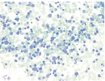
Figure 1. Bone marrow Aspirate showed megaloblastic erythroid hyperplasia with plasmacytosis (Leishman stain × 100X)
Figure 2. Bone marrow Aspirate showed nonspecific myeloid reaction with plasmacytosis (Leishman stain × 100X)
The plasma cell concentration ranged from 5% to 36%. The cases were arranged according to the plasma cell percentage, 88 cases (77.1%) had 5-9%, 16 cases (14.0%) had 10- 20% and 10 cases (8.7%) had > 20% plasma cells respectively. As far as the etiology is concerned, 96 cases (84.2%) were non-neoplastic and 18 cases (15.7%) had non-neoplastic eti-ology [Table 1] [Figure1, Figure 2, Figure 3, Figure 4].
Other significant findings on the bone marrow aspirates revealing plasmacytosis were lymphocy-tosis 47 cases (41.2%), prominence of reticulum cells 26 cases (22.8%) and increased eosinophilic precursors in 9 cases (7.8%).
M. Gupta, et al. Etiological profile of plasmacytosis 3
Dicle Tıp Derg / Dicle Med J www.diclemedj.org Cilt / Vol 43, No 1, 1-4
Figure 3. Bone marrow Aspirate showed normoblastic erythroid hyperplasia with plasmacytosis (Leishman stain x 100X)
Figure 4. Bone marrow Aspirate showed plasmacytosis (Leishman stain × 1000X)
Non neoplastic Clinical and/or bone marrow diagnosis n %
Anemia (n=77) Nutritional deficiency anemia 40 35.0
Hemolytic anemia 17 14.9
Hypoplastic marrow 20 17.5
Thrombocytopenia (n=6) Megakaryocytic 5 4.3
Amegakaryocytic 1 0.8
Infections (n=4) Non-specific myeloid reaction 13 11.4
Primary neoplastic (n=6)
Leukemia Acute LeukemiaChronic Myeloid Leukemia 41 3.50.8 Chronic Lymphoproliferative disorder 1 0.8
Plasma cell dyscrasias (n=11) Multiple Myeloma 10 8.7
Waldenstrom Macroglobinemia 1 0.8
Secondary (n=1) Metastasis 1 0.8
Table 1. Clinical pre-sentation and bone marrow findings in pa-tients with plasmacy-tosis
DISCUSSION
In recent years, during routine examination of bone marrow aspirates, an increased plasma cell per-centage has been noted in a good number of cases, which included both neoplastic and non-neoplastic diseases.
Reactive plasmacytosis is polyclonal character-ized by a diffuse distribution of mature plasma cells in the bone marrow. Morphological features sug-gesting a reactive nature of plasma cells, although not specific, are mature forms of plasma cells, peri-vascular location of plasma cells and plasmacytic satellitosis [5,6].
The mechanism of polyclonal proliferation of plasma cells is probably multifactorial and is con-sidered as a defense response mechanism to the un-derlying disease processes. Some types of infection and abnormal immune reactions also lead to marked proliferation of plasma cells [2]. Increased release of paracrine growth factors such as cytokines, IL-6 or IL-10 are known to stimulate plasma cell genera-tion, may also contribute to this process [5,6,7].
Reactive conditions such as chronic infection, autoimmune diseases, hypersensitivity states, meg-aloblastic anemia, hemolytic anemia, diabetes mel-litus, cirrhosis, drug related agranulocytosis strepto-kinase treatment, angio-immunoblastic
lymphade-M. Gupta, et al. Etiological profile of plasmacytosis 4
Dicle Tıp Derg / Dicle Med J www.diclemedj.org Cilt / Vol 43, No 1, 1-4
nopathy and multicentric Castleman’s disease are associated with bone marrow plasmacytosis [1,2,3].
Neoplastic processes are diagnosed based on morphological findings, protein electrophoresis and bone marrow immunohistological examination. In myelomatosis, the marrow is most often hypocellu-lar that is reduced hematopoietic elements, and the plasma cells mainly abnormal, shifted to the left, fo-cal in distribution, associated with reticulum cells, and usually more numerous than 20% [8].
In our study, we have tried to represent the data related to degree of plasmacytosis in various conditions. Less than 10% plasmacytosis has been chiefly found in various types of anemia, 10- 30% plasma cells were found infections and hypoplas-tic marrows. In reactive conditions, the percentage range was 5%- 24%. However, in cases of plasma cell dyscrasias the range varied from 20- 40%. In non-neoplastic conditions plasma cells were chiefly mature however, in plasma cell dyscrasias range of mature and immature plasma cells was found. The marrows in plasmacytic response were usually nor-mocellular for age, and the plasma cells were main-ly mature, uniformmain-ly distributed, often related to re-ticulum cells, and rarely more numerous than 20% of the total count. In our study, maximum cases (84.2%) with plasmacytosis had a benign etiology.
The mechanism of the significance of bone marrow plasmacytosis has not been addressed. Bone marrow plasmacytosis can present as diag-nostic dilemma and some time can be challenging to differentiate reactive from neoplastic condition as there is an overlap both in counts and morphol-ogy. Each case with plasmacytosis especially in the overlap range requires complete clinical evaluation,
individualized investigations and more specific tests like immunoelectrophoresis and bone marrow biop-sy with immunohistochemistry to arrive at a final diagnosis for patient management.
Declaration of Conflicting Interests: The
au-thors declare that they have no conflict of interest.
Financial Disclosure: No financial support
was received.
REFERENCES
1. Kojima M, Murayama K, Igarashi T, et al. Bone marrow plasmacytosis in idiopathic plasmacytic lymphadenopathy with polyclonal hyperimmunoglobinemia- A report of four cases. Pathology Research & Practice 2007;203:789-794. 2. Ozdemirkıran F, Gokgoz Z, Ozsan N, et al. (2013) Prominent
reactive plasmacytic proliferation presenting with transient pancytopenia: an unusual case. J Hematol Thromb Dis 2013:1:117-121.
3. Wei A, Juneja S. Bone marrow immunohistology of plasma cell neoplasms. J Clin Pathol 2003;56:406-411.
4. Bates I, Burthem J. Bone marrow biopsy. In Bain BJ, Bates I, Laffan MA, Lewis SM (eds). Dacie and Lewis Practi-cal Haematology. 11th ed. Elesvier Churchill Livingston. 2012:p130.
5. Ragan A, Arora B, Rangan, et al. Florid plasmacytosis in a case of acute myeloid leukemia: A diagnostic dilemma. In-dian J Med Paediatr Oncol 2010;31:36-38.
6. Dodhy MA, Habib M, Shaheen M, et al. Acute mono-cytic leukemia with reactive plasmacytosis. Int J Pathol 2014;12:94-95.
7. Ahsanuddin NA, Brynes RK, Li Shiyong. Peripheral blood polyclonal plasmacytosis mimicking plasma cell leukemia in patients with angioimmunoblastic T-cell lymphoma: re-port of 3 cases and review of the literature. Int J Clin Exp Pathol 2011;4:416-420.
8. Sukpanichnant S, Cousar JB, Leelasiri A, et al. Diagnostic criteria and histologic grading in multiple myeloma: his-tologic and immunohishis-tologic analysis of 176 cases with clinical correlation. Hum Pathol 1994;25:308-318.