Science
PAPER
Cite this:Biomater. Sci., 2018, 6, 1777
Received 15th March 2018, Accepted 8th May 2018 DOI: 10.1039/c8bm00311d rsc.li/biomaterials-science
Promotion of neurite outgrowth by rationally
designed NGF-
β binding peptide nanofibers†
Zeynep Okur,
a,bOya I. Senturk,
aCanelif Yilmaz,
a,bGulcihan Gulseren,
aBusra Mammadov,
aMustafa O. Guler
*
cand Ayse B. Tekinay
*
a,bPromotion of neurite outgrowth is an important limiting step for regeneration in nerve injury and depends strongly on the local expression of nerve growth factor (NGF). The rational design of bioactive materials is a promising approach for the development of novel therapeutic methods for nerve regeneration, and bio-materials capable of presenting NGF to nerve cells are especially suitable for this purpose. In this study, we show bioactive peptide amphiphile (PA) nanofibers capable of promoting neurite outgrowth by dis-playing high density binding epitopes for NGF. A high-affinity NGF-binding sequence was identified by phage display and combined with a beta-sheet forming motif to produce a self-assembling PA molecule. The bioactive nanofiber had higher affinity for NGF compared to control nanofibers and in vitro studies revealed that the NGF binding peptide amphiphile nanofibers (NGFB-PA nanofiber) significantly promote the neurite outgrowth of PC-12 cells. In addition, the nanofibers induced differentiation of PC-12 cells into neuron-like cells by enhancing NGF/high-activity NGF receptor (TrkA) interactions and activating MAPK pathway elements. The NGFB-PA nanofiber was further shown as a promising material to support axonal outgrowth from primary sensory neurons. These materials will pave the way for the development of new therapeutic agents for peripheral nervous system injuries.
Introduction
Nerve tissue is generally known to exhibit a low capacity for regeneration; however the peripheral nervous system (PNS) is able to partially recover from injuries caused by acute trauma or systemic disorders such as diabetes.1 The local microenvi-ronment of the site of injury greatly affects the success of the recovery process, and the absence of the essential environ-mental factors such as neurotrophic factors will often result in delayed healing, demyelination, axonal damage and/or neuro-nal cell death. Thus, materials capable of emulating the bio-chemical signals present in healthy neural tissue can be used to promote regeneration in peripheral nervous system injuries. Nerve growth factor (NGF), which is one of the neurotrophic factors, is important for the survival, maturation and di fferen-tiation of sensory neurons as well as the repair of injuries in the PNS. NGF production is upregulated by reactive and
de-differentiated Schwann cells (SCs) after injury,2 and plays an important role in the reconnection of injured nerve ends during the regeneration process. In particular, the alignment of axons is facilitated through a gradient of NGF at the site of injury, which allows undamaged neurons to bridge the gap that is created following axonal damage.3 In addition, the binding of NGF to its high-affinity cell surface receptor TrkA also activates intracellular signaling pathways that are essential for the expression of genes responsible for neuronal differentiation.4–6One of the major pathways activated by the NGF–TrkA interaction is the MAPK pathway, which involves the phosphorylation of extracellular signal-regulated kinase1/2 (ERK1/2) by MEK (MAPK kinase). This event allows phosphory-lated ERK1/2 ( pERK1/2) to translocate into the nucleus,7and activate the genes responsible for the generation of survival, differentiation and migration signals in nerve cells.8,9 Consequently, NGF is vital for cell survival and axonal out-growth following injury, and several studies are currently underway for its use in the treatment of peripheral nerve injuries.
The local delivery of soluble NGF at the injury site is a time-consuming and expensive method because of the requirement of repetitive injection of NGF. In addition, this method pro-vides little control over the NGF gradient created at the injury site, which can cause adverse effects for other cell types present in the region. In contrast, therapeutic materials that
†Electronic supplementary information (ESI) available. See DOI: 10.1039/ c8bm00311d
aInstitute of Materials Science and Nanotechnology and National Nanotechnology
Research Center (UNAM), Bilkent University, Ankara, 06800, Turkey. E-mail: [email protected]
bNeuroscience Graduate Program, Bilkent University, Ankara, 06800, Turkey
cInstitute for Molecular Engineering, University of Chicago, Chicago, IL 60637, USA.
E-mail: [email protected]
Published on 08 May 2018. Downloaded by Bilkent University on 2/25/2019 1:30:18 PM.
View Article Online
enhance the presentation of NGF can facilitate a more effective recovery process by mediating the interactions between neurons present at the injury site and NGF secreted by sur-rounding SCs.
High-affinity epitopes for growth factors can be identified using phage display libraries, which consist of a series of epi-topes that are displayed by genetically engineered bacterio-phages and can be tested against a target molecule to deter-mine the sequences that exhibit maximum affinity against it.10 Previous studies revealed that the phage display library is a promising method for the identification of growth factor binding epitopes. In one of these studies, NGF-binding fibrin matrices were developed, which were shown to enhance the axonal outgrowth of dorsal root ganglion (DRG) neurons.11In another study, a collagen-binding epitope was inserted into the sequence of human NGF-β, and the recombinant growth factor was shown to improve peripheral nerve regeneration by promoting NGF–collagen interactions in a rat sciatic nerve model.12 Although these studies revealed promising results, the effect of the developed NGF-binding materials on the molecular mechanism of NGF remains unclear. Also, the effect of the amino acid sequence on the affinity for NGF was not investigated. Therefore, a need to develop biomaterials that selectively bind to NGF and enhance the effect of NGF for neural differentiation and recovery from injury still exists.
Epitopes can be effectively incorporated into synthetic bio-materials in order to be displayed to neuronal cells at the site of injury. These biomaterials can be programmed to interact with growth factors through decoration of bioactive epitope sites for stability, increased local concentration and prolonged release of growth factors at the site of action.13Design of bio-active synthetic materials, which are able to both present the maximum number of epitopes and have control over epitope number, would be advantageous in the development of regen-erative medicine strategies. Peptide amphiphile (PA) nano-fibers are widely used synthetic biomaterials for regenerative purposes. Previous studies using laminin derived (LN-PA) and heparin mimetic (HSM-PA) nanofibers showed that functiona-lized nanofiber scaffolds enhance axonal outgrowth to a greater extent when compared to control PA nanofibers in vitro and facilitate recovery from sciatic nerve injury by inducing neurite outgrowth in vivo.15
In the present study, a 7-amino acid phage library was used in order to identify a high-affinity epitope for NGF-β, which was then incorporated into a PA nanofiber structure for the promotion of neurite growth. The affinity of the designed NGF binding PA molecule (NGFB-PA) for NGF-β was compared with a scrambled version (scrNGFB-PA) of the sequence in order to determine the role of the order of sequence on the strength of binding, and the effect of the PA scaffold on neurite outgrowth was evaluated using an in vitro model using PC-12 cells. PC-12 cells cultured on PA scaffolds were shown to differentiate into neuron-like cells by analysis of βIII-tubulin expression. Furthermore, the effect of the designed NGFB-PA scaffold on the MAPK pathway was investigated and the NGFB-PA nano-fiber was shown to enhance the MAPK pathway. Finally, the
efficiency of the developed NGFB-PA nanofiber system on the formation of neuronal connectivity among primary sensory neurons was investigated by axonal and synaptic protein marker analyses of isolated DRG neurons cultured on the nanofiber systems. Overall, our results suggest that the NGFB-PA nanofiber is able to bind to NGF with high affinity and enables differentiation of PC-12 cells into neuron-like cells by altering the essential differentiation pathway of NGF and supporting the generation of the neural connection of primary sensory neurons, which makes the NGFB-PA nanofiber a new, promising scaffold for regeneration in peripheral nerve injuries.
Materials and methods
Materials
The Ph.D.-7 Phage Display Peptide Library was purchased from New England Biolabs. The Echerichia coli strain XL1Blue (recA1 endA1 gyrA96 thi-1 hsdR17 supE44 relA1 lac [F′ proAB lacIq ZΔM15 Tn10 (Tetr)]) was courtesy of Prof. Alper Arslanoglu from Izmir Institute of Technology, Molecular Biology Department (Izmir, Turkey). Nerve growth factor (NGF-β) was purchased from Thermo Fisher Scientific. The HRP/anti-M13 monoclonal conjugate was purchased from GE Healthcare Life Sciences. LB broth, agar, NaCl, polyethylene glycol 8000 (PEG), Tris-HCl, sodium azide, sodium bicarbonate (NaHCO3), ethylene diamine tetra-acetic acid (EDTA), bovine
serum albumin (BSA) and other chemicals were purchased from Sigma-Aldrich. Glycine-HCl was purchased from Alfa Aesar. Isopropyl β-D-1-thiogalactopyranoside (IPTG) was
pur-chased from Amresco. 5-Bromo-4-chloro-3-indolyl-beta-D
-Galactopyranoside (X-Gal) was purchased from Biotium. 9-Fluorenylmethoxycarbonyl (Fmoc) and tert-butoxycarbo-nyl (Boc) protected amino acids, [4-[a-(20,40-dimethoxyphetert-butoxycarbo-nyl) Fmoc aminomethyl] enoxy] acetamidonorleucyl-MBHA resin (Rink amide MBHA resin) and 2-(1H-benzotriazol-1-yl)-1,1,3,3-tetramethyluronium hexafluorophosphate (HBTU) were pur-chased from NovaBiochem and ABCR. Poly-L-lysine (PLL) was
purchased from Trevigen. The Live/Dead Assay and other cell culture materials were purchased from Invitrogen as were ELISA reagents. Paired antibodies for NGF were purchased from R&D Systems. Antibodies for immunocytochemistry and western blot analyses were purchased from Abcam and Millipore. Other chemicals mentioned in the manuscript were purchased from Fisher, Merck, Alfa Aesar or Aldrich. All chemicals were used as provided.
Phage library screening against NGF
Library screening was performed against NGF-β according to the manufacturer’s instructions (New England Biolabs). NGF-β (10 µg mL−1 for each well) was dissolved in 100 µL of 0.1 M NaHCO3 ( pH 8.6), coated on the wells of a 96-well tissue
culture plate by swirling and incubated overnight at 4 °C with agitation in a sealed box with a damp paper. Then, the plate was blocked with blocking buffer [0.5% (w/v) bovine serum
albumin (BSA) in 0.1 M NaHCO3( pH 8.6)] for at least 1 h at
4 °C. The blocking buffer was washed 6 times with Tris-buffered saline pH 7.5 + 0.1% (v/v) Tween 20 (TBST). Then, the Ph.D.-7 Phage Display Library with a complexity of 1.9 × 10⁹ plaque forming units ( pfu) was added at approximately 1 × 10¹¹pfu in 0.1% TBST into each well and incubated at room temperature for 1 h. Unbound phages were washed 10 times with 0.1% TBST, while bound phages were eluted with 0.2 M glycine-HCl ( pH 2.2) containing 1 mg mL−1BSA for no more than 10–20 min at room temperature, and this reaction was neutralized by adding 1 M Tris-HCl ( pH 9.1). Eluates from the first two rounds were amplified and titered with XL1Blue. For the second and third rounds, the percentage of Tween-20 was increased to 5% (v/v) for washing steps. After the third round of screening, the eluates were not amplified but titered; and 24 random plaques from the third round of titering were selected for sequence identification.
DNA sequence analysis
For the identification of peptide sequences from selected phage plaques, DNA was isolated from each individual plaque as described in the manufacturer’s instructions (New England Biolabs). Each individual clone was amplified and precipitated in 20% (w/v) PEG/2.5 M NaCl. After centrifugation at 14 000 rpm for 10 min at 4 °C, the pellet was dissolved in an iodide buffer containing 10 mM Tris-HCl (pH 8.0), 1 mM ethylene diamine tetra-acetic acid (EDTA), and 4 M NaI. Phage DNA was subsequently precipitated with absolute ethyl alcohol and dis-solved in DNase/RNase free ultrapure water. DNA sequences were analyzed using an automated DNA sequencer (Refgen Biotechnology, Ankara, Turkey) with the primer sequence 5 ′-CCCTCATAGTTAGCGTAACG-3′ (purchased from Sentegen Biotechnology, Ankara, Turkey).
Phage ELISA for the identification of binding selectivity of candidate phage clones
After sequence analysis, 3 of the peptide sequences were observed to be displayed by more than two phage clones (Fig. S1A†). These sequences were selected for further analysis, and the affinity of one clone for each candidate peptide sequence was analyzed with phage ELISA by following the manufacturer’s instructions. 10 µg mL−1 NGF-β (120 µL per well) was dissolved in 0.1 M NaHCO3, immobilized on a
Maxisorb (NUNC, Thermo Scientific) ELISA plate and incu-bated overnight at 4 °C in a sealed box with a damp paper. Wells were washed with 0.5% TBST and then blocked with blocking buffer for at least 1 h at 4 °C. The same number of wells in a separate ELISA plate were coated with blocking buffer as a negative control. Additionally, the same number of wells in 96 well tissue culture plates were also coated with blocking buffer for phage dilution. After the blocking and washing steps, the phage diluents (serially diluted in 1 : 8 ratios) in 0.1% TBST (100 µL per well) were incubated for 2 h at room temperature with agitation. The wells were washed 10 times with 0.5% TBST and incubated with 1 : 5000 horseradish peroxidase-conjugated anti-M13 monoclonal antibody (200 µL
per well; GE Healthcare) in blocking buffer. After washing steps with 0.5%TBST, the HRP substrate (3,3′,5,5-tetramethyl-benzidine (TMB); 50 µL per well) was added. After 30 min incubation, the reaction was stopped with sulfuric acid. The change in absorbance resulting from color formation was measured by a Spectramax M5 microplate reader (Molecular Devices) at 450 nm wavelength. Then, this value was sub-tracted from the reference value measured at 650 nm.
Peptide synthesis
NERALTL-GEEGAVVK(Lauryl)-Am (NGFB-PA) and RNTLLAE-GEEGAVVK(Lauryl)-Am (scrNGFB-PA) peptide amphiphile molecules were synthesized via fluorenylmethyloxycarbonyl (Fmoc)-based reverse solid phase peptide synthesis. The lauryl-VVAGKK-Am peptide amphiphile (KK-PA) molecule was syn-thesized via Fmoc-based solid phase peptide synthesis. Amino acid couplings were performed using 2 equivalents of Fmoc protected amino acids, 1.95 equivalents of O-benzotriazole-N, N,N′,N′-tetramethyl-uronium-hexafluoro-phosphate (HBTU) and 3 equivalents of N,N-diisopropylethylamine (DIEA) for 3 h. After each coupling, the remaining free amine groups were blocked by treating the resin with 10% acetic anhydride/DMF solution for 30 min. Fmoc protecting groups were removed by shaking the PA with 20% piperidine/DMF solution for 20 min. Between each step, the resin was washed thoroughly with DMF, DCM and DMF, respectively. The Mtt groups on lysine residues were removed by shaking the resin with 5% TFA clea-vage cocktail containing TFA : TIS : H2O : DCM in the ratio of
5 : 2.5 : 2.5 : 90, 6 times in 5 min periods. Cleavage of NGFB-PA and scrNGFB-PA from the resin and deprotection of side groups were carried out by shaking the peptide with a mixture of TFA : TIS : phenol : H2O in the ratio of 88 : 5 : 5 : 2 for 2 h.
Cleavage of KK-PA from the resin and deprotection of side groups were carried out by shaking the peptide with a mixture of TFA : TIS : H2O in the ratio of 95 : 2.5 : 2.5 for 3 h. The resin
was washed with DCM after 2 h to collect the PA. DCM and TFA were removed by vacuum distillation. The remaining PA solution was triturated with ice-cold ether. The resulting white product was freeze-dried. The PA was then characterized by LC-MS. Large amounts of impurities were observed on LC-MS; therefore, the resulting peptide was purified further by the pre-parative HPLC technique using 0.1% NH4OH/water and 0.1%
NH4OH/acetonitrile solutions as eluents. After removing
aceto-nitrile, the peptide was freeze-dried and collected as a white powder.
Liquid chromatography–mass spectrometry
An Agilent Technologies 6530 Accurate-Mass Q-TOF LC-MS with ESI was used to perform liquid chromatography–mass spectrometry to characterize PAs. The concentration of the sample was adjusted to 1 mg mL−1. 0.1% NH4OH/water and
0.1% NH4OH/acetonitrile solutions were used as eluents.
SEM imaging of peptide nanofibers
Nanofiber networks formed by peptide amphiphiles were imaged by scanning electron microscopy (SEM). Oppositely
charged PA solutions (1 wt% for NGFB-PA and scrNBP/PA, 0.44 wt% for KK-PA) were mixed on a silicon wafer in a 1 : 1 volume ratio (final volume being 40 µL) to produce gels as used for cell culture experiments. Then, the gels were de-hydrated by transferring to 20%, 40%, 60%, 80% and 100% v/v ethanol sequentially. After one more dehydration step in 100% alcohol, the gels were critical point dried using Autosamdri®-815B equipment from Tousimis. For imaging, dried gels were coated with 6 nm Au/Pd and SEM (FEI Quanta 200 FEG) images were taken in the high vacuum mode with 15 keV beam energy using an Everhart–Thornley Detector (ETD). TEM imaging of peptide nanofibers
An FEI Tecnai G2 F30 transmission electron microscope (TEM) was used for TEM imaging. 1.5 mM PA solutions were pre-pared in ddH2O and aged overnight. The PA solutions were
mixed and incubated at room temperature for 1.5 h. PA gels were then diluted with water to a concentration of 2.0 × 10⁻⁴ M. 10 µL of the solutions were dropped on carbon-covered copper grids and incubated for 10 min. Then, 2% (w/v) uranyl acetate solution was used for staining and the grids were washed with water after staining. Finally, the carbon grids were dried in air overnight.
Circular dichroism (CD) spectroscopy
The secondary structures of PA solutions were analyzed with a JASCO J-815 CD spectrometer under an inert nitrogen atmo-sphere. PA solutions were prepared at 2 × 10⁻⁴ M in 300 µL final volumes. The samples were measured from 350 nm to 190 nm with 0.1 data pitch, 100 nm min−1 scanning speed, 1 nm bandwidth and 4 s D.I.T. The average of three measure-ments were adjusted and sensitivity was selected as standard.
Zeta potential measurements
A Nano-ZS Zetasizer (Malvern) was used for measuring the zeta potentials of PA solutions. Measurements were done at 25 °C and pH 7.0. PA solutions with 1.5 mM concentrations were pre-pared, mixed at a 1 : 1 ratio as necessary, and diluted with ddH2O to a final concentration of 5.0 × 10⁻⁵ M prior to the
measurements. A dip cell was used for the measurements. Formation of nanofiber scaffolds
Nanofiber scaffolds for in vitro experiments were formed by mixing oppositely charged PAs, which enables their self-assem-bly to form higher order nanofibers through the neutralization of the charges in the backbones of the PA molecules. In this study, 2 different types of nanofibers were used (Fig. S2†). NGFB-PA (the net charge is−2, Fig. S2†) and scrNGFB-PA (the net charge is−2, Fig. S2†) were separately mixed with KK-PA (+2 charge) at a 1 : 1 molar ratio. scrNGFB-PA was used in order to determine the effect of amino acid sequence order on the affinity of the epitope for NGF. All PAs were used at pH 7.4 for nanofiber formation.
Affinity analysis of PAs for NGF
The ELISA method was used in order to analyze and compare the binding affinity of NGFB-PA and scrNGFB-PA nanofibers. Maxisorb ELISA plates (NUNC, Thermo Scientific) were coated overnight at 4 °C with blocking buffer or the PA nanofibers were prepared by mixing negatively and positively charged PAs. For each PA group, 0.1 M, 0.25 M and 0.5 M negatively charged PAs were mixed with 0.1 M, 0.25 M and 0.5 M positively charged PAs in equal volumes (50 µL for each PA molecule per well). In order to prevent false positive results, separate wells were coated with human NGF-β antibody and serially diluted NGF-β concentrations (from 500 ng mL−1to 0 ng mL−1). On
the next day, the solutions were removed and the wells were washed with washing buffer (Tween 20 in 0.9% (w/v) NaCl solution, pH 7.4) and dried by tapping to remove excess solu-tion. The wells were then blocked with blocking buffer for 2 h and dried by tapping again. 3 empty plates were also blocked with blocking solution in order to preclude the possibility of nonspecific binding by anti-NGF-β. Then, 10 ng mL−1and 50 ng mL−1 NGF-β in blocking solution was added to separate wells and incubated overnight at 4 °C. Next day, the solution was removed and the wells were washed with washing buffer, dried and incubated for 30 min following the addition of bio-tinylated antibody against human NGF-β (R&D Systems), strepta-vidin-conjugated horseradish peroxidase (HRP) and HRP sub-strate (3,3′,5,5′-tetramethylbenzidine (TMB)). After 30 minutes of incubation, sulfuric acid was added to stop the reaction. The change in color was measured as absorbance change by a Spectramax M5 Microplate Reader (Molecular Devices) at 450 nm and subtracted from the reference value, which was measured at 650 nm.
Viability assay
96-Well plates were coated with PA scaffolds and 0.01% PLL solution as described in the neurite outgrowth assay protocol above. 1.5 × 10⁴PC-12 cells were seeded in each well in growth medium and cultured for 24 h and 72 h. The medium was dis-carded on the day of analysis (24 h or 72 h) and fresh medium containing 10% Alamar Blue reagent (Invitrogen; 100 µL per well) was added to the wells. As a control, in a separate 96 well plate, wells were coated with the same groups and the mixture was added into these wells in the absence of cells. The plates were incubated until a pinkish color was observed in experi-mental groups. Then, the medium in the wells was transferred into empty wells and absorbance was measured at 570 nm wavelength using a Spectramax M5 microplate reader. As a reference, the wells were read at 600 nm and this value was subtracted from the first measurement.
PC-12 neurite extension assay
Equal volumes of 1.5 mM NGFB-PA and KK-PA or 1.5 mM scrNGFB-PA and KK-PA were mixed to form gels at pH 7.4 in a 96-well plate. After mixing the PAs, the plates were incubated at 37 °C for 45 min and then dried overnight in a laminar flow hood at room temperature for solvent evaporation. Then, the
PA-coated plates were UV sterilized for 1 h in a laminar flow hood and 5 × 10³PC-12 cells were seeded in culture medium on these scaffolds or 0.01% poly-L-lysine (PLL) which was used
as positive control for cell culture experiments. Roswell Park Memorial Institute medium (RPMI) with 10% horse serum (HS), 5% fetal bovine serum (FBS), 2 mML-glutamine and 1%
penicillin/streptomycin (P/S) was used as the culture medium. On the day after seeding PC-12 cells, the culture medium was changed with neural induction medium consisting of MEM with 2% HS, 1% FBS, 2 mM L-glutamine and 1% P/S along
with 20 ng mL−1NGF-β. At day three, half of the medium was discarded and neural induction medium containing 40 ng mL−1 NGF-β (the final concentration was 20 ng mL−1) was added to the wells. At day six after neural induction with NGF, optical images of the cells were taken at 5 random points of each well at 200× magnification. Total neurite length in each image was quantified using ImageJ and normalized to cell numbers per image. Average neurite length was obtained from three replicate wells for each coating material. Neurite-extend-ing cells were also counted in the same images usNeurite-extend-ing ImageJ and the percentage of cells with neurites was calculated. Cellular extensions spanning at least one cell body length were considered as neurites for quantifications.
Immunocytochemistry analysis of PC-12 cells
13 mm glass coverslips placed in a 24-well plate were coated with equimolar concentrations of PA solutions as described above. 3 × 10⁴ PC-12 cells were seeded per well and the cells were induced for neural differentiation a day after with neural induction medium containing 20 ng mL−1NGF. Six days after neural induction, the cells were fixed with 4% paraformalde-hyde, permeabilized with 0.3% Triton X-100 in 1× PBS, blocked with 10% goat serum and incubated with antibodies againstβIII-tubulin (Abcam) overnight at 4 °C. After washing with PBS several times, the cells were incubated with mouse IgG (H + L) Alexa Fluor 488 (Millipore) secondary anti-body for 1 h at room temperature. After washing steps, the cells were incubated with 1 µM TO-PRO-3 (Invitrogen) in PBS for 15 min at room temperature in order to counter-stain nuclei, after which they were washed with PBS several times. Coverslips were then mounted with ProLong Gold Antifade Reagent (Invitrogen) and sealed with nail polish. Images were taken with confocal microscopy (Zeiss LSM510) at 200× magnification.
Immunoblot analysis
Cells were washed twice with ice-cold PBS and lysed in RIPA buffer (1 M Tris at pH 8, 5 M NaCl, 0.5 mM EDTA at pH 8, NP-40, 10% sodium deoxycholate, 10% SDS and ddH2O)
including freshly added protease inhibitor cocktail (Thermo Scientific) and phosphatase inhibitor cocktail (Sigma Aldrich) for 15 min on ice. The lysates were sonicated (Branson Digital Sonifier set at 50% amplitude) three times for two seconds. Then lysates were incubated for 15 min more on ice and then clarified by centrifugation at 13 000g for 5 min at 4 °C and the supernatants were collected in new microfuge tubes. The
protein concentration was determined with the Pierce BCA Assay (Thermo Scientific). Equal amounts of lysate proteins (40 µg mL−1) were subjected to electrophoresis on 12% acryl-amide gels. Proteins were electrophoretically transferred to PVDF membranes (Nycomed Amersham, Inc.) for 45 min and then the membranes were blocked in TBS containing 5% nonfat dry milk and 0.1% Tween 20 for 2 h at room tempera-ture before incubation with βIII-tubulin (Abcam), ERK1/2 or pERK1/2 (Abcam) antibodies in blocking solution. After washing three times in TBS with 0.1% Tween 20, the mem-branes were incubated for 1 h with HRP-conjugated goat anti-rabbit or anti-mouse IgG in blocking solution. After washing steps with TBS containing 0.1% Tween 20, immunoreactive bands were visualized using the ChemiDoc MP imaging system (BioRad). GAPDH and vinculin were used as the internal control and treated with the same protocol (Millipore). Gels were visualized using a ChemiDoc MP imaging system (BioRad) and protein concentrations in gel slabs were quantified using the ImageJ program. The intensi-ties of the bands were normalized either by GAPDH or by vinculin.
Immunocytochemistry analysis of DRG cells
13 mm glass coverslips placed in 24-well plates were coated with equimolar concentrations of PA solutions as described above. DRG neurons are isolated according to a previously described protocol16 from Sprague Dawley rats and were seeded on coverslips (3 × 10⁴per well). DRG neurons were cul-tured in DRG neuron culture medium as described in the iso-lation protocol. DRG neurons were fixed at 7 days of culture. Fixation was performed with 4% PFA in PBS. Fixed cells were then permeabilized with 0.3% Triton X-100, blocked with 10% goat serum and incubated with antibodies againstβIII-tubulin and/or synaptophysin (Abcam) overnight at 4 °C. After washing with PBS several times, the cells were incubated with anti-rabbit IgG-Cy3 and/or anti-mouse IgG Alexa 488 (H + L) (Chemicon-EMD Millipore) secondary antibody for 1 hour at RT. After washing steps, the cells were incubated with 1 µM TO-PRO-3 (Invitrogen) in PBS for 15 min at RT in order to counter-stain nuclei, after which they were washed with PBS several times. Coverslips were then mounted with ProLong Gold Antifade Reagent (Invitrogen) and sealed with nail polish. Samples were imaged with a Zeiss LSM510 confocal microscope.
Statistical analysis
Statistical analyses were performed using GraphPad Prism 6. One-way ANOVA was used to compare the differences between the groups and Bonferroni’s multiple comparisons test was used for post-hoc analyses. Error bars indicate ±SEM (standard error of mean). Two-way ANOVA was used to compare the differences between the groups in analysis of the affinity of individual phage clones for NGF-β and Bonferroni’s multiple comparisons test was used for post-hoc analysis. Error bars indicate ±SEM. At least three independent repeats were
performed for experiments, with a minimum of n = 3 for each repeat.
Results
Screening result of NGF-β binding epitope from a phage display peptide library
In this study, a 7-amino acid phage display library was screened against NGF-β in order to identify epitopes of varying affinity for NGF-β. In order to enhance the specificity of peptide binding, the screening process was repeated three times. At the end of the third round of biopanning, 24 ran-domly selected phage plaques were isolated for DNA sequen-cing. The peptide sequences obtained from DNA sequences are given in Fig. S1A.† Among 24 phage plaques, three of the peptide sequences were observed to be displayed in four dis-tinct plaques. These sequences were selected for further ana-lysis, and the affinity of these individual phage plaques for NGF-β was analyzed with phage ELISA. As shown in Fig. S1B,† the phage clone number 3 (NGF3) had higher affinity for NGF-β than blocking buffer (BSA 3) when compared to other clone numbers NGF1 and NGF2. In addition, the affinity of NGF3 to NGF-β was maintained even at lower concentrations of phage clones, and the solubility of this peptide sequence was higher than that of the others because of its increased number of hydrophilic amino acid residues, which is important for the purification of peptide amphiphile molecules.17Therefore, the peptide sequence displayed by NGF3 was chosen for the syn-thesis of NGF binding peptide amphiphile (NGFB-PA).
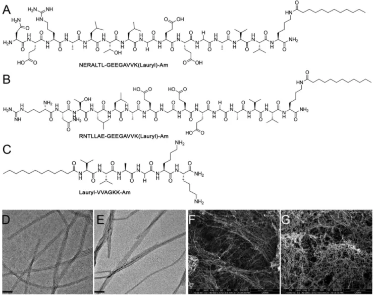
Design, synthesis and characterizations of PA molecules Two different peptide amphiphile molecules were designed and synthesized to study the NGF-β binding and neurite-out-growth-supporting ability of peptide amphiphile scaffolds con-taining the NGF-β binding epitope, NGF3. All PA molecules had a hydrophobic alkyl tail, which consists of lauric acid and aβ-sheet forming peptide sequence, VVAG. In order to increase the solubility, two glutamic acid residues were added to the peptide amphiphile backbone and glycine was used as a spacer amino acid between the hydrophilic part of the sequence and the epitope region.18,19Since the incorporated epitope region was obtained from phage structure and the epitope is displayed at the N terminus of the M13 phage, the peptide amphiphile synthesis was performed from the C to the N terminus by leaving the N terminus of the peptide as a free amine. NERALTL-GEEGAVVK(Lauryl)-Am (NGFB-PA) was designed as an NGF binding PA molecule (Fig. 1A) and RNTLLAE-GEEGAVVK(Lauryl)-Am (scrNGFB-PA; Fig. 1B) was designed with a scrambled version of the NGF binding epitope sequence in order to analyze whether the sequence order is important for the activity of the PA molecule. Lauryl-VVAGKK-Am (KK-PA; Fig. 1C) was synthesized with solid phase peptide synthesis as a filler PA in order to form nanofiber structures through charge neutralization.
Peptide molecules were characterized by LC-MS and puri-fied with preparative HPLC (Fig. S2†). TEM images were taken for each PA combination and these images showed that the combinations formed nanofibers with lengths ranging from a few hundred nanometers to several microns and diameters ranging from 10 to 20 nm (Fig. 1D and E). The SEM imaging of PA gels (Fig. 1F and G) showed the morphological similarity of nanofibrous PA scaffolds to the natural extracellular matrix structure that surrounds nerve cells in tissues.20The extracellu-lar matrix plays an important role in the differentiation of neural stem cells into mature neurons during development and the guidance of nerve ends for the appropriate formation of neural networks during the regeneration process.
Circular dichroism (CD) spectroscopy was employed to analyze the secondary structures of PA nanofibers, and the β-sheet motif was found to predominate in nanofibers formed by all PA combinations tested (Fig. 2A). Secondary structure formation by individual PA molecules was also analyzed with CD (Fig. S3B†) and, because of the presence of both negatively and positively charged amino acid residues, individual NGFB-PA and scrNGFB-PA molecules also formed β-sheet structures.
Zeta potential measurements were performed for both indi-vidual PA molecules and nanofiber structures to determine whether the experimental surface charges of PA scaffolds were in agreement with theoretical predictions (Fig. 2B and Fig. S3A†). The NGFB-PA nanofiber was formed by mixing bio-active NGFB-PA with the nonbiobio-active KK-PA at 1 : 1 molar ratio, and the scrNGFB-PA nanofiber was similarly formed by mixing scrNGFB-PA with the nonbioactive KK-PA at 1 : 1 molar ratio, resulting in neutral net charges for both nanofiber types (Fig. S3A†). Neutral or near-neutral surface charges were observed for all nanofibers (Fig. 2B).
NGFB-PA nanofiber binds to NGF-β with selective and strong affinity
The affinity of NGFB-PA and scrNGFB-PA nanofibers to NGF-β was tested with ELISA. Two different NGF-β concentrations (10 ng mL−1and 50 ng mL−1; Fig. 3) and three different nanofiber concentrations (0.1 mM, 0.25 mM and 0.5 mM) were used to determine whether the affinity of the NGFB-PA nanofiber to NGF-β is concentration dependent. The interaction between the NGFB-PA nanofiber and NGF-β was found to depend on the concentration of both the nanofiber and the growth factor, with higher concentrations of either resulting in higher absor-bance at 450 nm. In addition, for both NGF-β concentrations, the NGFB-PA nanofiber was observed to bind to NGF-β with significantly higher affinity than the scrNGFB-PA nanofiber. As a control, the NGF standard graph is provided in Fig. S3† and it is shown that human NGF-β antibody gives absorbance only in the presence of NGF-β.
NGFB-PA nanofiber provides a biocompatible environment for PC-12 cells
PC-12 cells, a cell line derived from rat adrenal gland pheo-chromocytoma, are a commonly used model system to analyze
Fig. 1 Chemical structures and imaging of self-assembling peptide amphiphile molecules at pH 7.4. Chemical structures of NGFB-PA, scrNGFB-PA and KK-PA are given in (A), (B) and (C), respectively. TEM images of the NGFB-PA nanofiber (D) and the scrNGFB-PA nanofiber (E) show individual nanofiber structures that were stained with uranyl acetate (scale bars are 50 nm). SEM images of NGFB-PA nanofiber (F) and scrNGFB-PA nanofiber (G) gels reveal the extracellular matrix-like structure formed by PA scaffolds. Scale bars are 1 µm. Peptides were dissolved in ddH2O and their pH was adjusted with NaOH for all experiments.
Fig. 2 Characterization of secondary structure formation by peptide scaffolds and analysis of charge properties of PA molecules. The NGFB-PA nanofiber and scrNGFB-PA nanofiber formed beta sheet secondary structures as analyzed by CD measurements (A). Charge properties of individual PA molecules and nanofiber scaffolds (NGFB-PA nanofiber and scrNGFB-PA nanofiber) are analyzed with zeta potential measurements (B).
the activity of different molecules on neural differentiation and axonal growth.21 These cells express the TrkA receptor, which is the high-affinity receptor of NGF. As such, these cells can be induced to terminally differentiate into neuron like cells in the presence of NGF.5,22The effect of peptide scaffolds on the cellular viability of neural progenitor PC-12 cells was analyzed with the Alamar Blue assay and representative images were taken after the staining of cells with calcein-AM and ethi-dium homodimer. PA nanofibers were observed to be biocom-patible with PC-12 cells after 24 h and 72 h of incubation (Fig. S5†).
NGFB-PA nanofiber induces differentiation of PC-12 cells into neuron-like cells
Materials developed for peripheral nervous system regener-ation should ideally promote neurite outgrowth, because this is one of the crucial steps of synapse formation during regen-eration. In addition, improving the presentation efficiency of neurotrophic factors such as NGF to regenerate nerve cells is another crucial factor of peripheral nervous system regener-ation. Here, we aimed to enhance the interaction of NGF with PC-12 cells using an NGF binding epitope on a PA scaffold for enhanced axonal outgrowth. In order to analyze the effect of the NGF-binding PA scaffold on neural differentiation, the lengths of neurites produced by PC-12 cells were measured. PC-12 cells cultured on NGFB-PA nanofibers were observed to have longer neurites than the scrNGFB-PA nanofiber group, and there were no significant differences between the cells cul-tured on the NGFB-PA nanofiber and PLL (Fig. 4A). In addition to neurite length, significantly higher number of cells
extended neurites on the NGFB-PA nanofiber when compared to the scrNGFB-PA nanofiber (Fig. 4B).
In order to further investigate that the NGFB-PA nanofiber enhances NGF-induced differentiation of PC12 cells into neuron-like cells on PA scaffolds, the expression of βIII-tubulin, a general neuronal marker and required in axon growth and guidance,23 was analyzed with immunostaining (Fig. 5).βIII-Tubulin is expressed almost exclusively in neurons and is used to separate neurons from glial cells, which do not express βIII-tubulin.24–27 As shown in Fig. 5, PC-12 cells cul-tured on the NGFB-PA nanofiber expressedβIII-tubulin in cell soma along with neurites. PC-12 cells cultured on the scrNGFB-PA nanofiber also expressedβIII-tubulin in cell soma but few and shorter neurites were observed.
In order to quantify the expression levels ofβIII-tubulin by PC-12 cells cultured on PA scaffolds, western blot analysis of βIII-tubulin expression at day 6 after differentiation was per-formed. As shown in Fig. 6, there is a significant increase in βIII-tubulin expression in PC-12 cells cultured on the NGFB-PA nanofiber, compared to the cells cultured on the scrNGFB-PA nanofiber. As such, it is likely thatβIII-tubulin expression is stimulated in cells differentiated on the NGFB-PA nanofiber scaffold.
NGFB-PA nanofiber enhances NGF-induced differentiation of PC-12 cells
The MAPK pathway is one of the major pathways implicated in the NGF-induced neural differentiation process. During the differentiation of PC-12 cells into neuron like cells, NGF binds to the TrkA receptor and induces the phosphorylation of the MEK1/2 protein complex ( pMEK1/2), which also induces the
Fig. 3 ELISA-based binding assay. Binding levels of 10 ng mL−1and 50 ng mL−1NGF-β to increasing concentrations of the NGFB-PA nanofiber and the scrNGFB-PA nanofiber nanofibers are compared. The difference between the NGFB-PA nanofiber and the scrNGFB-PA nanofiber was significant for both concentrations of NGF. *P < 0.05, **P < 0.01, ***P < 0.001 and ****P < 0.0001. Data presented are the mean OD values (±SEM) of triplicate samples and statistical analyses were performed with one-way ANOVA with Bonferronipost hoc analysis.
phosphorylation of ERK1/2.8,28 Then, the phosphorylated ERK1/2 protein complex ( pERK1/2) is activated and translo-cates to the nucleus and enables the transcription of genes responsible for the differentiation of PC-12 cells.7In order to
determine whether the NGFB-PA nanofiber scaffold enhances the MAPK pathway, PC-12 cells were cultured on PA scaffolds and induced for neural differentiation for 1 h in the presence of 20 ng mL−1 NGF. ERK1/2 and pERK1/2 levels were sub-sequently examined via western blotting. As shown in Fig. 7, PC-12 cells cultured on the NGFB-PA nanofiber were observed to express higher levels of pERK1/2 than cells cultured on the scrNGFB-PA nanofiber and PLL. Taken together, these results suggest that the NGF binding PA scaffold synergistically enhances the NGF-induced MAPK pathway to induce the differentiation of PC-12 cells.
NGFB-PA supports the formation of neural connectivity in cultured DRG neurons
Because the NGFB-PA nanofiber is developed for peripheral nervous system regeneration purposes, we further tested the biocompatibility of the nanofiber for isolated rat DRG neurons by culturing the isolated DRGs on peptide nanofibers and then visualizing by immunofluorescence staining for βIII-tubulin (microtubule marker) and synaptophysin (synaptic marker) protein markers. As shown in Fig. 8, NGFB-PA sup-ports the adhesion and viability of DRG neurons on the nano-fiber system and furthermore enables axon growth and branching from cultured DRG neurons.
Discussion
NGF is one of the most widely studied neurotrophic factors because of its importance in the survival and differentiation of sensory neurons. The role of NGF in the peripheral nervous system has been widely studied. It is well-known that Schwann cells produce NGF after injury, and the gradient of NGF enables broken nerve ends to extend their axons through the injury site to overcome degeneration. NGF binds to a high affinity receptor (TrkA) expressed by nerve cells and activates signaling pathways such as the MAPK pathway, Akt pathway and PKC pathway for the survival and differentiation of nerve cells. Because of these crucial roles of NGF in the peripheral nervous system, it is important to develop therapeutic strat-egies for the efficient presentation of NGF to nerve cells. Several controlled delivery studies were performed for NGF, but there are concerns about their safety.29–31 Furthermore, the development of a material, which is able to mediate the interactions between injured neurons and NGF that is natu-rally secreted by surrounding cells, would be safer and more effective than materials that release artificial NGF at the injury site.29,32In this study, we developed a synthetic peptide nano-fiber scaffold material that binds to NGF with high affinity and promotes axonal outgrowth in vitro. A phage display library containing randomly displayed seven-amino acid peptide sequences was used in order to find peptide sequences exhibit-ing strong affinity and sequence selectivity for NGF-β. After the most selectively binding sequence is identified, the selected epitope sequence was incorporated into the peptide
amphi-Fig. 4 The NGFB-PA nanofiber is biocompatible with PC-12 cells and the NGF-binding epitope supports the extension of much longer neurites than the scrambled version. Light microscope imaging of PC-12 cells cultured and differentiated on the NGFB-PA nanofiber, scrNGFB-PA nanofiber and PLL in the presence of 20 ng mL−1NGF at six days after induction are shown in (A), (B) and (C), respectively (scale bar = 50 µm). Images were taken at 200× magnification. Neurite length (D) and percentage of neurite-bearing PC-12 cells (E) quantified by ImageJ at day 6 after induction show that the incorporation of the NGF-binding epitope into the PA scaffold is able to induce neurite outgrowth. **P < 0.01 and ***P < 0.001. Statistical analyses were performed with one-way ANOVA with Bonferronipost hoc analysis, n = 3.
phile structure that is capable of self-assembling into stable, gel-like nanofiber scaffolds.33The reverse solid phase peptide
synthesis method was utilized for the synthesis of the bioactive PA molecule, as this method was previously reported to resemble the manner in which peptide sequences are dis-played on the M13 phage surface.34 NGFB-PA contains both hydrophobic and hydrophilic residues, which enable its self-assembly through electrostatic interactions and hydrophobic collapse in order to generate ordered structures. The order of the epitope sequence is critical for growth factor binding and in order to test this phenomenon, we designed and tested a scrambled version of the NGFB-PA nanofiber.35,36In our pre-vious study, the optimal charge condition for PC-12 cells was determined as neutral at pH 7.4.14Therefore, the net charge of the nanofibers used in this study was also adjusted at pH 7.4. The biocompatibility of the PA nanofiber scaffolds was
ana-lyzed with a viability assay and both the NGFB-PA nanofiber and scrNGFB-PA nanofiber combinations were shown to be biocompatible with PC-12 cells. In order to analyze whether the binding of the NGFB-PA nanofiber to NGF is concentration dependent, three different PA scaffold concentrations and two different NGF-β concentrations were tested and compared with the commercial NGF antibody. The bioactive NGFB-PA nano-fiber group was observed to exhibit a similar, concentration-dependent binding profile with the NGF antibody–NGF-β binding interaction. A significant difference was also observed between the affinities of NGFB-PA nanofiber and scrNGFB-PA nanofiber groups at both 10 ng mL−1and 50 ng mL−1NGF-β concentrations, suggesting that the strong affinity between NGF-β and NGFB-PA nanofiber is sequence-specific.
The effect of the affinity of the NGFB-PA nanofiber for NGF-β on neuronal differentiation was then analyzed in vitro using
Fig. 5 Immunostaining of PC-12 cells againstβIII-tubulin on NGFB-PA nanofiber, scrNGFB-PA nanofiber and PLL after six days of culture and induc-tion for neural differentiation in the presence of 20 ng mL−1NGF. Higher expression of the neural markerβIII-tubulin was obvious in PC-12 cells cul-tured on the NGFB-PA nanofiber and the positive control group (PLL). Nuclei were stained with TO-PRO-3 and images were taken with confocal microscopy at 200× magnification.
PC-12 cells by quantifying neurite length and percentages of neurite-bearing cells. The NGFB-PA nanofiber induced PC-12 cells to extend much longer neurites than the scrNGFB-PA nanofiber group, and the number of neurite-bearing cells was also increased in the NGFB-PA nanofiber group. These results were then confirmed by analyzing the expression of a general neural marker (βIII-tubulin) by immunostaining and western blot analyses, which confirmed the prior observation that the NGFB-PA nanofiber induces the differentiation of PC-12 cells into neuron-like cells.
NGF binding to the TrkA receptor is crucial for the initiation of the downstream signaling pathways for the
differ-entiation of cells.5,37The Ras pathway and the MAPK pathway are two distinct signaling cascades in PC-12 cells, which are induced by NGF for the survival and differentiation of PC-12 cells.38 It was previously shown that the MAPK pathway is necessary and sufficient for PC-12 differentiation when stimu-lated by NGF, while the Ras pathway is essential for cell survi-val.39,40In order to check whether PC-12 cells cultured on the NGFB-PA nanofiber are induced to differentiate into neuron-like cells by NGF stimulation, phosphorylation levels of ERK1/ 2 proteins were analyzed with western blotting. According to the results shown in Fig. 8, the expressions of endogenous ERK1/2 levels were maintained while phosphorylated ERK1/2
Fig. 6 βIII-Tubulin expression was enhanced in PC-12 cells after 6 days of culture and induction for neural differentiation on the NGFB-PA nanofiber in the presence of 20 ng mL−1NGF, whereas lower expression was observed in cells cultured on the scrNGFB-PA nanofiber. The density of the bands was evaluated by ImageJ and normalized to the GAPDH signal. Western blot analysis revealed thatβIII-tubulin expression in the NGFB-PA nanofiber was almost 2 fold higher than the scrNGFB-PA nanofiber. Data are presented as means ± SEM and ***P < 0.001.
Fig. 7 NGF-induced pERK1/2 levels are enhanced in PC-12 cells cultured on the NGFB-PA nanofiber. PC-12 cells were cultured on the NGFB-PA nanofiber, scrNGFB-PA nanofiber and PLL, and induced for neural differentiation with 20 ng mL−1of NGF. 1 hour after induction, the expression levels of downstream proteins of the NGF-induced MAPK pathway (ERK1/2 and pERK1/2) were analyzed with western blot. The densities of bands were evaluated by ImageJ and normalized to the vinculin signal. Western blot analysis revealed that pERK1/2 expression in the NGFB-PA nanofiber are significantly higher than both scrNGFB-PA nanofiber and positive control group (PLL). Data are presented as means ± SEM and *P < 0.05 and **P < 0.01.
protein levels were significantly increased in PC-12 cells cul-tured on the NGFB-PA nanofiber when compared to scrambled nanofiber and PLL groups, which only have charge inter-actions with NGF but no sequence-specificity. These findings suggest that the NGFB-PA nanofiber binds to NGF with a select sequence specificity and doesn’t inhibit the affinity of NGF for the TrkA receptor and hence enhances the neural differen-tiation pathway.
In addition to PC-12 cells, we further showed that the devel-oped system also supports the viability of sensory neurons. Since axon outgrowth and synaptic connectivity of neurons at the injury site are necessary for the regeneration and gain of function of periphery nerves, the development of proper axons from cultured DRGs in our system indicates that the NGFB-PA nanofiber system has the potential for supporting neural
out-growth and reconnection of sensory neurons after peripheral nerve injury.
Conclusions
In this study, we developed a synthetic NGF–affine peptide nanofiber scaffold, which can promote axonal outgrowth in PC-12 cells by enhancing the availability of NGF for the TrkA receptor. Furthermore, NGFB-PA was shown to promote the proper functioning of DRG neurons cultured on nanofibers by supporting axonal branching. Since NGF plays an important role in peripheral nervous system development and regener-ation, the developed NGF–affine synthetic scaffold can be used as a therapeutic agent for regeneration in peripheral nerve
Fig. 8 Immunostaining of DRG neurons againstβIII-tubulin and synaptophysin on the NGFB-PA nanofiber, scrNGFB-PA nanofiber and PDL/laminin after seven days of culture. DRG neurons cultured on the NGFB-PA nanofiber are shown to develop proper axons and synapse formation is also obvious. Nuclei were stained with TO-PRO-3 and cells were imaged with confocal microscopy. The scale bar is 50μm.
injuries (such as sciatic nerve injury) by binding to NGF secreted by Schwann cells and enhancing the neural regener-ation process. Overall, the NGF binding nanofiber is a promis-ing material for the development of novel therapeutic approaches and applicable for further in vivo peripheral nervous system regeneration studies.
Author contributions
Z. O., B. M., M. O. G. and A. B. T. designed the study. Z. O., O. I. S, C. Y., and G. G. performed research. Z. O. and C. Y. carried out cell culture experiments. Z. O., O. I. S. and G. G. performed peptide synthesis and characterization experiments. Z. O., M. O. G. and A. B. T. analyzed data and wrote the paper.
Con
flicts of interest
There are no conflicts to declare.
Acknowledgements
We would like to thank Prof. A. Arslanoglu from Izmir Institute of Technology, Izmir, Turkey, for his kind supplying of the XL1Blue bacteria strain. We would like to thank Z. Erdogan for help with LC-MS and M. Guler for TEM imaging. We also thank N. Gunduz for help with SEM imaging, M. Sever for her help with cell culture studies, and A. D. Ozkan for his valuable comments. This work was supported by the Scientific and Technological Research Council of Turkey (TUBITAK) Grant Number 215S770. Z.O. was supported by a TUBITAK BIDEB 2210-C fellowship. A.B.T. acknowledges support from the Science Academy Outstanding Young Scientist Award (BAGEP).
Notes and references
1 J. J. Duby, R. K. Campbell, S. M. Setter, J. R. White and K. A. Rasmussen, Diabetic neuropathy: an intensive review, Am. J. Health-Syst. Pharm., 2004, 61(2), 160–173; quiz 75-6. 2 R. Mirsky and K. R. Jessen, The neurobiology of Schwann
cells, Brain Pathol., 1999, 9(2), 293–311.
3 M. V. Sofroniew, C. L. Howe and W. C. Mobley, Nerve growth factor signaling, neuroprotection, and neural repair, Annu. Rev. Neurosci., 2001, 24, 1217–1281.
4 C. Wiesmann and A. M. de Vos, Nerve growth factor: struc-ture and function, Cell. Mol. Life Sci., 2001, 58(5–6), 748– 759.
5 S. O. Yoon, P. Casaccia-Bonnefil, B. Carter and M. V. Chao, Competitive signaling between TrkA and p75 nerve growth factor receptors determines cell survival, J. Neurosci., 1998, 18(9), 3273–3281.
6 C. F. Ibáñez, F. Hallböök, T. Ebendal and H. Persson, Structure-function studies of nerve growth factor:
func-tional importance of highly conserved amino acid residues, EMBO J., 1990, 9(5), 1477–1483.
7 R. H. Chen, C. Sarnecki and J. Blenis, Nuclear localization and regulation of ERK- and RSK-encoded protein kinases, Mol. Cell. Biol., 1992, 12(3), 915–927.
8 L. Pang, C. F. Zheng, K. L. Guan and A. R. Saltiel, Nerve growth factor stimulates a novel protein kinase in PC-12 cells that phosphorylates and activates mitogen-activated protein kinase kinase (MEK), Biochem. J., 1995, 307(Pt 2), 513–519.
9 Y. Xie, M. A. Tisi, T. T. Yeo and F. M. Longo, Nerve growth factor (NGF) loop 4 dimeric mimetics activate ERK and AKT and promote NGF-like neurotrophic effects, J. Biol. Chem., 2000, 275(38), 29868–29874.
10 G. P. Smith, Filamentous fusion phage: novel expression vectors that display cloned antigens on the virion surface, Science, 1985, 228(4705), 1315–1317.
11 S. M. Willerth, P. J. Johnson, D. J. Maxwell, S. R. Parsons, M. E. Doukas and S. E. Sakiyama-Elbert, Rationally designed peptides for controlled release of nerve growth factor from fibrin matrices, J. Biomed. Mater. Res., Part A, 2007, 80(1), 13–23.
12 W. Sun, C. Sun, H. Lin, H. Zhao, J. Wang, H. Ma, et al., The effect of collagen-binding NGF-beta on the promotion of sciatic nerve regeneration in a rat sciatic nerve crush injury model, Biomaterials, 2009, 30(27), 4649–4656.
13 K. Lee, E. A. Silva and D. J. Mooney, Growth factor delivery-based tissue engineering: general approaches and a review of recent developments, J. R. Soc., Interface, 2011, 8(55), 153–170.
14 B. Mammadov, R. Mammadov, M. O. Guler and A. B. Tekinay, Cooperative effect of heparan sulfate and laminin mimetic peptide nanofibers on the promotion of neurite outgrowth, Acta Biomater., 2012, 8(6), 2077–2086. 15 B. Mammadov, M. Sever, M. Gecer, F. Zor, S. Ozturk,
H. Akgun, et al., Sciatic nerve regeneration induced by glycosaminoglycan and laminin mimetic peptide nanofiber gels, RSC Adv., 2016, 6(112), 110535–110547.
16 T. H. Burkey, C. M. Hingtgen and M. R. Vasko, Isolation and culture of sensory neurons from the dorsal-root ganglia of embryonic or adult rats, Methods Mol. Med., 2004, 99, 189–202.
17 H. Cui, M. J. Webber and S. I. Stupp, Self-assembly of peptide amphiphiles: from molecules to nanostructures to biomaterials, Biopolymers, 2010, 94(1), 1–18.
18 S. Toksoz, R. Mammadov, A. Tekinay and M. Guler, Electrostatic effects on nanofiber formation of self-assem-bling peptide amphiphiles, J. Colloid Interface Sci., 2011, 356(1), 131–137.
19 C. Smith, J. Withka and L. Regan, A thermodynamic scale for the beta-sheet forming tendencies of the amino-acids, Biochemistry, 1994, 33(18), 5510–5517.
20 T. Nishida, K. Yasumoto, T. Otori and J. Desaki, The network structure of corneal fibroblasts in the rat as revealed by scanning electron-microscopy, Invest. Ophthalmol. Visual Sci., 1988, 29(12), 1887–1890.
21 L. A. Greene and A. S. Tischler, Establishment of a nor-adrenergic clonal line of rat adrenal pheochromocytoma cells which respond to nerve growth factor, Proc. Natl. Acad. Sci. U. S. A., 1976, 73(7), 2424–2428.
22 D. Vaudry, P. J. Stork, P. Lazarovici and L. E. Eiden, Signaling pathways for PC12 cell differentiation: making the right connections, Science, 2002, 296(5573), 1648–1649.
23 M. A. Tischfield and E. C. Engle, Distinct alpha- and beta-tubulin isotypes are required for the positioning, di fferen-tiation and survival of neurons: new support for the ‘multi-tubulin’ hypothesis, Biosci. Rep., 2010, 30(5), 319–330. 24 G. Karkavelas, C. D. Katsetos, J. F. Geddes, M. M. Herman,
S. A. Vinores, H. S. Cooper, et al., Class III beta-tubulin isotype (beta III) in the adrenal medulla: II. Localization in primary human pheochromocytomas, Anat. Rec., 1998, 250(3), 344–350.
25 C. D. Katsetos, M. M. Herman and S. J. Mörk, Class III beta-tubulin in human development and cancer, Cell Motil. Cytoskeleton, 2003, 55(2), 77–96.
26 C. D. Katsetos, A. Legido, E. Perentes and S. J. Mörk, Class III beta-tubulin isotype: a key cytoskeletal protein at the crossroads of developmental neurobiology and tumor neuropathology, J. Child Neurol., 2003, 18(12), 851–866; dis-cussion 67.
27 C. D. Katsetos, G. Karkavelas, M. M. Herman, S. A. Vinores, J. Provencio, A. J. Spano, et al., Class III beta-tubulin isotype (beta III) in the adrenal medulla: I. Localization in the developing human adrenal medulla, Anat. Rec., 1998, 250(3), 335–343.
28 M. Frödin, P. Peraldi and E. Van Obberghen, Cyclic AMP activates the mitogen-activated protein kinase cascade in PC12 cells, J. Biol. Chem., 1994, 269(8), 6207–6214.
29 S. C. Apfel, Neurotrophic factor therapy–prospects and pro-blems, Clin. Chem. Lab. Med., 2001, 39(4), 351–355.
30 S. J. Jhaveri, M. R. Hynd, N. Dowell-Mesfin, J. N. Turner, W. Shain and C. K. Ober, Release of nerve growth factor from HEMA hydrogel-coated substrates and its effect on
the differentiation of neural cells, Biomacromolecules, 2009, 10(1), 174–183.
31 P. R. Chen, M. H. Chen, J. S. Sun, C. C. Tsai and F. H. Lin, Biocompatibility of NGF-grafted GTG membranes for peri-pheral nerve repair using cultured Schwann cells, Biomaterials, 2004, 25(25), 5667–5673.
32 C. M. Valmikinathan, S. Defroda and X. Yu, Polycaprolactone and bovine serum albumin based nano-fibers for controlled release of nerve growth factor, Biomacromolecules, 2009, 10(5), 1084–1089.
33 A. Dehsorkhi, V. Castelletto and I. W. Hamley, Self-assem-bling amphiphilic peptides, J. Pept. Sci., 2014, 20(7), 453– 467.
34 R. N. Shah, N. A. Shah, M. M. Del Rosario Lim, C. Hsieh, G. Nuber and S. I. Stupp, Supramolecular design of self-assembling nanofibers for cartilage regeneration, Proc. Natl. Acad. Sci. U. S. A., 2010, 107(8), 3293–3298.
35 C. J. Newcomb, S. Sur, J. H. Ortony, O. S. Lee, J. B. Matson, J. Boekhoven, et al., Cell death versus cell survival instructed by supramolecular cohesion of nanostructures, Nat. Commun., 2014, 5, 3321.
36 K. Saar, M. Lindgren, M. Hansen, E. Eiríksdóttir, Y. Jiang, K. Rosenthal-Aizman, et al., Cell-penetrating peptides: a comparative membrane toxicity study, Anal. Biochem., 2005, 345(1), 55–65.
37 M. V. Chao, Neurotrophin receptors: a window into neuro-nal differentiation, Neuron, 1992, 9(4), 583–593.
38 L. J. Klesse, K. A. Meyers, C. J. Marshall and L. F. Parada, Nerve growth factor induces survival and differentiation through two distinct signaling cascades in PC12 cells, Oncogene, 1999, 18(12), 2055–2068.
39 S. Cowley, H. Paterson, P. Kemp and C. J. Marshall, Activation of MAP kinase kinase is necessary and sufficient for PC12 differentiation and for transformation of NIH 3T3 cells, Cell, 1994, 77(6), 841–852.
40 D. R. Kaplan and R. M. Stephens, Neurotrophin signal transduction by the Trk receptor, J. Neurobiol., 1994, 25(11), 1404–1417.