RESEARCH ARTICLE
The effects of L-carnitine on blood and tissue parameters of
male rats fed with different levels of fish oil
Halil Yavuz, Firuze Kurtoğlu*
Department of Biochemistry, Faculty of Veterinary Medicine, Selcuk University, Konya, Turkey Received: 22.02.1024, Accepted: 01.04.2014
Özet
Yavuz H, Kurtoğlu F. Farklı düzeylerde balık yağı ile beslenen erkek ratlarda L-karnitinin kan ve doku parametrelerine etkileri.
Amaç: Bu çalışmada, farklı oranlarda balık yağı içeren rasyonla beslenen erkek ratlarda L-karnitinin (300 mg/kg/gün) 30 gün süreli periton içi uygulamasının plazma L-karnitin, lipit hidro-peroksit (LPO), antioksidan enzimler (SOD ve CAT) ile glutasyon (GSH), trigliserit, kolesterol seviyeleri; doku SOD, CAT, GSH dü-zeyleri ve canlı ağırlık değerlerine etkileri araştırılmıştır. Gereç ve Yöntem: Araştırmada 72 adet Sprague-Dawley erkek rat kullanıldı. Ratlar 6 deneysel gruba ayrıldı;1. Kontrol, 2. Balık yağı (%1), 3. Balık yağı (%5), 4. L-karnitin (300 mg/kg/gün), 5. L-karnitin (300 mg/kg/gün) + balık yağı (%1) ve 6. L-karnitin (300 mg/kg/gün) + Balık yağı (%5). İlk 30 gün sonunda karnitin uygulamaları sonlandırılarak, her bir ratın canlı ağırlık değerleri elde edildi. Plazma ve doku parametreleri ticari test kitleri kulla-nılarak spektrofotometrik yöntemlerle tayin edildi.
Bulgular: Deneme ve arınma periyodu (30. ve 60. gün) sonunda plazma karnitin düzeyleri karnitin uygulamalarından etkilen-miştir. Tüm çalışma boyunca trigliserit ve kolesterol değerlerin-deki en belirgin düşüşler karnitin gruplarından özellikle 60. gün değerlerinde elde edildi. LPO değerleri hem dönemler arası hem de gruplar arası anlam taşıyan düşüşler gösterdi. GSH değerle-rinde 4. grupta, SOD ve CAT düzeyledeğerle-rinde ise 4. ve 5. gruplarda belirgin oranda artışlar elde edildi.
Öneri: Ekstra karnitin ve balık yağı alımları canlılarda lipidpro-filini düzenleyebilir, kan ve doku antioksidanlarını artırabilir-ken, LPO değerleri karnitin etkilerini değerlendirmede belirle-yici olarak değerlendirilebilir.
Anahtar kelimeler: L-karnitin, balık yağı, antioksidanlar, lipit profili, rat
Abstract
Yavuz H, Kurtoglu F. The effects of L-carnitine on blood and tis-sue parameters of male rats fed with different levels of fish oil.
Aim: The effects of L-carnitine (300 mg/kg/day) treated in-traperitoneally (i.p) to rats fed ration containing several pro-portions of fish oils for 30 days on plasma L-carnitine, lipid hydroperoxide (LPO), triglyceride, cholesterol, body weight; plasma-tissue antioxidant enzymes (SOD, CAT) and glutathione (GSH) levels were investigated.
Materials and Methods: Seventy two Sprague-Dawley male rats were used. Rats were divided into 6 groups as follows; 1-Control; 2-Fish oil (1%); 3-Fish oil (5%); 4-L-carnitine (300 mg/kg/day), 5-L-carnitine (300 mg/kg/day) plus fish oil (1%) 6-L-carnitine (300 mg/kg/day) plus fish oil (5%). All plasma and tissue pa-rameters were determined by spectrophometric methods using commercial kits.
Results: At the end of the experimental and washout period (day 30 and 60), the plasma carnitine concentrations were affected by carnitine administrations. Remarkable decreases in triglycer-ide and cholesterol values were obtained particularly at the end of day 60 in carnitine groups. LPO values were associated with significant differences between period and groups comparisons for carnitine administrations. The highest GSH values were ob-tained in group 4; CAT and SOD status in group 4 and 5. Conclusions: Extra-carnitine and fish oil additions may regulate plasma lipid profile and increase blood and tissue antioxidants, while LPO levels scan be display controlling effect by carnitine administration.
Keywords: L-carnitine, fish oil, antioxidants, lipid profile, rat
Eurasian J Vet Sci, 2014, 30, 3, 138-144
DOI:10.15312/EurasianJVetSci.201436513
Eurasian Journal
of Veterinary Sciences
www.ejvs.selcuk.edu.tr www.eurasianjvetsci.org
Introduction
Carnitine is a compound which has structure of “gamma-trimethyl amino beta hydroxy butyric acid” and it has D- and L-form but L-form, metabolically active form only, is endogenously synthesized in tissues (Da Torre et al 1991, Çitil 2002) such as brain, kidneys and liver via conversion from lysine and methionine (Krajcovicova-Kudlackova et al 2000, Hoppel 2003). It is mostly taken in diet and stored in skeletal muscle (Guarnieri et al 2001, Rebouche 2004). Basic function of this important metabolite is to ensure transportation of long-chain fatty acids from cytoplasm into mitochondria, resulting with availability of them for mitochondrial β-oxidation (Seline and Johein 2007). L-carnitine has vital function in intra-cellular energy metabolism and the function is exerted via two ways: First, it is involved in β-oxidation by transporting long-chain fatty acids (12 to 20 carbon atoms), which are energy sources in the form of acylcarnitine, into mitochondria. Second, it inhibits toxic effects resulting from release of free CoA, which develops when short-chain (4-6 carbon atoms) and medium-chain (6-12 carbon atoms) fatty acids are metabolized in mitochondria (Calabrese et al 2012).
Supplementation of L-carnitine to diets, feeds and rations, as an anti-oxidant substance, provided positive effects on overall metabolic activity and increase in mitochondrial function without any increase associated with oxidative stress (Arrigoni-Martelli and Caso 2001, Hagen et al 2002). In oxidative stress, increased intra-cellular calcium ion concentration causes conversion of xhantine dehydrogenase, which catalyzes conversion to superoxide anion (O2*), into xhantine oxidase by oxidizing xhantine (Annunziato et al 2003). Such effects lead to remarkable reduction in superoxide dismutase (SOD) activity secondary to further exposure to oxidative stress particularly at aging process. It was found that decreased SOD activities started to increase based on effect of L-carnitine and alpha lipoic acid supplementation (Muthuswamy et al 2006). Free oxygen radicals accumulate in cells and tissues, resulting with several types of damages. It is suggested that omega-3 fatty acids exert effects via 2 metabolic pathways to prevent the damage (Masters 1996, Özgöcmen et al 2000); in the first pathway, fatty acids increase catalase (CAT) level in intracellular peroxisomes and a stronger cellular defense is promoted against radicals. In the second metabolic pathway, omega-3 fatty acids are substituted with other polyunsaturated fatty acids (PUFAs) in cell membrane and they fight against superoxide anions, hydrogen peroxide and hydroxyl radicals, which may act to damage cell membranes. Anti-oxidant factors exert activities to balance others effects. For example, conversion of hydrogen peroxide (H2O2) into water and oxygen is suppressed as a result of decrease in activity of glutathione peroxidase (GPx), while CAT activity is promoted; however, researchers (Gomez-Amores et al 2006)reported that in the hypertensive rat models, GPx and CAT activities increased in combination in order to detoxify H2O2 when propionyl L-carnitine (PLC) was added and remarkable reduction was observed in lipid peroxidation rates (TBARS), triglyceride and cholesterol levels in normotensive and hypertensive groups of same experiment model (Gomez-Amores et al 2006). Antioxidative mechanism is activated secondary to
increasing lipid concentration in chronic hyperlipidemia, and GSH and vitamin B6 levels decrease, while peroxidation rate increases over time (Iwana and Okada 1982, Niki et al 1984). Carnitine also induces accumulation of thiol (SH) groups and methionine in plasma and tissues and it exerts indirect effects to sustain normal cellular functions under influence of those anti-oxidant substances (Khairallah and Wolf 1965). Also, carnitine protects low molecular-weight –SH groups (GSH, homocysteine and cysteine) and lipids against peroxynitrite (ONOO-) oxidation (Kolodziejczyk et al 2011). L-carnitine increases synthesis of phospholipids, which are necessary for cell membranes, and ensures reacylation of phospholipids, resulting with integrity of membrane structure or repair of damages (Kashiwagi et al 2001). This view is supported by the fact increased lipid peroxidation and reduced SOD, GSH and CAT enzymes and vitamin C and E, which have anti-oxidative effects, in old rats in comparison with young rats, and they may increase with carnitine supplementation (Schnackenberg and Wilcox 2001, Gülçin 2006). Rajasekar et al (2005) reported that L-carnitine supplementation decreased plasma VLDL-cholesterol and triglyceride levels in hyperlipidemic rats. Also other researcher (Yalçın et al 2007) observed that L-carnitine supplementation had positive effects on performance and decreased serum cholesterol and triglyceride levels. Siegner et al (2010) reported a significant reduction in triglyceride accumulation when they compared combined use of solution of lotus leaf extract and L-carnitine with alone administration of both compounds. They also reported that they have potential in treatment and prevention of obesity by stimulating different pathways of lipid storage. Similarly L-carnitine administration decreased serum or tissue lipids of obese rat (Amin and Nagy 2009), obese mice (Rajasekar and Anuradha 2007) and New Zealand rabbits (Elgazzar et al 2012).
In this study, effects of L-carnitine (300 mg/kg/day, IP) on plasma lipid hydroperoxide (LPO) and antioxidant enzymes (SOD, CAT), GSH, triglyceride, cholesterol levels, liver and muscle SOD, CAT, GSH levels and body weight of Sprague Dawley male rats received fish oil containing several long chain polyunsaturated fatty acids for 30 days were investigated. And also it was aimed to determine the effects of carnitine on all the plasma and tissue values by comparing the values obtained from second 30-day period so-called wash-out period (no carnitine addition).
Materials and Methods
Animals and experimental design
In this study, seventy two 5-6-months old male Sprague-Dawley rats were used as animal materials; rats were supplied from Experimental Animals Culture and Research Center, University of Baskent. The study protocol was approved by Local Ethics Committee of Experiment Animals Department, Faculty of Veterinary Medicine, University of Selcuk (Approval date and number: 13.01.2010-2010/01). Experimental groups were designed according to body weights of rats as homogeneous as possible and each group of 6 rats was kept in polyethylene rat cages at room temperature (22±1°C). Standard pelleted feed and tap water were available ad libitum throughout experimental period.
The feed was supplied from a local company (Kırıkkale Feed Anonim Company) and fatty acid content of fish oil (Menhaden fish oil, Sigma F-8020) added at concentration of 1% and 5% into feed. First 10-day period was reserved for adaptation of rats to conditions of experiment. After this adaptation period expired, body weight of each rat was measured and recorded at baseline. Blood samples were drawn from 8 rats using tail vein under superficial anesthesia in order to measure baseline plasma values. Stock carnitine solution was prepared by dissolving L-carnitine (Sigma C0283) in saline (0.9%); this solution was administered to each rat of relevant groups (group 4, 5 and 6) at dose of 300 mg/kg bw/day via i.p. for 30 days. Saline solution (carnitine free) was also administered via i.p. to rats of other three groups (group 1-control, group 2 and 3).
Sampling and determinations
At the end of first 30 days, carnitine administration was discontinued and body weights were measured for each rat. In each group of 12 rats, six rats were deprived of food during12 hours and blood samples were drawn into heparinised tubes, while spontaneous respiration was continued throughout the procedure during anesthesia with penthal sodium. After blood samples were drawn, rats survived were sacrificed with cervical dislocation; muscle and liver tissue samples were taken from homogeneous and connective tissue-free zones as much as possible; specimens were placed in phosphate buffer and immediately transferred to at -80oC. Remaining rats were underwent carnitine-free washout period, which lasted second 30 days. Blood and tissue samples were taken using same methods from thirty six rats, which survived at the end of experimental period. Blood samples, which were drawn into heparinized tubes at day 1, 30 and 60 of the experiment, were centrifuged (2500 rpm) at +4oC for 15 minutes. Plasma samples were stored at -80°C until they were analyzed. Plasma triglyceride and cholesterol levels were analyzed using Spinreact kit (Santa Coloma 7E 17176 Spain), while L-carnitine was analyzed with Biovision test kit (K642 CA 94043 USA); Cayman (Chemical Company 1180 E. Ann Arbor MU 48108) kits were used to analyze plasma LPO, plasma and tissue CAT, SOD and GSH values; all analyses were done using microplate reader (BiotekELx800 USA) with spectrophotometric method. The tissue specimens, which were stored at -80°C, were mechanically homogenized together with the phosphate buffer in dry ice using tissue homogenizer (Sartorius AG37070, Germany) for 2 (liver) and 5 (muscle) minutes at 1500 rpm and homogenate suspensions obtained were centrifuged (Universal 320 R Hettich Germany) at 6000 rpm for 30 minutes at +4°C.
Statistical analysis
Data was analyzed using SPSS 17 (Statistical Package for the Social Science, SPSS Inc., Chicago, IL, United States) software. One-way ANOVA was used for continuous variables with normal distribution to determine whether there was statistically significant difference between groups and periods (experimental period and washout period), while student’s t test was used to determine inter-period differences of tissues; groups leading to significant differences were analyzed with Duncan’s test. In the
experiment, day 1, day 30 (experiment period) and day 60 (washout period) values were obtained for each parameter, and one-way ANOVA was used to compare values between groups (control-group 1, group 2, 3, 4, 5 and 6) and periods (day 1, day 30 and day 60); while Duncan’s test was used to determine statistically significant difference; when tissue values were compared between two periods (no found tissue values for day 1) values obtained, student’s t test was used. ANOVA was used to analyze live body weight data, similar to plasma analyses.
Results
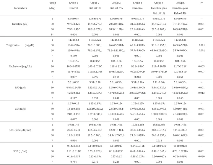
No statistically significant difference was observed between three blood drawing periods (day 1, day 30 and day 60) in all parameters of the control group, which was administered only saline (Table 1). At the end of the experiment period (day 30), where particular groups (group 4, 5 and 6) were administered carnitine at dose of 300 mg/kg/day via i.p. route, highest plasma carnitine concentrations were obtained in groups 4 and 6 (31.01, 31.14 µM, respectively) at day of 30th. Similar decreasing trend for carnitine levels in these groups were determined at day of 60th. Considering changes shown in Table 1, most remarkable decreases (P<0.001) in triglyceride levels were obtained at day of 30th and 60th especially in carnitine (group 4), carnitine plus fish oil 1% (group 5) and carnitine plus fish oil 5% (group 6) groups; similar differences for cholesterol values were determined between groups 4th and 6th at 30 and 60 (P<0.01) when compared the controls. But it was no found any statistical importance (P>0.05) between the periods in groups for cholesterol levels.
Lipid hydroperoxide values were associated with statistically significant differences as shown on inter-group comparisons especially in carnitine administered group (Table 1). The fish oil used in the study was rich in long-chain unsaturated fatty acids, resulting with vulnerability to lipid peroxidation. In comparison with LPO values of group 2 and 3, which were given only fish oil, it was found that carnitine supplementation (group 5 and 6) resulted with remarkable decrease in LPO levels at particular periods.
When plasma GSH values were examined (Table 1), highest values were obtained in group 4 in both experiment and washout periods, and it was noted that GSH values were higher in group 5 and 6, which were given carnitine in combination with fish oil, at the end of day 30 in comparison with the control group. Significantly increased anti-oxidant enzyme (CAT and SOD) values were noted in carnitine supplementation groups (P<0.001), and activity of two enzymes significantly increased in group 4 and 5, which were given carnitine and carnitine plus fish oil 1%, respectively. However, in comparison with the control group, CAT and SOD activities were significantly high only in group 4 and 5. Almost identically significant increases were observed in all enzyme levels between groups at day 30 and day 60 (P<0.001), while liver GSH and CAT values followed a trend of more significant increase in both periods.
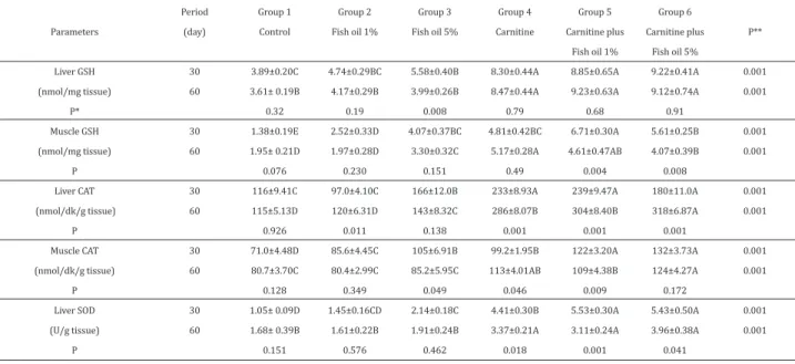
As shown Table 2, carnitine and fish oil additions significantly (P<0.001) effected all tissue antioxidant parameters (GSH,
CAT, SOD) between the experimental groups. It was found that the highest liver GSH and SOD values were found in groups 4, 5 and 6 at experimental and washout periods. Except for muscle GSH, other tissue parameters were determined as highest values in group 6, while the lowest tissue GSH, CAT and SOD values were found in control group in which no found detected any statistical significance between period of 30th and 60th day.
Statistically difference analysed in inter-group could be significant reached only for day-60 measurements with respect to body weight parameter (P<0.001), but a remarkable decrease was noted in group 4 (Table 3), which was administered carnitine, and group 5, which was given carnitine plus fish oil 1% (P<0.05).
Discussion
In the current study, carnitine administration at dose of 300 mg/kg bw/day for 30 days had positive influences on plasma carnitine levels, and highest plasma carnitine levels were obtained in group 4 and 6 in both experiment and washout periods; in the light of those findings, we concluded that the significant difference between plasma and tissue values of those groups may originate from carnitine and fish oil
administration. The plasma carnitine levels of those groups (31.01 and 31.14 µM, respectively) were within normal plasma carnitine ranges, and the lower mean concentration of baseline period (8.96 µM) was significantly different from mean values obtained from other groups (group 1, 2 and 3), which were not administered carnitine (P<0.001). A similar relation (P<0.001) was found between washout period groups, although it was at lower concentration. However, it is believed that in comparison with the control group, the increased in carnitine level of group 3, which was not administered carnitine, may be related with stimulation of carnitine synthesis secondary to high dietary fat (fish oil) content (5%) (Yavuz and Kurtoğlu 2012) The fact that a similar increase was noted in group 2, which was not administered carnitine and was only given fish oil at lower concentration (1%), at the end of washout period supports the view that carnitine synthesis can be induced by diet rich in fish oil.
In our study, carnitine administration resulted with decrease in triglyceride values particularly and significantly at experiment period. This effect of carnitine can be related with the fact that carnitine directs fatty acids over acyl carnitine to energy production, it decreases activity of lipoprotein
P** --0.001 0.001 0.001 0.001 --0.003 0.007 --0.001 0.013 --0.001 0.001 --0.001 0.001 --0.001 0.080 Group 6 Carnitine plus Fish oil 5% 8.96±0.57c 31.1±1.18A,a 16.9±0.78B,b 0.001 113±5.62a 76.3±6.52B,b 55.3±8.09C,c 0.001 104±3.56 91.7±2.21C 92.3±5.61D 0.051 5.31±0.30a 3.66±0.64BC,b 4.50±0.34A,ab 0.046 1.25±0.15c 3.80±0.48B,a 2.84±0.28C,b 0.001 19.8±1.48a 134±0.98C,b 16.0±1.06C,b 0.003 014±0.013c 0.29±0.023Ba 0.22±0.019b 0.001 Group 5 Carnitine plus Fish oil 1% 8.96±0.57b 24.9±2.01B,a 22.5±1.10A,a 0.001 113±5.62a 93.8±3.75A,b 68.3±4.22ABC,c 0.001 104±3.56 112±7.10AB 98.9±4.57BCD 0.209 5.31±0.30a 5.84±0.42A,a 2.29±0.24C,b 0.001 1.25±0.15c 4.65±0.49B,a 3.08±0.70BC,b 0.001 19.8±1.48b 28.6±3.01A,a 26.1±1.25A,a 0.013 0.14±0.013b 0.40±0.04A,a 0.36±0.017a 0.001 Group 4 Carnitine 8.96±0.57c 31.0±3.05A,a 22.1±0.84A,b 0.001 113±5.62a 65.5±4.34B,b 57.9±5.96C,b 0.001 104±3.56 96.8±1.86C 95.2±5.79CD 0.213 5.31±0.30a 2.64±0.36C,b 3.09±0.39BC,b 0.001 1.25±0.15b 5.97±0.35A,a 5.48±0.64A,a 0.001 19.8±1.48b 33.2±1.49A,a 24.6±1.07B,b 0.001 0.14±0.012b 0.41±0.02A,a 0.38±0.027a 0.001 Group 3 Fish oil 5% 8.96±0.57b 20.5±0.41B,a 18.9±1.12B,a 0.001 113±5.62a 76.6±5.99B,b 73.8±3.41ABC,b 0.001 104±3.56 118±4.81A 109±5.21ABC 0,116 5.31±0.30a 5.09±0.37A,a 4.07±0.37AB,b 0.047 1.25±0.15c 2.65±0.36C,b 4.41±0.42AB,a 0.001 19.8±1.48a 12.2±1.14C,b 14.5±1.29CD,b 0.003 0.14±0.013 0.11±0.009C 0.27±0.12 0.226 Group 2 Fish oil 1% 8.96±0.57c 11.9±1.27C,b 18.9±0.37B,a 0.001 113±5.62a 76.9±3.38B,b 79.1±8.45B,b 0.001 104±3.56 100±2.02BC 111±4.12AB 0.095 5.31±0.30 5.23±0.21A,a 4.21±0.33A,b 0.033 1.25±0.15b 1.95±0.23CD,a 2.37±0.30C,a 0.006 19.8±1.48a 13.5±0.74C,b 11.5±0.70D,b 0.001 0.14±0.013b 0.23±0.02B,a 0.22±0.03a 0.010 Group 1 Control 8.96±0.57 9.78±0.42C 7.96±1.47C 0.404 113±5.62 104±4.91A 120±4.83A 0.150 104±3.56 100±6.67BC 117±4.55A 0.087 5.31±0.30 4.89±0.54AB 4.20±0.41A 0.177 1.25±0.15 1.51±0.23D 2.02±0.35C 0.097 19.8±1.48 20.0±1.53B 19.6±1.03B 0.984 0.14±0.013 0.13±0.014C 0.14±0.015 0.764 Period (day) 1 30 60 P* 1 30 60 P 1 30 60 P 1 30 60 P 1 30 60 P 1 30 60 P 1 30 60 P Parameters Carnitine (µM) Triglyceride (mg/dL) Cholesterol (mg/dL) LPO (µM) GSH (µM) CAT (nmol/dk/mL) SOD (U/mL)
Table 1. Levels of biochemical and antioxidant parameters determined in plasma of experimental groups (mean ± SE).
n=6-8 rats. 1st day means are obtained by 8 rats and common data for all experimental groups. Statistical differences between periods. P*(Duncan a-c; P<0.05); and between groups P**; (Duncan A-D; P<0.05)
lipase and accordingly, triglyceride levels decrease (Kurban and Mehmetoglu 2006, Li et al 2007). The reduction is more notable in the L-carnitine supplementation group, while triglyceride levels of group 2 and 3, which were given extra fish oil, were lower in comparison with the control group. Similar to triglyceride levels, a trend towards reduction of cholesterol levels was found in group 3, but this trend was not as clear as the triglyceride values. Similarly, Erol and Yalcın (2009) supplemented the different amounts of L-carnitine (50, 100, 150 mg/kg) into turkey feeds, and they observed insignificant changes in triglyceride and total cholesterol values, while authors determined quantitative decrease in triglyceride, although it was not notable but no clear change was observed for cholesterol in the study. In the light of all those findings, it is concluded that influences of carnitine can be more clearly determined on HDL and LDL cholesterol profiles, which were not examined in our study. Considering cholesterol and triglyceride values, results of the current study are consistent with recent experimental animal models (Tanaka et al 2004, Rajasekar et al 2005, Gomez-Amores et al 2006, Mansour 2006, Sutken et al 2009), ruminant (Çitil et al 2009), quail (Yalçın et al 2007) and investigational and therapeutic (clinical) human studies (Vacha et al 1983, Özener et al 1995).
Fish oil (omega 3 fatty acids) regulates intrecellular methionine synthesis; methionine involved in endogenous
carnitine synthesis and accordingly the synergistic effect of carnitine and fish oil on most parameters can be related with this mechanism. On the other side, this mechanism is verified with the fact that plasma carnitine levels of group 2 and 3, which were only given fish oil, were closer to that of carnitine supplementation group and were significantly higher than that of the control group.
In the study, LPO were considered as a measure of lipid peroxidation and a decrease is observed, as expected, in LPO values of group 4, which was given carnitine supplementation. Considering elevating LPO values in group 2 and 3, which were given fish oil, most remarkable effect of carnitine was observed in group 5, which was given fish oil 1% plus carnitine supplementation, at washout period, and the value regressed to 2.29 µM most remarkable influence of carnitine was noted at experiment period in group 6 in comparison with the group which was given only 5% fish oil. Polyunsaturated fatty acids may stimulate or activate defense mechanisms to fight against oxidative process. If organism does not have sufficient anti-oxidant reserves, peroxidation products (MDA, TBARS and LPO) will increase since unsaturated fatty acids are vulnerable to oxidation, and the increase may turn into decrease in case of sufficient anti-oxidant support. Considering LPO values, the inter-period and inter-group differences as well as changes in
anti-P** 0.001 0.001 0.001 0.001 0.001 0.001 0.001 0.001 0.001 0.001 Group 6 Carnitine plus Fish oil 5% 9.22±0.41A 9.12±0.74A 0.91 5.61±0.25B 4.07±0.39B 0.008 180±11.0A 318±6.87A 0.001 132±3.73A 124±4.27A 0.172 5.43±0.50A 3.96±0.38A 0.041 Group 5 Carnitine plus Fish oil 1% 8.85±0.65A 9.23±0.63A 0.68 6.71±0.30A 4.61±0.47AB 0.004 239±9.47A 304±8.40B 0.001 122±3.20A 109±4.38B 0.009 5.53±0.30A 3.11±0.24A 0.001 Group 4 Carnitine 8.30±0.44A 8.47±0.44A 0.79 4.81±0.42BC 5.17±0.28A 0.49 233±8.93A 286±8.07B 0.001 99.2±1.95B 113±4.01AB 0.046 4.41±0.30B 3.37±0.21A 0.018 Group 3 Fish oil 5% 5.58±0.40B 3.99±0.26B 0.008 4.07±0.37BC 3.30±0.32C 0.151 166±12.0B 143±8.32C 0.138 105±6.91B 85.2±5.95C 0.049 2.14±0.18C 1.91±0.24B 0.462 Group 2 Fish oil 1% 4.74±0.29BC 4.17±0.29B 0.19 2.52±0.33D 1.97±0.28D 0.230 97.0±4.10C 120±6.31D 0.011 85.6±4.45C 80.4±2.99C 0.349 1.45±0.16CD 1.61±0.22B 0.576 Group 1 Control 3.89±0.20C 3.61± 0.19B 0.32 1.38±0.19E 1.95± 0.21D 0.076 116±9.41C 115±5.13D 0.926 71.0±4.48D 80.7±3.70C 0.128 1.05± 0.09D 1.68± 0.39B 0.151 Period (day) 30 60 30 60 30 60 30 60 30 60 Parameters Liver GSH (nmol/mg tissue) P* Muscle GSH (nmol/mg tissue) P Liver CAT (nmol/dk/g tissue) P Muscle CAT (nmol/dk/g tissue) P Liver SOD (U/g tissue) P
Table 2. Levels of tissue (liver and muscle) antioxidant of experimental groups (mean ± SE).
n=6 tissue samples, Statistical differences between periods of 30 and 60. day (independent t-test) P*; and between groups P**; (Duncan A-E; P<0.05).
Statistical differences between periods (Duncan a-b; P<0.05) P*; and groups (Duncan A-B; P<0.05) P**
P** 0.60 0.59 0.001 Group 6 Carnitine plus Fish oil 5% 264±19.1 253±21.3 216±12.5B 0.35 Group 5 Carnitine plus Fish oil 1 % 278±13.8a 241±12.6ab 217±3.58B,b 0.015 Group 4 Carnitine 276±18.5a 245±16.5ab 197±8.11B,b 0.03 Group 3 Fish oil 5 % 271±16.8 280±12.2 267±14.4A 0.831 Group 2 Fish oil 1 % 294±14.1 256±15.3 265±11.3A 0.15 Group 1 Control 252±15.7 260±15.2 292±24.0A 0.34 Periods (day) 1 30 60 P*
oxidant enzymes (CAT and SOD) and GSH values (Table 2) provide support to the explanation written above.
In experimental rat models with chronic renal disorder (Sener et al 2004) plasma and tissue (kidney, heart, aorta, corpus cavernosum) MDA, GSH, SOD, GPx and CAT values changed with addition of L-carnitine, while a significant reduction was noted in oxidative stress (MDA-LPO) values. In the same study, increase in use of GSH secondary to oxidative damage, resulting with decrease in GSH values, and similar decreases in SOD and CAT activities were noted in renal disorder group. It was found that L-carnitine supplementation restored such values closer to values obtained in group of healthy rats and other group of rats, which were administered carnitine. In the study, values of CAT and SOD, among anti-oxidant enzymes, were examined and it was determined that in comparison with baseline period, SOD enzyme increased in all groups, while CAT enzyme decreased in control and fish oil groups; CAT enzyme increased in group 4 (significant) and group 5, in comparison with other groups (P<0.001). In group 6, CAT enzyme values had reduction closer to that of control and non-carnitine groups, despite carnitine supplementation. Similar changes are observed in SOD enzyme and GSH values in all groups. In the light of a trend of increase in LPO values at washout period of group 6, it can be speculated that high fish oil addition led to metabolic and oxidative stress, which exceeded positive influences of carnitine supplementation, or carnitine supplementation is required for longer time and at higher dose in this circumstance. It should be reminded that anti-oxidant defense is a synergistic or interactive system and decreased activity of one enzyme is balanced with increased activity of other. In a similar study (Baskaya 2007), it was considered that the decreased activity of glutathione reductase (GR) enzyme in mice, which were given fish oil, was related with decreased requirement to GR enzyme by decreased amount of oxidized glutathione (GSSH). Similarly, Connor (1995) reported that GR enzyme is not influenced by dietary factors and synthesis of this enzyme is regulated according to amount of GSSG. Those changes were reflected by increases in mean values of both day 30 and day 60 in carnitine supplemented groups, particularly group 6, which was given carnitine and fish oil at concentration of 5 percent. It is remarkable that in addition to a trend towards decrease of plasma GSH, CAT and SOD values in group 6, which was given carnitine and 5% fish oil, same parameters had remarkable increase in liver and muscle tissue samples of the relevant group (Table 2). If washout values are carefully examined, it is observed that effect of carnitine, which was administered daily in 30-day experiment period, on anti-oxidant values continued largely in washout values and enzyme activities had significant increases or decreases at day 60 in comparison with that of day 30, and muscle GSH values decreased in 3 groups, which were not given carnitine supplementation. It can be speculated that rate of oxidative reaction increased and anti-oxidant reserves decreased in those groups due to high amount of dietary fatty acid and partially increased plasma LPO values, partially secondary to the former one. In addition, it is considered that carnitine supplementation is useful and efficient in restoring decreased reserves in group 4, 5 and 6.
Conclusions
Application of carnitine by 300 mg/kg/day to rats statistically affected the blood and tissue biochemical-antioxidant parameters and body weight of rats. It was also evaluated; extra-carnitine and fish oil (omega 3 fatty acids) may decrease triglyceride levels and increase blood and tissue antioxidant status, while lipid hydroperoxide level scan be display controlling effect by carnitine administration.
Acknowledgements
This doctoral thesis (Project number is 10202005) was financially supported by Scientific Research Organization of Selcuk University.
References
Amin KA, Nagy MA, 2009. Effect of Carnitine and herbal mix-ture extract on obesity induced by high fat diet in rats. Dia-betol Metab Syndr, 16, 1, 1, 17.
Annunziato L, Amoroso S, Pannacccione A, Cataldi M, Pig-nataro G, D'Alessio A, Sirabella R, Secondo A, Sibaud L, Di Renzo GF, 2003. Apoptosis induced in neuronal cells by oxidative stress: role played by caspases and intracellular calciumions. Toxicol Lett, 139,125-133.
Arrigoni-Martelli E, Caso V, 2001. Carnitine protects mitoc-hondria and removes toxicacylstrom xenobiotics. Drugs Exp Clin Res, 27, 1, 27-49.
Baskaya A, 2007. Determination of the effects of (menhaden fish oil) omega 3 fatty acid and olive oil supplements on lipid peroxidation and some biochemical parameters of male mice.Master Thesis. University of Selcuk, Institute of Health Sciences, Konya.
Calabrese V, Cornelius C, Dinkova-Kostova AT, Iavicoli I, Di Paola R, Koverech A, Cuzzocrea S, Rizzarelli E, Calabrese EJ, 2012. Cellular stress responses, hormeticphyto chemi-cals and vitagenes in aging and longevity. Biochim Biophys Acta, 1822, 5, 753-783.
Çitil M, 2002. Dascarnitin in der veterinarmedizin. KafkasU-niv Vet Med J, 8, 1, 77-82.
Çitil M, Karapehlivan M, Erdoğan HE, Yucayurt R, Atakisi E, Atakisi O, 2009. Effect of orally administered L-carnitine on selected biochemical indicators of lactating Tuj-ewes. Small Rumin Res, 81, 174.
Connor WE, 1995.Diabetes, fish oil and vascular diseases. Ann Int Med, 123, 950-954.
Da Torre SD, MH Creer, SM Progwizd, PB Corr, 1991. Amphi-pathic lipid metabolites and their relation to arrhythmoge-nesis in ischemic heart. J MolCel Card, 23, 1, 11-22. Elgazzar UB, Ghanema IIA, Kalaba ZM, 2012. Effect of dietary
L-carnitine supplementation on the concentration of circu-lating serum metabolites in growing New Zealand rabbits.
Aust J Basic Appl Sci, 6, 2, 80-84.
Erol H, Yalcın S, 2009. The effects of dietary supplementati-on of L-carnitine supplementati-on performance, blood parameters and immune system in broiler turkeys. Vet J Ankara Univ, 56, 4, 275-281.
Gómez-Amores L, Mate A, Revilla E, Santa-María C, Vázquez CM, 2006. Antioxidant activity of propionyl-L-carnitine in liver and heart of spontaneously hypertensive rats. Life Sci, 78, 17, 1945-1952.
Guarnieri G, Situlin R, Biolo G, 2001. Carnitine metabolism in uremia. Am J Kidney Dis, 38 (4 Suppl 1), 63-67.
Gülçin İ, 2006. Antioxidant and antiradical activities of L-carnitine. Life Sci, 78, 803-811.
Hagen TM, Moreau R, Suh JH, Visioli F, 2002. Mitochondrial decay in theaging rat heart: evidence for improvement by dietary supplementation with acetyl-L-carnitine and/or lipoic acid. Ann NY Acad Sci, 959, 491-507.
Hoppel C, 2003. The role of carnitine in normal and altered fatty acid metabolism. Am J Kidney Dis, 41, 4, S4-12. Iwana T, Okada M, 1982. Stimulation of cholesterol
metabo-lism in pyridoxine-deficient rats. J Nutr Sci Vitaminol (Tok-yo), 28, 2, 77-84.
Kashiwagi A, Kanno T, Arita K, Ishisaka R, Utsumi T, Utsumi K, 2001. Suppression of T (3)-and fatty acid-induced memb-rane permeability transition by L-carnitine. Comp Bioc-hem Physiol B BiocBioc-hem Mol Biol, 130, 411-418.
Khairallah EA, Wolf G, 1965. Growth promoting lipotropic effect of carnitine in rats fed diets limited in protein and methionine. J Nutr, 87, 469-476.
Kolodziejczyk J, Saluk-Juszczak J, Wachowicz B, 2011. L-carnitine protects plasma components against oxidative alterations. Nutrition, 27, 6, 693-699.
Krajcovicova-Kudlackova M, Simoncic R, Bederova A, Babins-ka K, Beder I, 2000. Correlation
of carnitine levels to methionine and lysine intake. Physiol Res, 49, 399-402.
Kurban S, Mehmetoglu İ, 2006. Conjugated linoleic acid me-tabolism and its physiological effects. J Turk Clin Biochem, 4, 2, 89-100.
Li Z, Yang D, Jiang L, Ji J, Ji H, Zeng X, 2007. Lipase-catalyzed esterification of conjugated linoleic acid with L-carnitine in solvent-free system and acetonitrile. Bioprocess Biosyst Eng, 30, 5, 331-336.
Mansour HH, 2006. Protective role of carnitine ester against radiation-induced oxidative stress in rats. Pharmacol Res, 54, 3, 165-71.
Masters C, 1996. Omega-3 fatty acids and the peroxisome. Mol Cell Biochem, 165, 2, 83-93.
Muthuswamy AD, Vedagiri K, Ganesan M, Chinnakannu P, 2006. Oxidative stress-mediated macromolecular damage and dwindle in antioxidant status in aged rat brain
regi-ons: Role of L- carnitine and DL-α-lipoic acid. Clin Chim Acta, 368, 84-92.
Niki E, Saito T, Kawakami A, Kamiya Y, 1984. Inhibition of oxidation of methyl linoleate in solution by vitamin E and vitamin C. J Biol Chem, 259, 4177.
Özener Ç, İlçöl B, Budak Y, Emerk K, Akooğlu E, 1995. Plas-ma carnitine levels and the effect of carnitine treatment on lipid profile in hemodialysis patients. Turk Neph Dial Transpl, 1, 33-36.
Özgöcmen S, Catal SA, Ardıçoğlu O, Kamanli A, 2000. Effect of omega-3 fatty acids in the management of fibromyalgia syndrome. Int J Clin Pharm Ther, 38, 362-363.
Rajasekar P, Anuradha CV, 2007. Effect of L-carnitine on ske-letal muscle lipids oxidative stress in rats fed high-fructose diet. Exp Diabetes Res, 727, 41.
Rajasekar P, Ravichandran MK, Anuradha CV, 2005. Intera-peritoneal L-carnitine regulates lipid metabolism and re-duces oxidative stress in fructose-induced hyperlipidemic rats. Diabetologia Croat, 34-3.
Rebouche C, 2004. Kinetics, pharmacokinetics and regulati-on of L-carnitine and acetyl-L-carnitine metabolism. Ann N Y Acad Sci, 1033, 30-41.
Schnackenberg CG, Wilcox C, 2001. The SOD mimetic tempol restores vasodilation in afferent arterioles of experimental diabetes. Kidney Intl, 59, 1859-1864.
Seline KG, Johein H, 2007. The determination of L-carnitine in several food samples. Food Chem, 105, 2, 793-804. Siegner R, Heuser S, Holtzmann U, Söhle J, Schepky A,
Rasc-hke T, Stäb F, Wenck H, Winnefeld M, 2010.Lotus leaf ext-ract and L-carnitine influence different processes during the adipocyte life cycle. Nutr Metab, 7, 66.
Sütken E, Uslu S, Aral E, Ozdemir F, Alataş O, 2009. Levels of L-carnitine, N-acetyl-β-D-glucosaminidase in diabetic rats and role of plasma fibronectin in early diabetic nephro-pathy. Anadolu Univ Journal of Science and Technology, 10, 2, 421-429.
Tanaka Y, Sasaki R, Fukui F, Waki H, Kawabata T, Okazaki M, Hasegawa K, Ando S, 2004. Acetyl-L-carnitine supplemen-tation restores decreased tissue carnitine levels and impa-ired lipid metabolism in aged rats. J Lipid Res, 45, 729-735. Vacha GM, Giorcelli G, Siliprandi N, Corsi M, 1983. Favorable
effects of L-carnitine treatment on hypertriglyceridemia in hemodialysis patients: decisive role of low levels of high density lipoprotein cholesterol. Am J ClinNutr, 38, 532-540. Yalcın S, Bugdaycı KE, Ozsoy B, Erol H, 2007. Effects of
L-carnitine supplementation to the diets containing diffe-rent levels of energy on performance and some blood pa-rameters of quails.Vet J Ankara Univ, 54, 127-132.
Yavuz H, Kurtoğlu F, 2012. Biyokimyasal özellikleri ile L-karnitin. Review. İstanbul Üni Vet Fak Derg, 38, 2, 207-218