Case report
Medial femoral condyle fracture after cementless unicompartmental knee
replacement: A rare complication
☆
Burak Akan
⁎
, Tugrul Yildirim, Dogac Karaguven
Department of Orthopedics and Traumatology, Ufuk University Faculty of Medicine, Ankara, Turkey
a b s t r a c t
a r t i c l e i n f o
Article history:
Received 15 September 2012
Received in revised form 18 October 2012 Accepted 23 October 2012
Keywords: Unicomparmental Complications Knee arthroplasty Unicondyler knee prothesis Fracture
This case report describes a rare complication of unicompartmental knee arthroplasty. Femoral fracture after TKR is a serious and relatively common problem, but to the best of our knowledge, only one case of femoral condylar fracture after UKA has been reported thus far.
© 2012 Elsevier B.V. All rights reserved.
1. Introduction
Unicompartmental knee replacement (UKR) has been used as an al-ternative surgical procedure to total knee replacement (TKR). It is pre-ferred by surgeons because of the minimal blood loss, reduced pain, better range of motion and early rehabilitation associated with UKR
[1–4]. The indications for UKR include medial compartment osteoar-thritis with intact anterior cruciate ligament, medial collateral ligament and correctable deformity. Good clinical and functional results have been reported in the literature[5]. The most commonly observed com-plications are aseptic loosening, polyethylene wear, polyethylene dislo-cation and periprosthetic fracture[6,7]. Distal femoral fracture after TKR is a serious and relatively common problem[8]. To the best of our knowledge, only one case of femoral condylar fracture after UKA has been reported thus far[9].
2. Case report
A 50-year-old woman who was suffering from medial right knee pain was carefully examined, and a diagnosis of medial compartment osteoarthritis with intact ligaments was made. The patient's preopera-tive Oxford knee score was 23, and her range of motion was 130°. UKR was performed successfully using the cementless Oxford partial knee
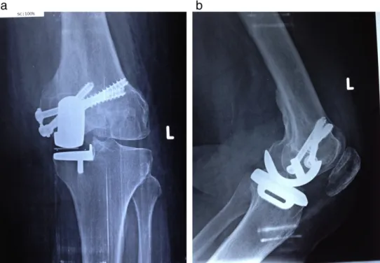
phase 3 (Biomet Orthopedics). After surgery, anterior–posterior and lateral X-rays were taken, and there was no evidence of periprosthetic fracture (Fig. 1). The patient was cleared for full-weight mobilization, and the standard rehabilitation program was initiated. One year after surgery, the patient fell while walking on the street. A minimally displaced medial femoral condyle fracture with well-fixed femoral and tibial components was observed on X-ray imaging. There was no liga-mentous instability. Prior to the fracture, the patient's Oxford knee score was 46. We successfully performed a closed reduction and percu-taneousfixation with 6.5 mm cannulated cancellous lag screws. After the second surgery, active and passive range-of-motion exercises were implemented (Fig. 2). A brace was used, and weight bearing during mo-bilization was restricted for 6 weeks. No major complications occurred. At her 3-month follow-up, union had been achieved, and the patient was pain free, with a range of motion of 130°. At her 6-month follow-up, her Oxford knee score was 42.
3. Discussion
Osteoarthritis (OA) of the knee is one of the most common causes of disability in the elderly. Currently, several treatment modalities, includ-ing surgical and non-surgical options, are available. Determininclud-ing which of these methods is appropriate for a specific patient depends on the stage and location of the osteoarthritis as well as the severity of the pa-tient's symptoms. Over the last two decades, UKR has become a popular treatment option for medial osteoarthritis of the knee[10]. Reasons for the popularity of UKR are the fact that it is a minimally invasive surgical technique with rapid recovery, preservation of bone stock, more normal knee kinematics and lower morbidity with excellent medium- and long-term results[1,3,4,6,11,12]. Complications of UKR are polyethylene
The Knee 20 (2013) 295–297
☆ Each author certifies that he or she has no commercial associations (e.g., consultancies, stock ownership, equity interest, and patent/licensing arrangements) that might pose a conflict of interest in connection with the submitted article.
⁎ Corresponding author at: Dikmen C Parkpinar Evleri 9/B No 28 Keklikpinari Cankaya, Ankara, Postcode 06420, Turkey. Tel.: +90 5055025327.
E-mail address:[email protected](B. Akan).
0968-0160/$– see front matter © 2012 Elsevier B.V. All rights reserved.
http://dx.doi.org/10.1016/j.knee.2012.10.023
Contents lists available atSciVerse ScienceDirect
wear, polyethylene insert dislocation, aseptic loosening, contralateral os-teoarthritis, limited range of motion and periprosthetic fractures[6,7]. We have treated 300 patients with UKR in our clinic between 2008 and 2012. In addition, we have treated three postoperative displaced medial tibial plateau fractures that converted to total knee arthroplasty. Periprosthetic tibial plateau fracture is a rare complication of UKR, and Pandit et al.[13]
reported the incidence as less than 1% out of 1000 cases. But only one case of medial femoral condyle fracture in UKR has been reported thus far[9]. The incidence of distal femoral fractures following TKR is higher that ranges from 0.5 to 2.2%[14,15]. Risk factors for periprosthetic fractures are rheumatoid arthritis, osteoporosis, osteomalacia, Paget's disease, osteopetrosis, osteomalacia, osteogenesis imperfecta, use of uncemented
prosthetic components and technical errors[16]. Our patient did not have any patient-related risk factors, and we did not identify any technical errors during the operation. We have two theories related to the reasons of the fracture. Firstly, the use of an uncemented prosthesis and axial loading with valgus force may be the reason for this fracture. Secondly, during the surgery of UKR, we used an intramedullary alignment rod in order to orientate the femoral component. Using intramedullary rod may create a stress riser and this stress riser may be the reason for the fracture. Minimally displaced femoral condyle fractures with good bone stock can be treated with 6.5 mm cancellous lag screws[17]. In our case, the patient's bone quality was good, components were intact and we used three 6.5 mm cannulated cancellous lag screws forfixation.
Fig. 1. a) Preoperative anterioposterior X-ray view. b) Preoperative lateral X-ray view.
Fig. 2. a) Anterioposterior X-ray view 6 weeks after surgery. b) Lateral X-ray view 6 weeks after surgery.
We required a short operation time, with minimal blood loss and risk of infection and without any damage to the components and soft tissue. Treatment of distal femoral fractures after TKR has been controversial. Complication rates of these fractures are as high as 30% with non-operative and non-operative treatments[8]. Closed reduction and immobili-zation have been recommended for nondisplaced fractures but were as-sociated with loss of motion, prolonged immobilization and malunion or nonunion. Displaced fractures that treated surgically have nonunion rates between 0 and 50%. Poor clinical results of internalfixation after distal femoral fracture following TKR have been reported in elderly patients[18].
This report shows that periprosthetic distal femoral fracture fol-lowing UKR has less morbidity and better results as compared with periprosthetic distal femoral fracture following TKR.
4. Conclusion
Medial femoral condyle fracture is a rare complication of UKR, and only one case has been reported in the literature. Management of these fractures depends on the displacement of the fracture, the bone quality and the condition of the implants. Minimally displaced fractures with intact components and good bone quality can be treated with closed re-duction and percutaneousfixation.
Conflict of interest statement
The authors have no proprietary,financial, professional or other per-sonal gains of any nature regarding any product, service and/or company that could influence the results and views presented in this article. References
[1] Deshmukh RV, Scott RD. Unicompartmental knee arthroplasty, long-term results. Clin Orthop 2001;392:272.
[2] Murray DW. Mobile bearing unicompartmental knee replacement. Orthopedics 2005;28:985.
[3] Berger RA, Nedeff DD, Barden RM, Sheinkop MM, Jacobs JJ, Rosenberg AG. Unicompartmental knee arthroplasty: clinical experience at 6-to 10-year follow-up. Clin Orthop Relat Res 1999;367:50-60.
[4] Murray DW, Goodfellow JW, O'Connor JJ. The Oxford medial unicopartmental arthroplasty: a ten year survival study. J Bone Joint Surg Br 1998;80:983-9. [5] Svard UC, Price AJ. Oxford medial unicompartmental knee arthroplasty. A survival
analysis of an independent series. J Bone Joint Surg Br 2001;83:191-4. [6] Kim KT, Lee S, Park HS, Cho KH, Kim KS. A prospective analysis of Oxford phase 3
unicompartmental knee arthroplasty. Orthopedics 2007;30:15-8.
[7] Vince KG, Cyran LT. Unicompartmental knee arthroplasty. New indications, more complications? J Arthroplasty 2004;19:9–16.
[8] Chen F, Mont MA, Bachner RS. Management of ipsilateral supracondyler femur fractures following total knee arthroplasty. J Arthroplasty 1994;9:521-6. [9] Kim KT, Lee S, Cho KH, Kim KS. Fracture of the medial femoral condyle after
unicompartmental knee arthroplasty. J Arthroplasty 2009;24:1143.e21-4. [10] Ritter MA, Faris PM, Thong AE, Davis KE, Neding JB, Berend ME. Intra-operative
findings in varus osteoarthritis of the knee. An analysis of pre-operative align-ment in potential candidates for unicompartalign-mental arthroplasty. J Bone Joint Surg Br 2004;86:43-7.
[11] Goodfellow JW, O'Connor J, Murray DW. The Oxford meniscal unicompartmental knee. J Knee Surg 2002;15:240-6.
[12] Pandit H, Jenkins C, Barker K, Dodd CA, Murray DW. The Oxford medial unicompartmental knee replacement using a minimally-invasive approach. J Bone Joint Surg Br 2006;88:54-60.
[13] Pandit H, Murray DW, Dodd CAF, Deo S, Waite J, Goodfellow J, et al. Medial tibial pla-teau fracture and the Oxford unicompartmental knee. Orthopedics 2007;30:28-31. [14] Ayers DC, Dennis DA, Johanson NA, Pellegrini JWD. Instructional course lecture
The American Academy of Orthopaedic Surgeons— common complications of total knee arthroplasty. J Bone Joint Surg Am 1997;79:278-311.
[15] Meek RM, Norwood T, Smith R, Brenkel IJ, Howie JR. The risk of peri-prosthetic fracture after primary and revision total hip and knee replacement. J Bone Joint Surg Br 2011;93:96–101.
[16] Haddad FS, Masri BA, Garbuz DS, Duncan CP. The prevention of periprosthetic frac-tures in total hip and knee arthroplasty. Orthop Clin North Am 1999;30(2):191-207. [17] Whittle AP. Fractures of the lower extremity. In: Candle ST, Beaty JH, editors.
Campbell's operative orthopaedics. Philadelphia: Mosby; 2008. p. 3085. [18] Hoffman MF, Jones CB, Sietsama DL, Koenig SJ, Tornetta P. Outcome of periprosthetic
distal femoral fractures following knee arthroplasty. Injury 2012;43:1084-9. 297 B. Akan et al. / The Knee 20 (2013) 295–297