DOI: 10.5455/annalsmedres.2019.05.240 2019;26(9):2045-52
Comparative effects of atorvastatin 80 mg versus
rosuvastatin 40 mg on the neutrophil to lymphocyte
ratio, platelet to lymphocyte ratio and monocyte to
HDL-cholesterol ratio in patients with acute myocardial
infarction
Abdullah Tuncez
Selcuk University Faculty of Medicine, Department of Cardiology, Konya, Turkey Copyright © 2019 by authors and Annals of Medical Research Publishing Inc.
Abstract
Background: Previous studies have shown the association between the elevated levels of hematological markers like Neutrophil to Lymphocyte ratio (NLR), Platelet to lymphocyte ratio (PLR) and Monocyte to High density lipoprotein cholesterol (HDL-C) ratio (MHR) and increased risk of the existence of cardiovascular disease, increased risk of acute coronary syndromes and severity of cardiovascular disease. One of the most commonly used drugs in atherosclerotic cardiovascular diseases are statins and we know that statins have beneficial effects in addition to LDL-lowering effects known as pleiotropic effects. However the effects of statins on the hematological markers are unclear. We performed this investigation to clarify and compare the effects of maximum-dose of atorvastatin and rosuvastatin on hematological biomarkers in patients with acute myocardial infarction.
Methods: Statin or other anti-lipid drugs naive patients with either ST-segment elevation myocardial infarction or Non-ST elevation myocardial infarction were enrolled to our study. Biochemistry parameters, lipid parameters, blood-count parameters and NLR, PLR and MHR levels were measured at baseline and 30 days after discharge. Baseline characteristics and results of 2 groups after one-month treatment were compared.
Results: Among the 128 statin-naive patients included, 65 patients received atorvastatin (80 mg/day) and 63 patients recieved rosuvastatin (40 mg/day). Baseline clinical characteristics of groups were similar. Atorvastatin 80 mg significantly decreased the levels of NLR (p=0.001) and MHR (p=0.024) at the end of one-month therapy. Rosuvastatin 40 mg also significantly decreased the levels of NLR (p=0.001) and MHR (p=0.006) at the end of one-month therapy. Both statins were ineffective on the levels of PLR. Percent and absolute changes of NLR, MHR and PLR were similar and there were no statistically significant differences between both groups. The percent and absolute changes of lipid parameters were also similar among both treatment arms.
Conclusion: Our results showed that atorvastatin 80 mg and rosuvastatin 40 mg decreased the NLR and MHR levels significantly at the end of one-month therapy. However, both statins have no effects on PLR levels.
Keywords: Acute myocardial infarction, atorvastatin, monocyte-to-HDL-cholesterol ratio, neutrophil-to-lymphocyte ratio, platelet-to-lymphocyte ratio, rosuvastatin.
INTRODUCTION
Coronary artery disease (CAD) and acute coronary syndrome (ACS) are the primary causes of death in worldwide. CAD killed approximately 9 million people, which account for 15.5% of all deaths worldwide in 2015 (1). A lot of research has been carried out to reduce morbidity and mortality in patients with CAD and ACS.
In recent years many researches especially focused on the association between hematologic markers and atherosclerotic cardiovascular disorders. Baseline and follow-up hematologic parameters can easily be measured routinely and can provide crucial prognostic data in patients with acute myocardial infarction (AMI) and stable CAD.
Received: 04.05.2019 Accepted: 10.07.2019 Available online: 01.10.2019
Corresponding Author: Abdullah Tuncez, Selcuk University Faculty of Medicine, Department of Cardiology, Selcuklu, Konya, Turkey, E-mail: [email protected]
The GRACE (Global Registry of Acute Coronary Events) registry, one of the most important studies on ACS, demonstrated that elevated admission leukocyte count is associated with hospital death and heart failure among patients with ACS (2). Neutrophils, play a significant and important role in the process of atherosclerosis and atherothrombosis (3) An association was found in a previous study between the low lymphocyte count (lymphocytopenia) and a significantly higher risk of major adverse cardiac events (MACE) in patients with unstable angina (UA) and ACS (4). Neutrophil to lymphocyte ratio (NLR) recently emerged as an important biomarker which has been associated with in-hospital, one-month and long-term MACE and contrast induced nephropathy in patients with stable CAD, ACS and AMI (5-9).
Elevated blood platelet count is associated with adverse cardiac outcomes (10,11). On the other hand low lymphocyte count predicts future MACE (4,12). Platelet to lymphocyte ratio (PLR) is an inflammatory biomarker and was initially used in cancer population to predict mortality (13,14) In previous studies, PLR was found to be associated with CAD severity and complexity, in-hospital and long-term mortality in patients admitted to hospital with ACS, Non-ST segment elevation myocardial infarction (NSTEMI) and ST-segment elevation myocardial infarction (STEMI) (15-17).
Monocytes are an important part of the immune system and one of the major source of pro-inflammatory and pro-oxidant cytokines at inflammatory sites. On the other hand differentiation of activated monocytes to lipid-laden macrophages is a key event for the formation of atherosclerotic lesions. Peripheral blood monocyte count was found to be associated with cardiac events (18). High density lipoprotein cholesterol (HDL-C) have various favorable functions like cholesterol efflux from tissues and macrophages, anti-inflammatory and anti-oxidant effects and inhibition of macrophage migration (19). HDL-C level is inversely correlated with cardiovascular morbidity and mortality. Monocyte to HDL-C ratio (MHR) is a novel parameter and increased levels of MHR was found to be associated with increased risk of morbidity and mortality in patients with STEMI and was found to be associated with the severity of CAD in stable CAD patients and was found to be associated with the presence of coronary artey ectasia (20-22).
Statins are associated with reduced morbidity and mortality when used after ACS and current guidelines recommends high-dose potent statin therapy for the patients with AMI (23). But there is lack of evidence about the effects of high-dose potent statins on above-mentioned biomarkers; NLR, PLR and MHR. Our aim in this study was to investigate and compare the effects of atorvastatin 80 mg and rosuvastatin 40 mg on NLR, PLR and MHR in patients with AMI.
MATERIALS and METHODS
Patient Population of StudyPatients hospitalized with STEMI and NSTEMI and eligible
for our study were enrolled. STEMI is a clinical syndrome defined by typical symptoms of myocardial ischemia lasting at least 30 minutes or more with persistent electrocardiographic ST elevation and subsequent release of biomarkers of myocardial necrosis. ST elevation was defined as a new ST elevation at the J point in at least 2 contiguous leads of ≥ 2mm (0.2mV) in men or ≥1.5mm (0.15mV) in women in leads V2–V3 and/or of ≥1mm (0.1mV) in other contiguous chest leads or the limb leads (24). The diagnosis and definiton of NSTEMI was performed according to current clinical practical guidelines (25).
Eligible patients for our study were as follows: age >18 years; fasting low density lipoprotein cholesterol (LDL-C) >100 mg/dl and AMI in last 24 h. All eligible patients were statin-naive. Patients with cardiogenic shock, serum creatinine >2,5 mg per deciliter, current statin, fibrate or other antilipid drug users, contraindication to statin therapy or an unexplained creatine kinase elevation to 2,5 fold to upper normal limits, chronic muscle disease, blood transfusion within 3 months, active infection or sepsis, presence of any active or inactive chronic inflammatory-autoimmune disease, malignancy, presence of obstructive hepatobiliary disease, presence of rheumatological disorders and cirrhosis were excluded from the study. After coronary angiography and/or percuteneous coronary intervention treatment, eligible patients were randomized to highest dose of atorvastatin (80 mg/day) or highest dose of rosuvastatin (40 mg/day) immediately. In addition to statins, aspirin (300 mg/oral loading dose and 100 mg/oral maintenance dose), clopidogrel (or ticagrelor/prasugrel), ACE inhibitors or ARB’s and ß-blockers were prescribed according to current guideline recommendations. Use of GpIIb/IIIa antogonists left the operators discretion.
Venous blood samples were drawn when the patient initially presented to the emergency department or the coronary care unit before randomization and at the end of four-week period of the therapy by a cubital venipuncture avoiding venous stasis to an evacuated serum separator tube. Dry tubes were used for biochemical analysis, and EDTA tubes were used for the hematological tests. Whole blood counting parameters were analyzed by a autoanalyzer (Coulter Gen-S Hematology Analyzer, Beckman Coulter Corp, Hialeah, Florida) within 5 min of blood sampling. The levels of total cholesterol (TC), triglyceride (TG), HDL-C, were measured by chemistry autoanalyzer (ARCHITECT c16000, Abbott Diagnostics, USA) via enzymatic colorimetric methods. Friedewald Formula was used to calculate the levels of LDL-C. The results of laboratory parameters were collected by using electronic database of the hospital. NHR was calculated by dividing neutrophil count to lymphocyte count, PLR was calculated by dividing platelet count to lymphocyte count and MHR was calculated by dividing monocyte count to HDL level from the same blood sample obtained before randomization. All patients provided written informed
consent. Local ethics committee of Selçuk University has approved the protocol of this investigation.
Statistical Analysis
SPSS for Mac version 20.0 (SPSS Inc., Chicago, Illinois) was used to perform the statistical analyses. Normal distribution of continuous variables was tested using Kolmogorov-Smirnov test. Student’s t test was used to compare the continuous variables with normal distribution and those without normal distribution were compared using Mann-Whitney’s U test. The chi-square test was used for comparing categorical variables. Continuous variables were defined as means ± standard deviation (SD) or median (interquartile range, IQR) and categorical variables were given as percentages. Paired sample t test or Wilcoxon Signed Ranks test were used to compare the baseline clinical characteristics of the patients and post-treatment changes between groups and within groups. We substracted the baseline values from after-treatment values to calculate the absolute change and we divided the absolute difference after statin treatment by pre-treatment value to calculate the percent change. P < 0.05
was considered as statistically significant for all tests.
RESULTS
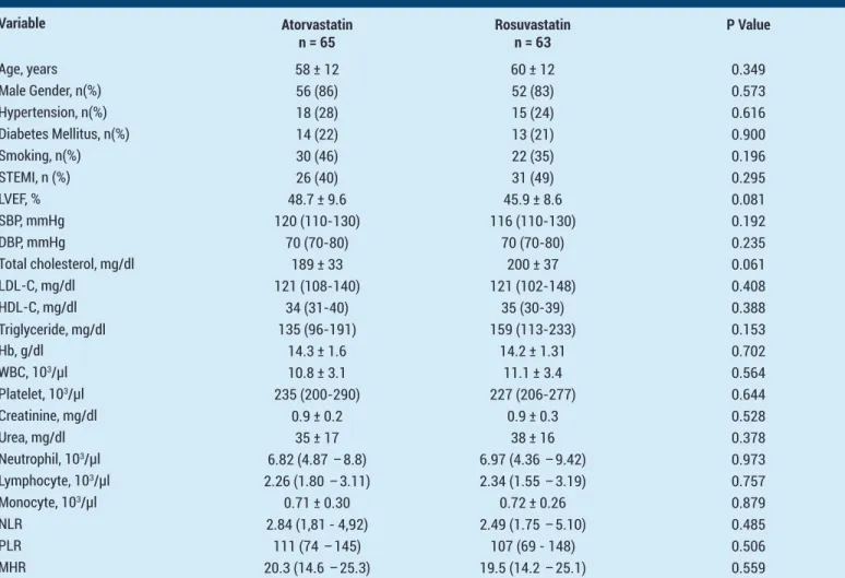
A total of 131 eligible patients were enrolled to our study. At the end of one-month treatment, clinical follow-up had been completed in 63 patients in the rosuvastatin 40 mg group (96,92%) and 65 patients in the atorvastatin 80 mg group (98.48%). Two patients in rosuvastatin group and one patient in the atorvastatin group had lost the follow-up. The final analysis included a total of 128 patients, of which 65 were from the atorvastatin group and 63 were from the rosuvastatin group. Table 1 shows the baseline demographic characteristics of the 128 patients and baseline clinical characteristics of two treatment groups were similar. Baseline hematological parameters (NLR, PLR and MHR) were similar between groups and there was no statistically significant difference between the groups. Baseline lipid profiles were also comparable between groups (Table 1).
Lipid Parameter Outcomes
Table 2 shows laboratory values obtained during the
Table 1. Comparison of the Baseline Clinical Characteristics and Laboratory Parameters of the Patients
Variable Atorvastatin n = 65 Rosuvastatinn = 63 P Value Age, years 58 ± 12 60 ± 12 0.349 Male Gender, n(%) 56 (86) 52 (83) 0.573 Hypertension, n(%) 18 (28) 15 (24) 0.616 Diabetes Mellitus, n(%) 14 (22) 13 (21) 0.900 Smoking, n(%) 30 (46) 22 (35) 0.196 STEMI, n (%) 26 (40) 31 (49) 0.295 LVEF, % 48.7 ± 9.6 45.9 ± 8.6 0.081 SBP, mmHg 120 (110-130) 116 (110-130) 0.192 DBP, mmHg 70 (70-80) 70 (70-80) 0.235 Total cholesterol, mg/dl 189 ± 33 200 ± 37 0.061 LDL-C, mg/dl 121 (108-140) 121 (102-148) 0.408 HDL-C, mg/dl 34 (31-40) 35 (30-39) 0.388 Triglyceride, mg/dl 135 (96-191) 159 (113-233) 0.153 Hb, g/dl 14.3 ± 1.6 14.2 ± 1.31 0.702 WBC, 103/µl 10.8 ± 3.1 11.1 ± 3.4 0.564 Platelet, 103/µl 235 (200-290) 227 (206-277) 0.644 Creatinine, mg/dl 0.9 ± 0.2 0.9 ± 0.3 0.528 Urea, mg/dl 35 ± 17 38 ± 16 0.378 Neutrophil, 103/µl 6.82 (4.87 – 8.8) 6.97 (4.36 – 9.42) 0.973 Lymphocyte, 103/µl 2.26 (1.80 – 3.11) 2.34 (1.55 – 3.19) 0.757 Monocyte, 103/µl 0.71 ± 0.30 0.72 ± 0.26 0.879 NLR 2.84 (1,81 - 4,92) 2.49 (1.75 – 5.10) 0.485 PLR 111 (74 – 145) 107 (69 - 148) 0.506 MHR 20.3 (14.6 – 25.3) 19.5 (14.2 – 25.1) 0.559
Abbreviations: BUN: Blood Urea Nitrogen, DBP: Diastolic blood pressure, HDL-C: High density lipoprotein cholesterol, Hb: Hemoglobine, LDL-C: Low density lipoprotein cholesterol, LVEF: Left ventricular ejection fraction, NLR: Neutrophil to lymphocyte ratio, MHR: Monocyte to HDL ratio, TC: Total cholesterol, PLR: Platelet to lymphocyte ratio, SBP: Systolic blood pressure, STEMI: ST segment elevation myocardial infarction, WBC: White blood cell. Data given as mean±SD or number (%)
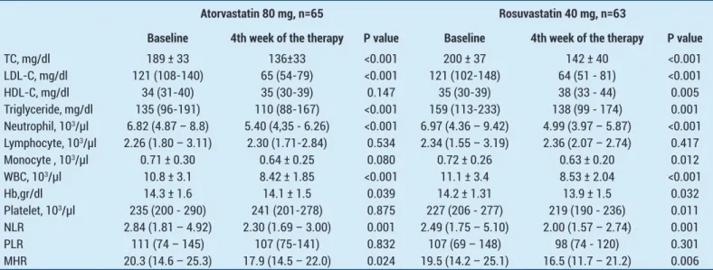
study. The mean (SD) baseline TC level of 189 (33) mg/ dL declined to 136 (33) mg/dL, representing a mean reduction of 26.4% (p < 0.001) with atorvastatin and baseline TC level of 200 (37) mg/dl declined to 142 (40) mg/dl, representing a mean reduction of 28.3% (p < 0.001) with rosuvastatin respectively. The mean (IQR) baseline LDL-C level of 121 (108-140) mg/dL declined to 65 (54-79) mg/dL, representing a mean reduction of 42.1% (p < 0.001) with atorvastatin and baseline LDL-C level of 200 (37) mg/dl declined to 142 (40) mg/dl, representing a mean reduction of 47.7% (p < 0.001) with rosuvastatin. Table 3 summarizes the absolute and percent changes of lipid parameters after high dose atorvastatin and rosuvastatin. Atorvastatin 80 mg and rosuvastatin 40 mg resulted in similar reductions in TC, LDL-C and TG levels and there were no statistically significant differences between the two groups in terms of absolute and percentage changes of TC, LDL-C and TG (Table 3).
HDL-C levels did not changed with atorvastatin, HDL-C level of 34 (31–40) mg/dl increased to 35 (30–39) mg/ dl representing a mean increasing of 2,6%, p=0.147. But
baseline HDL-C levels of 35 (30 – 39) mg/dl increased to 38 (33 – 44) mg/dl with rosuvastatin 40 mg representing a mean increase of 8.7%, p=0.005, Table 2 and Table 3). On the other hand, when the increase in HDL-C levels was examined in terms of absolute and percent change, there was no statistically significant difference between two groups (Table 3).
Hematologic Parameter Outcomes
Baseline hematologic parameters were similar between 2 groups (Table 1). At the end of 4-week therapy, there was a significant decrease in NLR [from 2.84 (1.81-4.92) to 2.30 (1.69–3.00) in atorvastatin group, p=0.001 and from 2,49 (1.75–5.10) to 2,00 (1.57- 2.74) in rosuvastatin group, p=0.001] and MHR [from 20.3 (14.6 – 25.3) to 17,9 (14.5-22.0)in atorvastatin group, p=0.024 and from 19,5 (14.2 – 25.1) to 16.5 (11.7 – 21.2) in rosuvastatin group, p=0.006] levels in both groups respectively (Table 2). Rosuvastatin 40mg provided significant decrease in platelet count (from 227 (206 - 277) to 219 (190 – 236) *103, p=0.011) and monocyte count (from 0,72 (0.26) to 0,63 (0.20) *103, p=0.012), whereas the effect of atorvastatin 80 mg on
Table 2. Effects of Atorvastatin 80 mg and Rosuvastatin 40 mg on laboratory parameters after 4-week treatment.
Atorvastatin 80 mg, n=65 Rosuvastatin 40 mg, n=63
Baseline 4th week of the therapy P value Baseline 4th week of the therapy P value
TC, mg/dl 189 ± 33 136±33 <0.001 200 ± 37 142 ± 40 <0.001 LDL-C, mg/dl 121 (108-140) 65 (54-79) <0.001 121 (102-148) 64 (51 - 81) <0.001 HDL-C, mg/dl 34 (31-40) 35 (30-39) 0.147 35 (30-39) 38 (33 - 44) 0.005 Triglyceride, mg/dl 135 (96-191) 110 (88-167) <0.001 159 (113-233) 138 (99 - 174) 0.001 Neutrophil, 103/µl 6.82 (4.87 – 8.8) 5.40 (4,35 - 6.26) <0.001 6.97 (4.36 – 9.42) 4.99 (3.97 – 5.87) <0.001 Lymphocyte, 103/µl 2.26 (1.80 – 3.11) 2.30 (1.71-2.84) 0.534 2.34 (1.55 – 3.19) 2.36 (2.07 – 2.74) 0.417 Monocyte , 103/µl 0.71 ± 0.30 0.64 ± 0.25 0.080 0.72 ± 0.26 0.63 ± 0.20 0.012 WBC, 103/µl 10.8 ± 3.1 8.42 ± 1.85 <0.001 11.1 ± 3.4 8.53 ± 2.04 <0.001 Hb,gr/dl 14.3 ± 1.6 14.1 ± 1.5 0.039 14.2 ± 1.31 13.9 ± 1.5 0.032 Platelet, 103/µl 235 (200 - 290) 241 (201-278) 0.875 227 (206 - 277) 219 (190 - 236) 0.011 NLR 2.84 (1.81 – 4.92) 2.30 (1.69 – 3.00) 0.001 2.49 (1.75 – 5.10) 2.00 (1.57 – 2.74) 0.001 PLR 111 (74 – 145) 107 (75-141) 0.832 107 (69 – 148) 98 (74 - 120) 0.301 MHR 20.3 (14.6 – 25.3) 17.9 (14.5 – 22.0) 0.024 19.5 (14.2 – 25.1) 16.5 (11.7 – 21.2) 0.006
Abbreviations: HDL-C: High density lipoprotein cholesterol, Hb: Hemoglobine, LDL-C: Low density lipoprotein cholesterol, NLR: Neutrophil to lymphocyte ratio, MHR: Monocyte to HDL ratio, TC: Total cholesterol, PLR: Platelet to lymphocyte ratio, WBC: White blood cell.
Table 3. Comparison of Atorvastatin 80 mg and Rosuvastatin 40 mg by means of absolute and percent change on laboratory parameters
Absolute change Percent change, %
Atorvastatin Rosuvastatin P value Atorvastatin Rosuvastatin P value TC, mg/dl -52.4 ± 37.1 -58.3 ± 40.2 0.391 -26.4 ± 18.7 -28.3 ± 18.3 0.554 LDL-C, mg/dl -52 (-71 / -35) -55 (-79 / -34) 0.225 -42.1 (-58.4 / -26.7) -47.7 (-58.5 / -32.4) 0.368 HDL-C, mg/dl 1.0 (-2.0 / 4.0) 3.0 (-3.0 / 7.0) 0.117 2.6 (-5.7 / 14.3) 8.7 (-7.7 / 18.2) 0.168 TG, mg/dl -29 (-72 / 13) -28 (-72 / -2) 0.903 -22.6 (-41 / 10.6) -15.6 (-34.3 / -2.4) 0.808 NLR -0.40 (-2.68 / 0.45) -0.47 (-2.53 / 0.22) 0.687 -15.2 (-56.6 / 36.2) -24.8 (-55.8 / 15.1) 0.748 PLR 0.92 (-30.8 / 33.9) -2.6 (-37.8 / 21.2) 0.347 1.2 (-26.7 / 33.3) -4.9 (-29.1 / 25.8) 0.459 MHR -2.67 (-6.78 / 2.84) -3.58 (-9.23 / 1.98) 0.592 -13.36 (-37.21 / 16.9) -21.35 (-38.72 / 23.79) 0.553
Abbreviations: HDL-C: High density lipoprotein cholesterol, Hb: Hemoglobine, LDL-C: Low density lipoprotein cholesterol, NLR: Neutrophil to lymphocyte ratio, MHR: Monocyte to HDL ratio, TC: Total cholesterol, PLR: Platelet to lymphocyte ratio, TG: Triglyceride, WBC: White blood cell.
platelet (p=0.875) and monocyte count (p=0.080) couldn’t reached to the statistically significant level (Table 2). Our results showed that both statins have limited effects on the PLR levels (from 111 (74–145) to 107 (75-141)in atorvastatin group, p=0.832 and from 107 (69 -148) to 98 (74–120) in rosuvastatin group, p=0.301, Table 2). The absolute and percentage changes of NLR, PLR and MHR at the end of four-weeks in two groups are listed in Table 3 and no statistically significant difference were detected between the atorvastatin and rosuvastatin groups.
DISCUSSION
Atherosclerosis is a progressive disease and main feature of atherosclerosis is the accumulation of lipids and fibrous elements in the large arteries. Atherosclerosis constitutes the most important and integral contributor to global burden of cardivascular disease (26). Until the 1970’s, the relationship betweeen lipids and atherosclerosis was the most popular topic for researchers. In 1970’s and 1980’s researchers focused on growth factors and vascular smooth muscle cell proliferation. The key role of inflammation on atherosclerosis was demonstrated in 1990’s (26). Inflammation plays an important role in the development, progression, and devastating results of atherosclerosis. Previos studies comfirmed that most foam cells arise from mononuclear phagocytes (27). Statins are indispensable for the patients with CAD and AMI (28). Main function of statins is lowering the LDL-C. On the other hand statins have favorable effects beyond LDL-C reduction which called as pleiotropic effects (29). However the exact mechanism of the pleiotropic effects of statins is not clear. Various theroies have been proposed to explain the pleiotropic effects of statins; antiinflammatory effects, anti-oxidative effects and endothelial-function healing properties. NLR, PLR and MHR emerged as novel biomarkers of inflammatory status and increased levels of these biomarkers found to be associated with various cardiovascular diseases (CVD), AMI, heart failure and cardiovascular death. Present study shows and compares the effects of high-dose potent statins on these biomarkers in patients with AMI for the first time and our findings may provide evidence for the mechanisms of pleiotropic effects of statins. Our findings showed that both of these high-dose potent statins significantly reduced the levels of NLR and MHR which are indirect markers of inflammatory status. In real-life clinical setting the decreases in NLR and MHR values may be an indirect indicator of the pleiotropic effects of statins. On the other hand, the ineffectiveness of these potent statins on the other important biomarker the PLR, must be evaluated in large-scale future studies. The value of our study is that it is the first study in the current literature to reveal data on the effects of most-used and most-potent statins on the hematological biomarkers NLR, PLR and MHR.
a. Evaluating and comparing the effects of atorvastatin (80mg/day) and rosuvastatin (40mg/day) therapy on Lipid Parameter Outcomes
Current guidelines recommends high-dose statins early
after admission in all ACS patients without contra-indication regardless of initial LDL values (23). In our study, we selected the most potent statins with the highest doses, atorvastatin 80 mg and rosuvastatin 40mg, for the patients with AMI included in this study.
In our study, rosuvastatin 40 mg resulted in further reductions in LDL-C levels when compared with atorvastatin 80 mg. Atorvastatin 80 mg led to %42.1 and rosuvastatin 40 mg led to %47,7 reductions of LDL-C levels from baseline respectively at the end of 4 weeks. This finding is consistent with previous studies. In the LUNAR study, while the atorvastatin 80 mg provided %42 reduction in LDL-C levels, rosuvastatin 40 mg provided %46.8 reduction in LDL-C levels at the end of 6 weeks (30). In the LUNAR study atorvastatin 80 mg provided %18 reduction in TG levels and rosuvastatin 40mg provided %15 reduction in TG levels. Similarly, atorvastatin 80 mg provided %22.6 reduction in TG levels, while rosuvastatin 40mg decreased the serum TG levels by %15.6 in our study (p=0.808). Although both statins have comparable effects on the levels of HDL-C in terms of absolute and percentage changes, our study showed that rosuvastatin 40mg increased HDL-C levels, whereas atorvastatin 80 mg had no effect on HDL-C levels according to baseline. Our findings are consistent with previous studies. LUNAR study showed that rosuvastatin 40mg increased HDL-C levels more effectively than atorvastatin 80mg (30). In another study, which conducted by Aydın et al. showed that atorvastatin 80 mg decreased the HDL-C while rosuvastatin 20 mg increased the HDL-C according to baseline (31).
b. The effects of atorvastatin (80mg/day) and rosuvastatin (40mg/day) therapy on NLR and the comparison of these effects
Inflammation plays a pivotal role in the pathogenesis of the CVD and previous studies showed that statins have anti-inflammatory effects. In the JUPITER trial, apperently healthy subjects without hyperlipidemia but with elevated high-sensitivity C-reaktive protein levels randomized to rosuvastatin 20 mg and placebo. The trial was stopped early, after a median follow-up of 1,9 years and rosuvastatin significantly reduced cardiovascular events (32). In a substudy of MIRACL, atorvastatin 80 mg resulted in a significant decline in inflammation (33). There are very limited studies investigating the effects of atorvastatin and/or rosuvastatin on hematological markers and there are no studies involving patients with myocardial infarction. A study conducted by Gungoren et al. investigated the effects of statins on NLR and mean platelet volume (MPV) in patients with hypercholesterolemia and they showed that statins were ineffective on NLR and MPV (34). However dose of statins were not fixed in this study and only %4 of patients were on atorvastatin 80 mg and %6 of patients were on rosuvastatin 40 mg at the end of study (34). On the other hand, Akın et al. demonstrated a reduction in NLR and MPV levels with atorvastatin treatment in patients with hypercholesterolemia (35). Our findings are consistent
with the results obtained in this study. In our trial high dose atorvastatin (80 mg) and high dose rosuvastatin (40 mg) provided significant reductions in NLR levels and this reduction were similar between the two groups in terms of absolute and percent changes. To the best of our knowledge, our study is the first trial showing the positive effects of high-dose potent statins on NLR and comparing these positive effects in patients with AMI.
c. The effects of atorvastatin (80mg/day) and rosuvastatin (40mg/day) therapy on PLR and the comparison of these effects
Increased platelet count and low lymphocyte count were found to be associated with adverse cardiac outcomes (4,10-12). Platelets are highly active cells and secrete various cytokines and chemokines. Megakaryocytic proliferation and relative thrombocytosis are consequences of an ongoing inflammatory response and increased platelet count closely correlated with acute phase reactants and proinflammatory cytokines like high-sensitivity C-reactive protein, IL-1, IL-6 and tumor necrosis factor alpha that results in a prothrombotic state (36). Contrarily lymphocytes represent a more appropriate immune response and suppress the aggravated inflammatory state. PLR is a novel biomarker, representing both inflammatory and prothrombotic status and calculated as the ratio of the platelet count to lymphocyte count. PLR is associated with various CVD and predicting worse cardiovascular outcome in patients with ACS, NSTEMI and STEMI (15-17).Ozcan et al. showed an association between PLR and increased risk of in-hospital and long term MACE in patients with STEMI (15). Yıldız et al. demonstrated that high preprocedural PLR and NLR are predicting the no-reflow phenomenon significantly and independently in patients with STEMI (37). Metabolic syndrome includes several cardiovasular risk factors like hypertension, dyslipidemia, central obesity and impaired glucose metabolism (38). Akboğa et al. showed that PLR is correlated with the presence and severity of metobolic sydrome (39). Statins have been shown to be associated with reduced morbidity and mortality in patients with CVD, diabetes mellitus or dyslipidemia. Our study has shown that, high-dose potent statins have no effect on PLR. Although platelet counts decreased with rosuvastatin 40 mg significantly and rosuvastatin seems to be more effective than atorvastatin on PLR levels, effects of rosuvastatin couldn’t reached to statistically significant level. Although, in a previous study nicotinic acid provided a statistically significant decrease in platelet count in patients with primary hyperlipidemia there is limited data about effects of statins and other lipid-lowering agents on platelets, platelet count and PLR and further large scale randomized studies are needed (40).
d. The effects of atorvastatin (80mg/day) and rosuvastatin (40mg/day) therapy on MHR and the comparison of these effects
Monocytes play an important role in the pathogenesis of atherosclerosis and atherom plaque formation. Circulating monocytes becomes an intimal macrophage
after migrating into intima. Intimal macrophages express scavenger receptors that bind to internalized lipoproteins and transformed into foam cells which was a hallmark of the arterial lesion (26). Foam cells secretes cytokines and pro-inflammatory mediators and accelerating the atherosclerosis (26). Contrarily HDL-C collects cholesterol from tissues and macrophages, inhibits macrophage migration and exhibits anti-inflammatory and anti-oxidant effects (19). MHR is a novel biomarker and increased levels of MHR found to be associated with several cardiovascular disease (20,21). Karataş et al. demonstrated an independent correllation between admission MHR levels and in-hospital MACE in patients with STEMI (41) A study by Canpolat et al. showed that elevated levels of preablation MHR is a strong predictor of the atrial fibrillation recurrence after cryoballoon-based catheter ablation (42). Previous studies have investigated the effects of statins on monocytes. Tani et al. investigated the association of leukocyte subtypes counts with the coronary plaque regression following pravastatin treatment and they found a decrease in monocyte count with pravastatin treatment and this decrease was an independent predictor of coronary plaque regression (43). Fildes et al. demonstrated depletion in classical and non-classical monocyte subtypes with statin treatment in patients with heart transplantation (44). On the other hand, previous studies have shown that statins have incremental effects on HDL-C. A large randomized controlled study (The ASTEROID trial) showed that intensive statin therapy increased the HDL-C levels by about %15 and the authors suggested that increase in HDL-C may also have an important impact on coronary plaque regression (45). Our investigation is the first research evaluating and comparing the possible effects of maximum approved doses of atorvastatin and rosuvastatin on MHR in patients with AMI and our results showed that atorvasatin and rosuvastatin similarly and significantly decreased the MHR levels.
Limitations
Our study has some limitations. First, our study population is relatively small. Second, we couldn’t show and compare the effects of these potent statins on other inflammatory markers like hs-CRP, TNF-α, IL-1 and IL-6. Third, our study was designed to investigate and compare the effects of potent statins on hematological parameters at the end of the first month. For this reason, we do not have any information whether there are any long-term effects of statins on hematological parameters, and whether these effects are associated with hard endpoints such as death and myocardial infarction. On the other hand, because of our study population consists patients with AMI, our results cannot be extrapolated for patients with stable CAD.
CONCLUSION
Our study showed that atorvastatin 80 mg and rosuvastatin 40 mg have similar effects on LDL-C and the other lipid parameters. Moreover, while both statins
reduced NLR and MHR levels in a similar manner, our results indicate that they were ineffective on PLR level. At the same time, results of our study may provide important evidence about the mechanisms of the pleiotropic effects of statins. Further large scale studies are needed to clarify the clinical significance of high dose intensive statin therapy on hematological parameters and its association with clinical outcomes.
Competing interests: The authors declare that they have no competing interest.
Financial Disclosure: There are no financial supports
Ethical approval: Local ethics committee has approved the protocol of our investigation.
Abdullah Tuncez: ORCID:0000-0002-6512-1327
REFERENCES
1. The top 10 causes of death. http://www.who.int/ mediacentre/factsheets/fs310/en/. accessed date 07.03.2017
2. Furman MI, Gore JM, Anderson FA, et al. Elevated leukocyte count and adverse hospital events in patients with acute coronary syndromes: findings from the Global Registry of Acute Coronary Events (GRACE). Am Heart J 2004;147:42-8.
3. Horne BD, Anderson JL, John JM, et al. Which white blood cell subtypes predict increased cardiovascular risk? J Am Coll Cardiol 2005;45:1638-43.
4. Zouridakis EG, Garcia-Moll X, Kaski JC. Usefulness of the blood lymphocyte count in predicting recurrent instability and death in patients with unstable angina pectoris. Am J Cardiol 2000;86:449-51.
5. Cho KH, Jeong MH, Ahmed K, et al. Value of Early Risk Stratification Using Hemoglobin Level and Neutrophil-to-Lymphocyte Ratio in Patients With ST-Elevation Myocardial Infarction Undergoing Primary Percutaneous Coronary Intervention. American Journal of Cardiology 2011;107:849-56.
6. Azab B, Zaher M, Weiserbs KF, et al. Usefulness of Neutrophil to Lymphocyte Ratio in Predicting Short- and Long-Term Mortality After Non-ST-Elevation Myocardial Infarction. Am J Cardiol. 2010;106:470-6.
7. Nunez J, Nunez E, Bodi V, et al. Usefulness of the neutrophil to lymphocyte ratio in predicting long-term mortality in ST segment elevation myocardial infarction. Am J Cardiol 2008;101:747-52.
8. Gul M, Uyarel H, Ergelen M, et al. Predictive Value of Neutrophil to Lymphocyte Ratio in Clinical Outcomes of Non-ST Elevation Myocardial Infarction and Unstable Angina Pectoris A 3-Year Follow-Up. Clin Appl Thromb-Hem 2014;20:378-84.
9. Tanık VO, Çınar T, Velibey Y, et al. Neutrophil-to-lymphocyte ratio predicts contrast-induced acute kidney injury in patients with ST-elevation myocardial infarction treated with primary percutaneous coronary intervention. J Tehran Univ Heart Cent 2019;14:59-66.
10. Thaulow E, Erikssen J, Sandvik L, et al. Blood-platelet count and function are related to total and cardiovascular death in apparently healthy-men. Circulation 1991;84:613-7. 11. Iijima R, Ndrepepa G, Mehilli J, et al. Relationship between
platelet count and 30-day clinical outcomes after percutaneous coronary interventions - Pooled analysis of
four ISAR trials. Thromb Haemostasis 2007;98:852-7. 12. Bian C, Wu Y, Shi Y, et al. Predictive value of the relative
lymphocyte count in coronary heart disease. Heart Vessels 2010;25:469-73.
13. Smith RA, Bosonnet L, Raraty M, et al. Preoperative platelet-lymphocyte ratio is an independent significant prognostic marker in resected pancreatic ductal adenocarcinoma. Am J Surg 2009;197:466-72.
14. Proctor MJ, Morrison DS, Talwar D, et al. A comparison of inflammation-based prognostic scores in patients with cancer. A Glasgow Inflammation Outcome Study. Eur J Cancer 2011;47:2633-641.
15. Cetin EHO, Cetin MS, Aras D, et al. Platelet to lymphocyte ratio as a prognostic marker of in-hospital and long-term major adverse cardiovascular events in ST-segment elevation myocardial infarction. Angiology 2016;67:336-45. 16. Azab B, Shah N, Akerman M, et al. Value of platelet/ lymphocyte ratio as a predictor of all-cause mortality after non-ST-elevation myocardial infarction. J Thromb Thrombolys 2012;34:326-34.
17. Kurtul A, Murat SN, Yarlioglues M, et al. Association of platelet-to-lymphocyte ratio with severity and complexity of coronary artery disease in patients with acute coronary syndromes. Am J Cardiol 2014;114:972-8.
18. Olivares R, Ducimetiere P, Claude JR. Monocyte count: a risk factor for coronary heart disease? Am J Epidemiol 1993;137:49-53.
19. Ansell BJ, Navab M, Hama S, et al. Inflammatory/ antiinflammatory properties of high-density lipoprotein distinguish patients from control subjects better than high-density lipoprotein cholesterol levels and are favorably affected by simvastatin treatment. Circulation 2003;108:2751-6.
20. Cicek G, Kundi H, Bozbay M, et al. The relationship between admission monocyte HDL-C ratio with short-term and long-term mortality among STEMI patients treated with successful primary PCI. Coronary Artery Dis 2016;27:176-84.
21. Kundi H, Kiziltunc E, Cetin M, et al. Association of monocyte/ HDL-C ratio with SYNTAX scores in patients with stable coronary artery disease. Herz 2016;41:523-9.
22. Tosu A, Çinar, T , Güler, A , et al. The usefulness of monocyte to high density lipoprotein cholesterol ratio in prediction for coronary artery ectasia. Turk J Clin Laborat 2019;10:68-73. 23. Catapano AL, Graham I, De Backer G, et al. 2016 ESC/EAS
Guidelines for the Management of Dyslipidaemias The Task Force for the Management of Dyslipidaemias of the European Society of Cardiology (ESC) and European Atherosclerosis Society (EAS) Developed with the special contribution of the European Assocciation for Cardiovascular Prevention & Rehabilitation (EACPR). Atherosclerosis 2016;253:281-344. 24. Thygesen K, Alpert JS, Jaffe AS, et al. Third Universal
Definition of Myocardial Infarction. Circulation.
2012;126:2020-25. Amsterdam. 2014 ACC/AHA Guideline for the management of patients with non-st-elevation acute coronary syndromes: executive summary: a report of the american college of cardiology/american heart association task force on practice guidelines. Circulation. 2014;130:431-2.
26. Libby P. Inflammation in atherosclerosis. Nature 2002;420:868-74.
27. Aqel NM, Ball RY, Waldmann H, Mitchinson MJ. Identification of Macrophages and smooth-muscle cells in human atherosclerosis using monoclonal-antibodies. J Pathol 1985;146:197-204.
28. Baigent C, Keech A, Kearney PM, et al. Efficacy and safety of cholesterol-lowering treatment: prospective meta-analysis of data from 90,056 participants in 14 randomised trials of statins. Lancet 2005;366:1267-78.
29. Almuti K, Rimawi R, Spevack D, Ostfeld RJ. Effects of statins beyond lipid lowering: potential for clinical benefits. Int J Cardiol 2006;109:7-15.
30. Pitt B, Loscalzo J, Monyak J, et al. Comparison of Lipid-Modifying Efficacy of Rosuvastatin Versus Atorvastatin in Patients With Acute Coronary Syndrome (from the LUNAR Study). Am J Cardiol 2012;109:1239-46.
31. Aydin MU, Aygul N, Altunkeser BB, et al. Comparative effects of high-dose atorvastatin versus moderate-dose rosuvastatin on lipid parameters, oxidized-LDL and inflammatory markers in ST elevation myocardial infarction. Atherosclerosis 2015;239:439-43.
32. Ridker PM, Danielson E, Fonseca FAH, et al. Rosuvastatin to Prevent Vascular Events in Men and Women with Elevated C-Reactive Protein. New Engl J Med 2008;359:2195-207. 33. Kinlay S, Schwartz GG, Olsson AG, et al. High-dose
atorvastatin enhances the decline in inflammatory markers in patients with acute coronary syndromes in the MIRACL study. Circulation 2003;108:1560-6.
34. Gungoren F, Besli F, Caliskan S, et al. Statin Therapy May not Effect NLR and MPV Levels in Patients With Hypercholesterolemia: A Retrospective Study. Angiology 2016;67:536-40.
35. Akin F, Ayca B, Kose N, et al. Effect of atorvastatin on hematologic parameters in patients with hypercholesterolemia. Angiology 2013;64:621-625
36. Alexandrakis MG, Passam FH, Moschandrea IA, et al. Levels of serum cytokines and acute phase proteins in patients with essential and cancer-related thrombocytosis. Am J Clin Oncol-Canc 2003;26:135-40.
37. Yildiz A, Yuksel M, Oylumlu M, et al. The Utility of the
Platelet-Lymphocyte Ratio for Predicting No Reflow in Patients With ST-Segment Elevation Myocardial Infarction. Clin Appl Thromb Hemost 2015;21:223-8.
38. Expert Panel on Detection E, Treatment of High Blood Cholesterol in A. Executive Summary of The Third Report of The National Cholesterol Education Program (NCEP) Expert Panel on Detection, Evaluation, And Treatment of High Blood Cholesterol In Adults (Adult Treatment Panel III). JAMA 2001;285:2486-97.
39. Akboga MK, Canpolat U, Yuksel M, et al. Platelet to lymphocyte ratio as a novel indicator of inflammation is correlated with the severity of metabolic syndrome: A single center large-scale study. Platelets 2016;27:178-83.
40. Kei A, Elisaf M. Nicotinic acid/laropiprant reduces platelet count but increases mean platelet volume in patients with primary dyslipidemia. Arch Med Sci 2014;10:439-44. 41. Karatas MB, Canga Y, Ozcan KS, et al. Monocyte to
high-density lipoprotein ratio as a new prognostic marker in patients with STEMI undergoing primary percutaneous coronary intervention. Am J Emerg Med 2016;34:240-4. 42. Canpolat U, Aytemir K, Yorgun H, et al. The role of
preprocedural monocyte-to-high-density lipoprotein ratio in prediction of atrial fibrillation recurrence after cryoballoon-based catheter ablation. Europace 2015;17:1807-15. 43. 43. Tani S, Nagao K, Anazawa T, et al. Association of
leukocyte subtype counts with coronary atherosclerotic regression following pravastatin treatment. Am J Cardiol 2009;104:464-9.
44. Fildes JE, Shaw SM, Mitsidou A, et al. HMG-CoA reductase inhibitors deplete circulating classical and non-classical monocytes following human heart transplantation. Transpl Immunol 2008;19:152-7.
45. Nissen SE, Nicholls SJ, Sipahi I, et al. Effect of very high-intensity statin therapy on regression of coronary atherosclerosis: the ASTEROID trial. JAMA 2006;295:1556-65.