ARAŞTIRMA
MRI IN GERİATRİC PATIENTS WITH VERTIGO
VERTİGOSU OLAN GERİATRİK HASTALARDA
TEMPORAL MRG’DE VESTİBULOKOKLEAR
SİNİRİN NÖROVASKÜLER KOMPRESYONU
Introduction: This study aimed to assess the neurovascular compression of the vestibulocochlear nerve using temporal magnetic resonance imaging in geriatric patients with vertigo.
Materials and Method: This study included 92 patients with vertigo [50 (54.3%) women; mean age, 53.6±14.2 years (min–max, 20–84 years)] and 85 healthy volunteers [46 (54.1%) women; mean age, 52.2±15 years (min–max, 23–79 years)]. Individuals aged ≥65 years included 52 of 92 patients and 45 of 85 healthy volunteers. All participants underwent temporal magnetic resonance imaging to detect neurovascular compression of vestibulocochlear nerve. Results: The presence of neurovascular compression was more prominent in geriatric patients with vertigo than in patients aged <65 years with vertigo (p=0.001). Compression was detected on one side of vestibulocochlear nerve in patients with vertigo (p=0.007). Vestibulocochlear nerve compression was more common ingeriatric patients than in non-geriatric patients.
Conclusion: The study results describe the role of neurovascular compression of vestibulocochlear nerve in elderly patients with episodic peripheral vertigoin whom other causes were excluded.
Key Words: Vertigo; Vestibulocochlear Nerve; Geriatrics; Magnetic Resonance Imaging
Giriş: Bu çalışmada vertigo şikayeti olan geriatrik hastalarda, temporal manyetik rezonans görüntüleme kullanarak vestibülokohlear sinirin nörovasküler kompresyonunun değerlendirilmesi amaçlanmıştır.
Gereç ve Yöntem: Çalışmaya, vertigosu olan 92 hasta [50 (% 54.3) kadın; yaş ortalaması: 53.6±14.2 yaş (min-max, 20-84 yaş)] ve 85 sağlıklı gönüllü [46 (% 54.1) kadının yaş ortalaması 52.2±15 yaş (min-max, 23-79 yaş)] dahil edildi. Doksaniki hastadan 52’si ve 85 sağlıklı gönüllüden 45’i, ≥65 yaş bireylerden oluştu. Tüm katılımcılar vestibülokohlear sinir nörovasküler kompresyonunun saptanması amacıyla temporal manyetik rezonans görüntüleme ile değerlendirildi.
Bulgular: Nörovasküler kompresyon varlığı, vertigosu olan geriatrik hastalarda vertigosu olan <65 yaş hastalardan daha belirgindi (p=0.001). Vertigo hastalarında vestibülokohlear sinirin bir tarafında kompresyon tespit edildi (p=0.007). Vestibülokohlear sinir kompresyonu, geriatrik hastalarda geriatrik olmayan hastalara göre daha sıktı.
Sonuç: Çalışma sonuçları, diğer nedenlerin dışlandığı epizodik periferik vertigosu olan yaşlı hastalarda vestibülokoklear sinir nörovasküler kompresyonunun rolünü tanımlamaktadır.
Anahtar Sözcükler: Vertigo; Vestibülokoklear sinir; Geriatri; Manyetik rezonans görüntüleme
A
BSTRACTÖ
ZTurkish Journal of Geriatrics 2017;20 (3):164-171
Ebru OZAN1 Hande ARSLAN2
Correspondance Ebru OZAN
Ufuk University, Faculty of Medicine, Department of Radiology, ANKARA
Phone: 5334309528 e-mail: [email protected]
Received: 24/04/2017 Accepted: 19/07/2017
1 Ufuk University, Faculty of Medicine,
Department of Radiology, ANKARA
2 Ankara Dışkapı Yıldırım Beyazıt Training and
Research Hospital, Department of Otolarynglogy ANKARA
INTRODUCTION
Vertigo is a common complaint in geriatric patients and has significant negative impacts on the quality of life in this population (1,2). It may also contribute to fall-related fractures, which increase the need for nursing care, and eventually lead to death of the elderly (3). Several recent clinical and imaging studies have focussed on neurovascular compression of the vestibulocochlear nerve (VCN), which is defined as vestibular paroxysmia (VP) (4-6). Vestibular paroxysmia is an episodic peripheral vestibular disorder that leads to acute, short and recurrent attacks of vertigo and is responsive to anti-epileptics (7). Magnetic resonance imaging (MRI) with high-resolution three-dimensional (3D) T2-weighted sequencesis useful for demonstrating neurovascular compression in patients with VP (4,6,8).
Vascular variations present at birth may form curves and loops around the cranial nerves, leading to neurovascular compression owing to degenerative alterations during ageing (9, 10). No study describing the association between neurovascular compression of VCN and vertigo in geriatric patients was foundin the literature. Therefore, this study specifically assessed neurovascular compression of VCN using temporal MRI in geriatric patients with vertigo. We grouped patients with vertigo and healthy volunteers according to their age (<65 years and ≥65 years) and compared their MRI results with regard to the means of neurovascular compression of VCN. In patients in whom MRI revealed neurovascular compression of VCN, the age andanatomical site of the compression were compared.
MATERIALS AND METHOD
This study was approved by the institutional review board (decision number: 04012017-8) and all patients signed informed consent forms.
Patient Selection
Patients presenting with short durationepisodic peripheral vertigoand who underwent temporal MRI were included in this study. Patients with clinical and/or imaging findings of Ménière’s disease, benign paroxysmal positional vertigo, labyrinthitis, vestibular neuritis, vestibular schwannoma, vertebrobasilar insufficiency and cervical spondylosis were excluded. Recent acoustic trauma, upper airway infection, history of migraine, use of ototoxic medications, stroke, malignancy, central nervous system disease, type 1 or 2 diabetes mellitus, hypertension and atherosclerotic diseasewere other exclusion criteria. Patients and age- and sex-matched healthy volunteers were evaluatedfor the presence of neurovascular compression of VCNusing temporal MRI.Cases in whichMRI revealed neurovascular compression of VCN, the age and anatomical site of the compression were compared.
The geriatric group included patients and healthy volunteers aged ≥65 years, whereas the non-geriatric group included participants aged <65 years. Subjects were divided into four groups. Group 1 comprised patients with vertigoaged ≥65 years (52 patients; mean age, 71.3±4.8 years). Group 2 comprisedpatients with vertigoaged <65 years (40 patients; mean age, 38.1±10.8 years). Group 3comprised healthy volunteers aged ≥65 years (45 patients; mean age, 69.3±3.5 years). Group 4 comprisedhealthy volunteers aged <65 years (40 patients; mean age, 38.2±9.7 years). Groups 1 and 2 formed the study groups, and groups 3 and 4 formed the control groups.
Imaging Technique
All scans were performed using the General Electric Signa 1.5-T MRI scanner (GE Medical systems, Milwaukee, WI, USA) with an
eight-channel head coil. The standard temporal MRI protocol included axial T2-weighted images of the whole brain (TR/TE, 4500–5500/100 ms; NEX, 2; section thickness, 5 mm; intersection spacing, 1.5 mm; matrix size, 352 × 352) and coronal and axial T1-weighted images of the cerebellopontine angle before and after intravenously administering a contrast material (TR/TE, 12/5 ms; NEX, 2; section thickness, 0.8 mm; intersection spacing, 0.4 mm; matrix size, 256 × 256, field of view, 200 × 200 mm). For high-resolution MR images of the internal acoustic canal (IAC), axial 3D fast imaging employing steady-state acquisition (3D-FIESTA) sequence was used, andthe scanning parameters were as follows: TR/TE, 5.5/2.1 ms; bandwidth, 244 Hz/px; section thickness, 0.8 mm; intersection spacing, 0.6 mm; matrix size, 256 × 256; NEX, 4; flip angle, 65°; field of view, 220× 220 mm.
Image Analysis
Imaging studies wereretrieved from the Picture Archiving and Communication System (Centricity PACS, GE Healthcare) and loaded onto a dedicated workstation with three 3MP high-resolution monitors (Barco, Inc., Kortrijk, Belgium). To visualise the path of VCN from the brain stem to IAC and to depict the neurovascular compression, sagittal, sagittal and semi-coronal reconstructions were created from axial 3D-FIESTA images using the scanner console. The absence of a detectable cerebrospinal fluid plane between VCN and any neighbouring vessel was defined as neurovascular compression (8). All vessels that could be followed to their origin were anatomically classified (contrast-enhanced images were also evaluated when required). The anatomical site of the neurovascular compression was also evaluated. Compressions at IAC or near the porusacusticus were classified
as peri-intrameatal, whereas those at the cerebellopontine angle cistern or near pons were classified as non-meatal.
Statistical Analysis
Results were analysedusing IBM SPSS Statistics Version 21.0 software for Windows (Armonk, New York, USA). Data were tested for normal distribution using the Kolmogorov–Smirnov test. Datawith continuous normal distributionsare described as means and standard deviations, and categorical variables are described by frequency. Comparisons between the groups were performed using the Kruskal–Wallis test for nonparametric variables with more than two groups. Chi-square testswere performed for categorical variables. Statistical significance was defined as p < 0.05.
RESULTS Subjects
After excluding 23 patients (two patients had vestibular neuritis, two had Ménière’s disease, four had vestibular schwannoma, six had diabetes mellitus, five had hypertension and/or atherosclerotic disease and four had severe cervical spondylosis and vertebrobasilar insufficiency), 92 patients [50 (54.3%) women; mean age 53.6±14.2 years (min–max, 20–84 years)] were included.The control group included 85 healthy volunteers [46 (54.1%) women; mean age,52.2±15 years (min–max, 23–79 years)] with no history of vertigo or vestibulardiseases. There was no significant difference between the study and control groups in terms of sex and age (p=0.42 and p=0.56, respectively).
MRI Results
Neurovascular compression
In the study groups (groups 1 and 2), 40 (43.5%) patients had neurovascular compression of VCN using temporal MRI, whereas neurovascular compression was absent in 52 patients (56.5%). In the control groups (groups 3 and 4), 24 (28.2%) had neurovascular compression of VCN,
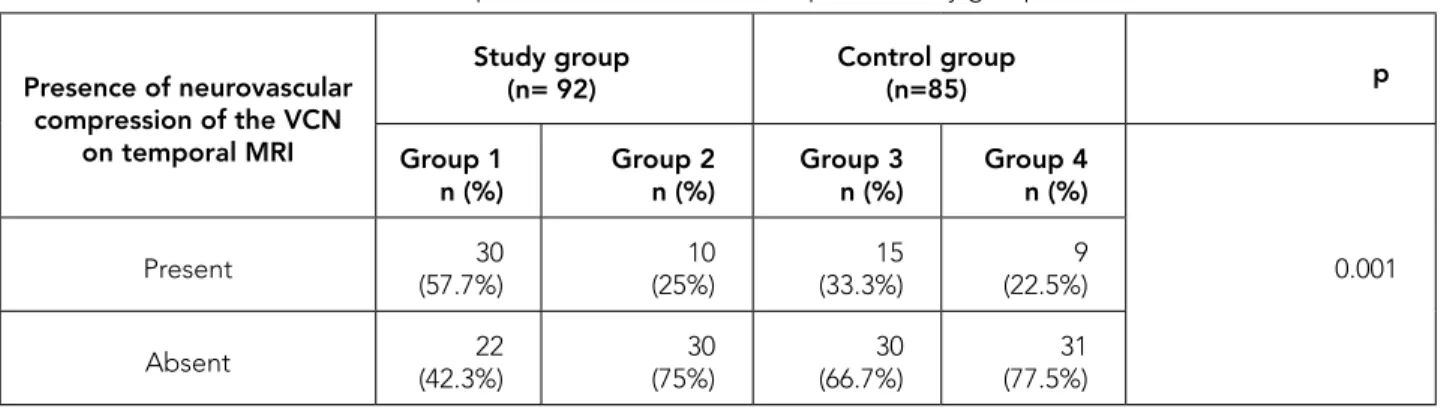
whereas neurovascular compression was absent in 61 (71.8%). There was a significant increase in neurovascular compression among elderly patients in the study group compared with that in the control group (p=0.001; Table 1). Among geriatric patients, vascular contact occurred in 58% of patients with vertigo versus 33% of patients without vertigo (p = 0.024)
Table 1. Anatomical sites of the neurovascular compression in the study and control groups. Anatomical sites of the neurovascular compression on temporal MRI Study group (n=92) Control group (n=85) p Group 1
n (%) Group 2n (%) Group 3n (%) Group 4n (%)
0.007 Peri-intrameatal
compression (60%)18 (10%)1 (33.3%)5 (11.1%)1
Nonmeatal
compression (40%)12 (90%)9 (66.7%)10 (88.9%)8
Table 2. Presence of neurovascular compression of the VCN on temporal MRI by groups. Presence of neurovascular compression of the VCN on temporal MRI Study group (n= 92) Control group (n=85) p Group 1
n (%) Group 2n (%) Group 3n (%) Group 4n (%)
0.001 Present (57.7%)30 (25%)10 (33.3%)15 (22.5%)9
Absent (42.3%)22 (75%)30 (66.7%)30 (77.5%)31
MRI: Magnetic resonance imaging VCN: vestibulocochlear nerve
Anatomical site of neurovascular compression
The anatomical sites of neurovascular compression in the study and control groups are shown in Table 2. When temporal MRI revealed
neurovascular compression, a comparison of the anatomical site of compression revealed a significant difference between the groups (p=0.007). Peri-intrameatal compression was more
common in geriatric patients in the study group than those in group 2 (60% and 10%, respectively; p=0.007).
Anatomical classification of compressing vessels
All vessels that could be followed to their origin were anatomically classified (contrast-enhanced images were also evaluated when required). Of 40 patients in whom MRI revealed neurovascular compression, 33 patients had arterial compression and the anterior inferior cerebellar artery (AICA) was the compressing vessel in 30 (75%) patients. In three patients the compressing vessels could not be differentiated as AICA or PICA definitely.
DISCUSSION
Neurovascular compression of VCN was more prominent in elderly patients with vertigo than inhealthy volunteers. Moreover, intrameatal compression was more prominent in elderly patients with vertigo compared with that in young patients with vertigo. Therefore, our study supports the hypothesis that variations in AICA and the posterior inferior cerebral artery branchespresent at birthlead to neurovascular compression because of degenerative alterations during ageing (9,10). To the best of our knowledge, this study is the first to describe the association between neurovascular compression of VCN and vertigo in geriatric patients.
According to the Global Health and Aging report of the World Health Organization, population ageing will continue and even acceleratebecause of the remarkable increases in life expectancy and decreasing fertility rates (11). The number of people aged>65 years willbemore than double, from 524 million in 2010 to nearly 1.5 billion in 2050, with most of the increase occurring in developing countries.
Vertigo, the sensation of motion in the absence of motion relative to Earth’s gravity, is a complaintof
approximately 30% of geriatric patients (12,13). Consequences of vertigo include postural instability, limitations in daily activities and social participation and fall-related injuries and fractures, and it represents an important public healthcare issue (14-17). Given its significant prevalence and negative effects in the elderly, vertigo deserves special attention, particularly with accelerating growth in the elderly population worldwide.
Vestibular paroxysmia, which is caused by neurovascular compression of VCN, leads to spontaneous, paroxysmal and short attacks of vertigowith or without auditory and vegetative symptoms (18). Vestibular paroxysmia accounts for 4% ofvertigo and balance disorders and may be effectively treated with low-dose sodium channel-blocking anti-epileptics such as carbamazepine. Surgical decompression should be reserved for refractory cases (7).
Temporal MRI, including high-resolution 3D T2-weighted sequences such as 3D-FIESTA, revealsneurovascular compression by providing an accurate visualisation of the cisternal and intracanalicularportion of VCN, as shown in recent studies (8, 19). Neurovascular contacts are frequent MRI findings in asymptomatic patients. However, the type of compressing vessel and the anatomical site of the compression are the two main factors that determine whetherneurovascular contacts become symptomatic. Accordingly, neurovascular compressions are most likely to cause symptoms when they are located at IAC or near the porus acusticus, also known as the transitional zone, which corresponds to this anatomical site of the nervebetween the central and peripheral myelin and is the most vulnerable part of VCN. Compressing vessels most likely to cause symptoms are arteries because of stronger pulsations (6,8). In contrast, De Ridder et al. (20) reported that compression causes symptoms only when contact occurs along the central nervous system segment of the nerve, which is the portion between the brainstem and root entry–exit zone. We found that peri-intrameatal
compressions were more prominent in elderly patients with vertigo than in young patients. The majority of compressions in patients with vertigo were arterial, andAICA was the compressing vessel in 75% of cases.
Many referring physicians, including otorhinolaryngologists, neurologists, neurosurgeons and radiologists, disregard VP in the differential diagnosis of vertigo. This may be because of the unclear clinical diagnostic criteria and frequent
MRI findings of neurovascular compressions in asymptomatic individuals. Nevertheless, recent studies have clarified theclinical and radiological diagnostic criteria of VP. In addition, anti-epileptics such as carbamazepine are reportedly highly effective in most patients with VP (5,18). Thus, both clinicians and radiologists should consider VP while evaluating geriatric patients with vertigo. To exclude VP in such patients, temporal MRI with high-resolution 3D T2-weighted sequences such as 3D-FIESTA should be implemented.
a
c
b
d
-Figure 1. Neurovascular compression of the vestibulocochlear nerve (VCN) in a 72-year- old woman with vertigo. Axial (a, b), coronal (c) and sagittal (d) reformatted 3D-FIESTA images show the neurovascular compression of right VCN (black arrows) by right anterior inferior cerebellar artery (AICA) (white arrows) originating from basilar artery (BA). Note the indentation of VCN by tortuous AICA.
There are some limitations to our study. First, it was a single-centreretrospective study. Second, before this study, patients were not evaluated on the basis of strict clinical or radiological diagnostic criteria of VP. Furthermore, their medical therapy and response totherapy were not considered. This study excluded cases of vertigo that occurred because of any other causes based on clinical and/or imaging findings. We also excluded patients with vascular diseases and related risk factors such as diabetes mellitus, hypertension and atherosclerotic diseases. Although there were no definitive studies on VP before this one, we believethat most patients with vertigo, particularly geriatric patients, have VP. This study may serve as a starting point for developing a prospective study in cases whereincontrolling
clinical variables would enable us to make useful conclusions.
In conclusion, we evaluated neurovascular compression of VCN using MRI in geriatric patients with vertigo. The study results highlight the role of neurovascular compression of VCN in elderly patients with vertigo in whom other causes were excluded. Vestibular paroxysmia should be considered in the differential diagnosis of every vertigo case, and temporal MRI, including high-resolution 3D T2-weighted sequences, should be performed, particularly in geriatric patients with vertigo.
Conflict of Interest
The authors have no conflict of interest to declare.
REFERENCES
1. Hsu LC, Hu HH, Wong WJ, Wang SJ, Luk YO, Chern CM. Quality of life in elderly patients with dizziness: analysis of the Short-Form Health Survey in 197 patients. Acta Otolaryngol 2005;125(1):55-9. (PMID:15799575).
2. Mueller M, Strobl R, Jahn K, Linkohr B, Peters A, Grill E. Burden of disability attributable to vertigo and dizziness in the aged: results from the KORA-Age study. Eur J Public Health 2014;24(5):802-7. (PMID:24213583).
3. Kim EK, Lee JC, Eom MR. Falls risk factors of inpatients. J Korean Acad Nurs 2008;38(5):676-84. (PMID:19114756).
4. Donahue JH, Ornan DA, Mukherjee S. Imaging of Vascular Compression Syndromes. Radiol Clin North Am 2017;55(1):123-38. (PMID:27890181).
5. Ward BK, Gold DR. Tinnitus, Oscillopsia, and Hyperventilation-Induced Nystagmus: Vestibular Paroxysmia. Open J Clin Med Case Rep 2016;2(7). pii: 1100. (PMID:27158666).
6. Haller S, Etienne L, Kövari E, Varoquaux AD, Urbach H, Becker M. Imaging of Neurovascular Compression Syndromes: Trigeminal Neuralgia, Hemifacial Spasm, Vestibular Paroxysmia, and Glossopharyngeal Neuralgia. AJNR Am J Neuroradiol 2016;37(8):1384-92. (PMID:26892985).
7. Brandt T, Strupp M, Dieterich M. Vestibular paroxysmia: a treatable neurovascular cross-compression syndrome. J Neurol 2016;263(Suppl 1):S90-6. (PMID:27083889).
8. Best C, Gawehn J, Krämer HH, et al. MRI and neurophysiology in vestibular paroxysmia: contradiction and correlation. J Neurol Neurosurg Psychiatry 2013;84(12):1349-56. (PMID:24006051). 9. Brunsteins DB, Ferreri AJ. Microsurgical anatomy
of VII and VIII cranial nerves and related arteries in the cerebellopontine angle. Surg Radiol Anat 1990;12(4):259-65. (PMID:2096460).
10. Borghei-Razavi H, Darvish O, Schick U. Disabling vertigo and tinnitus caused by intrameatal
compression of the anterior inferior cerebellar artery on the vestibulocochlear nerve: a case report, surgical considerations, and review of the literature. J Neurol Surg Rep 2014;75(1):e47-51. (PMID:25083388). 11. WHO Global health and ageing. NIH Publication no.
11–7737 2011 October (http://www.who.int/ageing/ publications/global_health.pdf).
12. Committee on Hearing and Equilibrium guidelines for the diagnosis and evaluation of therapy in Meniere’s disease. Otolaryngol Head Neck Surg 1995;113(3):181-5. (PMID:7675476).
13. Colledge NR, Wilson JA, Macintyre CC, MacLennan WJ. The prevalence and characteristics of dizziness in an elderly community. Age Ageing 1994;23(2):117-20. (PMID:8023718).
14. Gopinath B, McMahon CM, Rochtchina E, Mitchell P. Dizziness and vertigo in an older population: The Blue Mountains prospective cross-sectional study. Clin Otolaryngol 2009;34(6):552-6. (PMID:20070765). 15. Gassmann KG, Rupprecht R; IZG Study Group.
Dizziness in an older community dwelling population: a multifactorial syndrome. J Nutr Health Aging 2009;13(3):278-82. (PMID: 19262968).
16. Agrawal Y, Carey JP, Della Santina CC, Schubert MC, Minor LB. Disorders of balance and vestibular function in US adults: data from the National Health and Nutrition Examination Survey, 2001-2004. Arch Intern Med 2009;169(10):938-44. (PMID:19468085). 17. O’Loughlin JL, Robitaille Y, Boivin JF, Suissa S.
Incidence of and risk factors for falls and injurious falls among the community-dwelling elderly. Am J Epidemiol 1993;137(3):342-54. (PMID:8452142). 18. Hüfner K, Barresi D, Glaser M, et al. Vestibular
paroxysmia: diagnostic features and medical treatment. Neurology 2008;71(13):1006-14. (PMID:18809837).
19. Markowski J, Gierek T, Kluczewska E, Witkowska M. Assessment of vestibulocochlear organ function in patients meeting radiologic criteria of vascular compression syndrome of vestibulocochlear nerve-diagnosis of disabling positional vertigo. Med Sci Monit 2011;17(3):CR169-73. (PMID:21358605). 20. De Ridder D, Møller A, Verlooy J, Cornelissen M,
De Ridder L. Is the root entry/exit zone important in microvascular compression syndromes? Neurosurgery 2002;51(2):427-33. (PMID:12182781).