NOBEL MEDICUS 22 | CİLT: 8, SAYI: 1 110
T
HE
P
ERIODONTAL
M
ANAGEMENT
OF A
P
ATIENT WITH
A
CUTE
M
YELOMONOCYTIC
L
EUKEMIA
Esra Güzeldemir PhD,
1Hilal Uslu Toygar PhD,
2Nazım Emrah Koçer MD,
3Ebru Kızılkılıç MD
4 1 Kocaeli University, Faculty of Dentistry, Department of Periodontology, Kocaeli, Turkey2 Medipol University, Faculty of Dentistry, Department of Periodontology, Istanbul, Turkey 3 Ba kent University, Faculty of Medicine, Department of Pathology, Ankara, Turkey
4 SB Istanbul Göztepe Training and Research Hospital, Department of Hematology, Istanbul, Turkey
AKUT MYELOMONOSİTİK LÖSEMİSİ OLAN BİR HASTANIN PERİODONTAL TEDAVİSİ
ÖZET
Akut miyelomonositik lösemi (AML), kemik iliğinin kötü huylu, hemotopoetik klonal hastalığı olup, nor-mal kan hücrelerinin üretimi hatalı olarak meydana gelmektedir. Lösemik hücre infiltrasyonuna, anemi ve trombositopenide eşlik edebilir. Hastalığın oral bulgu-larını, gingival ülserasyonlar, mukositis, dişeti kana-ması ve dişetlerinde büyüme oluşturmaktadır.
AML olarak teşhis edilen 47 yaşındaki, erkek hasta, dişetlerindeki büyüme ve dişeti kanamalarının tedavi-si için, hematologu tarafından periodontoloji kliniğine yönlendirildi. Hastanın periodontoloji kliniğindeki ikinci randevusunda diffüz dişeti ülserasyonları mev-cuttu. Aynı seansta, antibiyotik profilaksisi altında, hastanın iki dişi çekildi. Çekim bölgesinden toplanan
dişeti dokusu, patolojik değerlendirme için patolo-ji bölümüne yönlendirildi. Dişeti dokusunda yapılan histokimyasal ve kimyasal/immunohistokimyasal ana-lizler, AML tanısını doğruladı. Periodontal tedavisi bi-ten hastaya kemoterapi uygulandı ve allojenik kemik iliği transplantasyonu yapıldı. Hasta ilk teşhisi takiben 9. ayda vefat etti.
Bu vaka da, AML’nin oral bulguları olan dişeti büyümesi, dişeti ülserasyonları ve dişeti kanamaları açıkça izlendi. Bu bulguların seyri, hastalığın akut ilerleme safhaları ile paralellik göstermekteydi. Diş hekimi ile yapılan kon-sültasyonlar ve dental tedaviler, hastanın medikal duru-munda düzelmeyle sonuçlanmıştır. Sistemik hastalıklar, uygun koşullar sağlandığında, dental ve periodontal giri-şimler için bir engel oluşturmamaktadır.
Anahtar Kelimeler: Akut myeloid lösemi, dişeti
bü-yümesi, kemoterapi, oral bulgular. Nobel Med 2012; 8(1): 110-113
ABSTRACT
Acute myelomonocytic leukemia (AML) is a malignant-hematopoetic clonal disease of bone marrow and impaired production of normal blood cells. The leukemic cell infiltration may be companied by anemia and thrombocytopenia. Oral manifestations of the disease are gingival ulcerations, mucositis, gingival bleeding and gingival enlargement. A 47-year-old-man with AML-M4 referred to periodontology clinic for treatment of gingival hyperplasia and bleeding. At the second appointment, diffuse gingival ulcerations were seen. Two teeth were extracted under antibiotic prophylaxis. Gingival specimens were sent to pathology. Histochemical/immunohistochemical analysis showed AML. His medical therapy continued with
chemotherapy and allogeneic bone marrow transplantation. He died 9 months after initial diagnosis.
This case report represents not only manifestations of AML such as gingival hyperplasia, ulcerations and bleeding, but also reflects the acute course of the disease in the oral cavity. Dental consultation is essential for diagnosis and improvement of medical conditions. Systemic diseases are not obstacles for dental/periodontal procedures under the proper circumstances.
Key Words: Acute myeloid leukemia, gingival hyperplasia,
chemotherapy, oral manifestations. Nobel Med 2012; 8(1):
NOBEL MEDICUS 22 | CİLT: 8, SAYI: 1
111
INTRODUCTION
Acute leukemias are rare diseases and highly malignant neoplasms responsible for large number of cancer-related deaths. The most common type of leukemia in adults is acute myeloblastic leukemia (AML). AML in adults has a slight male predominance in most countries.1, 2 AML is a malignant hematopoietic clonal,
disease of bone marrow and tissue that is characterized by proliferation of blastic cells in the bone marrow and impaired production of normal blood cells. The leukemic cell infiltration in marrow is companied by anemia and thrombocytopenia.3
While anemia is a constant feature, thrombocytopenia is nearly always present at the time of diagnosis.3
The cause of anemia is inadequate production of red cells; this causes weakness, pallor, fatigue, palpations, dyspnea on exertion. Petechia, epistaxis, posterior palate hemorrhage,gingival bleeding, gingival diffuse enlargement,prolonged bleeding time upon tissue injuries reflect thrombocytopenia.4-6 Gingival
ulcerations may occur as a result of infection by normal oral flora in the setting of neutropenia.4
These symptoms are frequent early manifestations of the disease. Major infections are uncommon until chemotherapy is begun, however respiratory system infections may be present as a finding. Lymphadenopathy is extremely uncommon.
Many systemic diseases associated with or predisposing to severe changes in periodontium have defective numbers of neutrophils and/or defective neutrophil functions as a common finding. The oral cavity is a frequent site of complications associated with chemotherapy and radiation therapy.7 Oral mucositis
is the most significant oral disorder associated with anticancer therapy. Gingival ulcerations, gingival hyperplasia and gingival bleeding are the most common events in these patients. Leukemic cells can infiltrate to gingiva and, less frequently, alveolar bone. Leukemic gingival infiltration creates pathologic pockets where bacterial plaque accumulates and initiates inflammation. The bacterial load of the mouth is among the greatest of any site in the body.7
Gingiva presents both inflammatory and leukemic compounds.
Untreated AML has an aggressive course and is a uniformly fatal disease. AML patients die of the leading complications associated with bone marrow failure such as infection, anemia, and bleeding.1,4 Treatment
begins with rapid-induction chemotherapy with cytosine arabinoside and idarubicine. Other regimens may inclued 6-thioguanine or etoposide. Induction produces severe marrow failure and cytopenia
THE PERIODONTAL
MANAGEMENT OF A
PATIENT WITH ACUTE
MYELOMONOCYTIC
LEUKEMIA
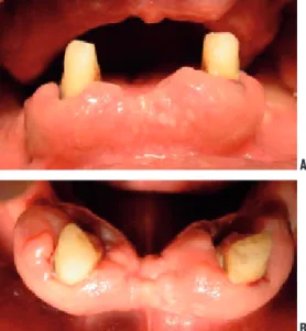
Figure 1A-B. First oral examination on September 2006. Gingival hyperplasia was
detected.
Figure 2A-B. Ten days after initial examination. Gingival ulcerations and gingival
bleeding were noted.
Figure 3. The lower two canine teeth were extracted and sutured.
NOBEL MEDICUS 22 | CİLT: 8, SAYI: 1 112
lasting 3-4 weeks. Platelet transfusions will minimize spontaneous bleeding.4 We report a newly diagnosed
acute myelomonocytic leukemia patient who was referred to periodontology clinic with periodontal complaints and oral manifestations. This case report also shows the oral and periodontal tissue alterations due to aggressive course of the disease within 20 days.
CASE REPORT
On September 8, 2006, a 47-year-old man who was preliminary diagnosed as acute myelomonocytic leukemia was referred by another hospital to Başkent University Hematology Clinic. He was suffering from weakness, fatigue and loss of sense of well being. He diagnosed as AML in hematology department. While his examination was continuing at the hematology clinic in Başkent University, he was also referred to periodontology clinic for the treatment of his gingival enlargement and gingival bleeding. The patient’s major complaint was being unable to use his prosthesis. He had only two lower canines, and adjacent gingiva was enlarged without pain during the last month (Figures 1A and B). He had severe periodontal destruction around these teeth. The extractions of them were planned, and he administered for antibiotic prophylaxis (amoxicillin-clavulanate-potassium 1000 mg twice daily for 1 week). Platelet transfusions were administered for minimizing the gingival bleeding. The chemotherapy was begun with rapid-induction chemotherapy with cytosine arabinoside (100mg/m2/
day), idarubicin (12mg/m2/day), ondansetron ampule
(1x1, IV) and granulocyte-macrophage-stimulating factor (during the neutropenia).
Ten days later, entire adjacent gingiva of teeth had excessive enlargement, necrosis, mucositis, ulceration and pseudomembrane formation; the view was more aggressive contrary to initial examination (Figures 2A and B). These teeth were extracted and conservative nonaggressive debridement and curettage of the soft tissues were performed after the platelet transfusion admission. Surgery area was sutured (Figure 3), chlorhexidine gluconate 0.12% and nystatin rinse were
prescribed. Antimicrobial therapy is instituted on the basis of neutropenia. Obtained tissue specimens were sent to pathological examination. His hematologic changes during the dental treatment are shown at Table 1. Ten days after the extraction, wound healing was uneventful. The patient experienced no pain around extraction sites and oral cavity. On November, neither periodontal and dental infection sources nor pain were detected (Figure 4).
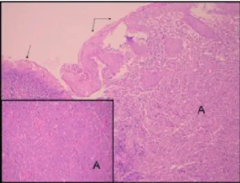
In histopathological evaluation, heavy infi ltrations that caused ulceration in the squamous epithelium and composed of atypical myeloid cells were observed. Immunohistochemically myeloperoxidase and CD117 were positive. There was biclonal staining with lambda and kappa. The diagnosis was AML infi ltration in gingiva (Figure 5).
Long-term survival rests with consolidation high-dose chemotherapy or bone marrow transplantation.5 Our
patient received consolidation therapy for 6 months. Then bone marrow transplantation was performed from his younger brother, however, the patient died three months later, on June 2007.
DISCUSSION
This case clearly demonstrated how periodontal tissues and oral mucosa were affected with the disease progression and side effects of the chemotherapy in a limited time. In this case, two involved teeth were extracted, abnormal tissues and potential infection sources in oral cavity were removed, healing was uneventful. He begun to be nourished, and infection risks were minimized. Dental consultation is essential and important for diagnosis or improvement of medical conditions of the patients.
Patients undergoing chemotherapy for hematologic malignancies are considered to be at high risk group. Complications of cancer chemotherapy could be seen in oral cavity. Pretreatment strategies include evaluation, treatment of preexisting dental and periodontal disease, patient and family education and counseling, prevention of oral mucosal infections, interventions to modify salivary gland dysfunction, reduction of iatrogenic and disease-related neutropenia, and prevention of mucositis.8
Oral mucosa and gingival tissue refl ect the cytotoxic, hemorrhagic, infectious, nutritional and neurologic signs of the chemotherapeutic drug toxicity and prolonged myelosuppression. The incidence and severity of oral complications associated chemotherapy are dependent upon the degree of stomatotoxicity and myelosuppressive nature of the therapy.9
Table 1: Total blood counts at the initial examination, surgery day and 30 days after the surgery.
HGB (12.00-16.00 gr/dl) HCT (40-50%) RBC (4.5-5.9 10e6/mm3) WBC (4.5-11.0 10e3/mm3) PLT (130-400 10e3/mm3) Initial Examination 8.25 23 2.63 1.05 29.9 Surgery Day 6.46 18.5 2.03 34.8 16.4 30 days Later 7.69 21.7 2.48 0.62 22.6 HGB: hemoglobin; HCT: hematocrit; RBC: red blood cells; WBC: white blood cells; PLT: platelet
NOBEL MEDICUS 22 | CİLT: 8, SAYI: 1
113 Myelosuppression alone is associated with mucositis that parallels the severity of neutropenia. Breakdown of oral mucosal integrity in neutropenic patients will be resulted with bacterial, viral or fungal infections.7
These problems have several different clinical presentations including: mucosal inflammation and ulceration of varying etiologies, oral candidiasis, viral and bacterial infections, dental or periodontal infections, and mucosal bleeding. The infectious source of septic episodes in these patients demonstrates a 25-54% relationship to oral infections.9
In the present case, since the changes in the oral cavity were aggressive type, hence this situation would affect his nutrition and general health, dental and periodontal treatment could not be postponed until the end of the chemotherapy. The patient was treated while he was undergone chemotherapy. However, wound healing was uneventful.
Oral hygiene and frequent oral care is essential in these patients. Patients and their relatives should be educated for dental and oral care. Oral care is facilitated by mechanical therapy by a periodontist and alternated rinses of sodium bicarbonate with saline solution and 0.12% chlorhexidine gluconate and nystatin.4 Acute
oral complications occurring during treatment are related to type of cancer and forms of therapy.
Acute or ongoing dental and periodontal problems should be managed before chemotherapy. Teeth which are under risk for infection should be extracted. Acute periodontal conditions should be managed nonsurgically until the platelet is at least 75,000/mL and the neutrophil count is at least 1,000 to 1,500/mL, and only with concurrent administration of prophylactic antibiotics.4
The neutrophil and platelet count must be considered before any patient undergoes an invasive procedure.
CONCLUSION
This case report showed that oral evaluation and treatment of existing diseases decreased the septicemia due to oral cavity in patients who received chemotherapy. On the other hand, oral tissues reflect systemic changes in the body. So, the dentist must be vigilant in detecting abnormal oral tissues and tissue alterations between subsequent visits which would be an initial sign of the most leukemic disorders and collaboration with a hematologist is required to ensure a good outcome of treatment for patients with leukemic disorders. The use of well-supervised treatment protocols, the dental management of individuals with cancer can be effective and safe.
Figure 5. The ulceration (single arrow) in the squamous epithelium (double arrows) and
atypical myeloid cells beneath the epithelium (A) are seen in the photomicrograph. Inner frame reveals atypical myeloid cells in higher magnification. (Main frame:HE x100, Inner frame: HE x200)
REFERENCES
1. Deschler B, Lubbert M. Acute myeloid leukemia: epidemiology and
etiology. Cancer 2006; 107: 2099-2107.
2. Jemal A, Siegel R, Ward E, et al. Cancer statistics, 2006. CA Cancer J
Clin 2006; 56: 106-130.
3. Lichtman MA, Liesveld Jane L. Acute Myelogenous Leukemia. In:
Beutler E, Lichtman, Marshall A, Coller Barry S, Kipps Thomass J, Seligsohn Uri, (eds). Williams Hematology. (6th Ed). New York: McGraw-Hill Medical Publishing Division; 2001; 1047-1084.
4. McKenna SJ. Leukemia. Oral Surg Oral Med Oral Pathol Oral Radiol
Endod 2000; 89: 137-139.
5. Chapple IL, Saxby MS, Murray JA. Gingival hemorrhage, myelodysplastic
syndromes, and acute myeloid leukemia. A case report. J Periodontol 1999; 70: 1247-1253.
6. Wu J, Fantasia JE, Kaplan R. Oral manifestations of acute myelomonocytic
leukemia: a case report and review of the classification of leukemias. J Periodontol 2002; 73: 664-668.
7. Sonis ST, Fey EG. Oral complications of cancer therapy. Oncology
(Williston Park) 2002; 16: 680-686.
8. Kocher T, Fanghanel J, Schwahn C, Ruhling A. A new ultrasonic device
in maintenance therapy: perception of pain and clinical efficacy. J Clin Periodontol 2005; 32: 425-429.
9. Barker GJ. Current practices in the oral management of the patient
undergoing chemotherapy or bone marrow transplantation. Support Care Cancer 1999; 7: 17-20.
CORRESPONDING AUTHOR: Esra Guzeldemir, PhD Kocaeli Univ., Faculty of Dentistry, Department of Periodontology, Kocaeli [email protected]
DELIVERING DATE: 16 / 08 / 2009 • ACCEPTED DATE: 18 / 03 / 2010
THE PERIODONTAL
MANAGEMENT OF A
PATIENT WITH ACUTE
MYELOMONOCYTIC