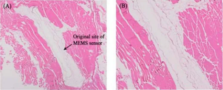
Development and biocompatibility characterization of a biomems sensor for monitoring the progression of fracture healing
Tam metin
Şekil
Benzer Belgeler
Sarıg¨ol, The space bv θ k and matrix transformations, In: 8th International Eurasian Conference on Mathematical Sciences and Applications (IECMSA-2019), Baku, August 27-30,
Biz meme kanserli hastalarda, aromataz inhibitörü tedavisinin serum total siyalik asit düzeyine etkisini ortaya koymayı amaçladık.. MATERYAL
Conclusion: Although patients with and without AF did not significantly differ with regard to blood and tissue magnesium levels, the coincidence of an early postoperative reduction
Proteins encoded by proto-oncogenes are shown in red ; tumor suppressor gene products are shown in blue.. Morphogenesis and cancer:
he comparative analysis and monitoring of related researches of talented youth on the example of the Republic of Tatarstan has allowed to identify positive dynamics
Introduction: Malignant tumors of the skin include basal cell carcinoma, squamous cell carcinoma, malignant melanoma and tumors of the skin appendages.Skin lesions in the head and
In this respect, it is seen that there was no significant criticism against the government of the period based on the publications between 1924- 1928 in Zümrüd-ü Anka and Akbaba
We performed soft tissue repairs by using flaps with a local random circulation pattern and a regional axial pattern in the patients included in our current study. All
