Current Medical Imaging Reviews, 2017, 13, 000-000 1 RESEARCH ARTICLE
1573-4056/17 $58.00+.00 ©2017 Bentham Science Publishers
Evaluation of Osteomeatal Complex Anomalies and Maxillary Sinus
Diseases Using Cone Beam Computed Tomography
Oğuzhan Demirel
1,*, Cemile Özlem Üçok
2and Meryem Toraman Alkurt
21Medipol University Faculty of Dentistry Department of Dentomaxillofacial Radiology, Atatürk Bulvarı Unkapanı Fatih,
Istanbul, Turky; 2Dr. Gazi University Faculty of DentistryDepartment of Dentomaxillofacial Radiology, Emek Mahallesi 83. Sokak Çankaya/Ankara, Turky
A R T I C L E H I S T O R Y Received: July 07, 2016 Revised: January 15, 2017 Accepted: February 07, 2017 DOI: 10.2174/1573405613666170322161655
Abstract: Introduction: Although obstruction of osteomeatal area was not accepted as an
impor-tant factor in the pathogenesis of sinus infections for years, recent studies point out the importance of this area. For the maintenance of normal functions of paranasal sinuses, ventilation and drainage of this area is necessary.
The purpose of this study was to determine the effect of anomalies of the osteomeatal area on sinus diseases.
Methods: This study included cone beam computed tomography scans of 200 patients. Osteomeatal
area anomalies were classified as concha bullosa, septal deviation, paradox middle concha, devia-tion of uncinate process, uncinate bulla, Haller cells and Agger nasi cells. Ethmoid infundibulum and presence of sinus pathologies were recorded.
Results: Pathologies were not determined in 243 (60.8 %) of the evaluated 400 sinuses while
de-tected in 157 (39.3 %) sinuses. Pathologies were found to be more frequent in males than females (p<0.05). Sinus diseases were more prevalent in patients with an obstructed ethmoid infundibulum (p<0.05). Septum deviation was found in 64.5 %, concha bullosa in 47 %, Haller cells in 17 %, paradox middle concha in 8 %, deviation of uncinate process in 5.5 %, over pneumatisation of ag-ger nasi in % 5, and uncinate bulla in 5 % of the investigated patients. Although we found a statisti-cally significant link between ethmoid infundibulum clarity and pathology presence; there was no statistically significant relationship between sinus pathologies and evaluated anomalies.
Conclusion: Although we found no relationship between these anomalies and sinus diseases,
fur-ther studies including the dimensions of the osteomeatal area anomalies will be helpful to determine the effect of these anomalies on sinus diseases.
Keywords: CBCT, osteomeatal complex, paranasal sinus, nasal septum, middle concha, anomaly. INTRODUCTION
Lateral wall of nasal cavity consists of inferior, middle and sometimes superior conchae and their related meatus. Middle meatus, the most important of these structures, gen-erates the drainage pathways of paranasal sinuses with hiatus semilunaris. Middle concha and meatus, hiatus semilunaris, maxillary sinus ostium, uncinate process and infundibulum are the major anatomic formations in the osteomeatal area (Fig. 1) [1]. Although obstruction of osteomeatal area has not been thought to be an important factor in the pathogene-sis of paranasal sinus diseases, recent studies point to the importance of this area [2]. Ventilation and drainage of this area is necessary for the maintenance of normal functions of paranasal sinuses. Especially drainage of maxillary, frontal and ethmoid sinuses are in a close relationship with o
*Address correspondence to this author at the Medipol University Faculty of Dentistry Department of Dentomaxillofacial Radiology, Atatürk Bulvarı Unkapanı Fatih, Istanbul, Turky; Tel: 090 212 5214966; Fax: 090 212 5210443; E-mail: [email protected]
Fig. (1). a: lamina papyrecea b: ostium c: infundibulum and hiatus
steomeatal area [3]. Anatomic variations are frequently seen in middle meatus and lateral nasal wall and these variations may be the primal factor of the underlying sinus disease [4]. These variations can be classified according to originating tissues as middle concha, uncinate process, ethmoid bulla and nasal septum anomalies. Concha bullosa is the pneuma-tisation of middle concha by ethmoid air cells [1]. Paradox middle concha is the lateral convexity of middle concha [5]. Uncinate process deviation is the dislocation of the tip of the uncinate process medially or laterally [1]. Uncinate bulla is the pneumatisation of uncinate process [3]. Haller cells are infraorbital ethmoid air cells located at the roof of maxillary sinus and lateral wall of infundibulum [6]. Agger nasi cells are the most anterior ethmoid air cells [7]. Deviation of the nasal septum is the inclination of structures that build the nasal septum as a result of various reasons [8]. All the previ-ously mentioned anomalies are thought to be related with the obstruction of ethmoid infundibulum and hiatus semilunaris and underlying sinus disease.
Cone beam computed tomography can be an alternative of the conventional tomography for the imaging and evalua-tion of pathologies seen in head and neck area with its low radiation dose and cost. Cone beam computed tomography has been used in previous studies and defined as a useful tool for the interpretation of paranasal sinus diseases and parana-sal sinus anatomy [9-13].
In this study our goal was to define if there is a relation-ship between anomalies seen in osteomeatal complex and maxillary sinus diseases.
MATERIALS AND METHODS
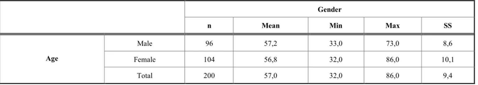
This study included cone beam computed tomography scans of maxillary posterior edentulous 200 patients who had undergone cone beam computed tomographic examination between June 2011 and March 2013 for various reasons such as, evaluation before implant surgery, sinonasal assessment for sinus augmentation processess or any kind of surgical process, from the archives of Gazi University Faculty of Dentistry Department of Dentomaxillofacial Radiology. Age and gender distribution of the patients are given in Table 1. Cone beam computed tomography images were taken using Planmeca Promax 3D with exposure parameters 90 kVp, 10 mA and 14 sn. and Romexis software was used for image reconstruction. Study included maxillary posterior edentu-lous patients in order to keep the effects of maxillary poste-rior teeth to sinus mucosa. The inclusion criteria were having no sign of recent tooth extraction from the maxillary poste-rior area and paranasal sinus surgery. Imaging field
con-tained paranasal sinuses, osteomeatal complex and maxillary alveolar bone.
Pathologies were classified as ‘no pathology, mucosal thickening (Fig. 2), polypoid mucosal thickening (Fig. 3), air-fluid level (Fig. 4), retention pseudocyst (Fig. 5) and si-nonasal polyposis (Fig. 6)’. Anomalies seen in osteomeatal complex area were defined as ‘nasal septum deviation (Fig.
7), concha bullosa (Fig. 8), overpneumatised agger nasi cell
(Fig. 9), uncinate bulla (Fig. 10), uncinate process deviation (Fig. 11), paradox middle concha (Fig. 12) and Haller cells (Fig. 13). Clarity of ethmoid infundibulums were recorded.
Any deviation from midline was considered as nasal sep-tum deviation. Radiolucencies inside middle concha were taken as pneumatization and these lesions were diagnosed as concha bullosa. Agger nasi cells are the most anterior eth-moid air cells and for this diagnosis overpneumatization or asymmetric pneumatizations of Agger nasi cells which can cause obstruction of the osteomeatal area were taken in to
Fig. (2). Bilateral mucosal thickening.
Fig. (3). Bilateral polypoid mucosal thickening. Table 1. Age and gender distribution of patients.
Gender
n Mean Min Max SS
Male 96 57,2 33,0 73,0 8,6
Female 104 56,8 32,0 86,0 10,1
Age
Fig. (4). Air-fluid level in right maxillary sinus.
Fig. (5). Retention pseudocyst in left maxillary sinus.
Fig. (6). Sinonasal polyposis.
Fig. (7). Right nasal septum deviation.
Fig. (8). Bilateral concha bullosa.
Fig. (9). Agger nasi cell.
Fig. (10). Uncinate bulla.
Fig. (12). Bilateral paradox middle concha.
consideration. Pneumatization of uncinate process appears as a radiolucency on this bony structure and any type of radio-lucency involving uncinate process are called as uncinate bulla. Uncinate process deviation was diagnosed as medial or lateral deviation from the regular uncinate process posi-tion. Normally convexity of middle concha is directed medi-ally, and lateral convexity of middle concha was diagnosed as paradox middle concha. Pneumatizations inferior to the orbit, along the lateral wall of infundibulum and at the roof of maxillary sinus were recorded as Haller cells.
SPSS 20 package software was used for data analysis. Descriptive statistics, Mann Whitney U test and Chi-Square tests were uperformed for statistical analysis of the obtained data.
RESULTS
While no pathologies were seen in 243 (60.8 %) of the evaluated 400 sinuses, 157 (39.3) showed pathologies. Mu-cosal thickening was the most frequent pathology with a percentage of 19 % and followed by retention pseudocysts (7.7%), polypoid mucosal thickening (5.5 %), air-fluid level (4.2 %) and sinonasal polyposis (2.7 %), respectively.
Pathologies were found more frequent in male patients than females for left and right sinuses (p<0.05). There was
no statistically significant relationship between age and pa-thologies (p>0.05). Table 2 and 3 demonstrates relationships between pathologies and gender and age.
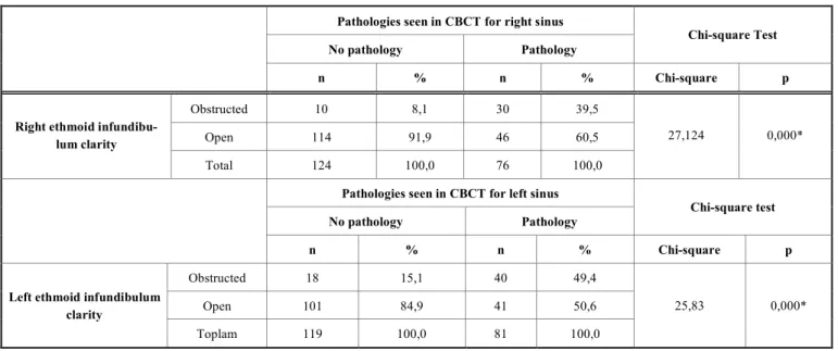
Ethmoid infundibulum was obstructed in 40 (20 %) of si-nuses in right and 58 (29 %) in left. There was a statistically significant relationship between ethmoid infundibulum ob-struction and pathology presence (p<0.05). Table 4 shows the relationship between ethmoid infundibulum clarity and for left and right maxillary sinus pathologies.
Deviation of nasal septum was found in 129 (64.5%) of the evaluated 200 patients. There was no statistically signifi-cant relationship between nasal septum deviation and age and gender (p>0.05). As the connections between nasal sep-tum deviation and presence of pathology were estimated; 55.6 % of patients with a right nasal septum deviation showed no sign of pathology in right and left maxillary si-nuses and 56% of patients with a left nasal septum deviation had no pathologies. No statistically significant relationship between nasal septum deviation and pathology was found (p>0.05). Beside these findings we found a link between left nasal deviation left ethmoid infundibulum clarity. Left eth-moid infundibulums of patients with a left nasal septal devia-tion seem to be more obstructed than patients without left
Table 2. Relationship between gender groups and pathologies for left and right maxillary sinuses.
Gender Male Female Chi-square Test n % n % Chi-square p No pathology 48 50,0 76 73,1 Pathology 48 50,0 28 26,9 Pathologies seen in CBCT for right sinus
Total 96 10,.0 104 100,0
11,284 0,001*
No pathology 42 43,8 77 74,0
Pathology 54 56,3 27 26,0
Pathologies seen in CBCT for left sinus
Total 96 100,0 104 100,0
19,005 0,000*
*p<0.05: there is a statistically significant relationship between gender and pathology. Pathologies are seen more frequent in males than females for both right and left maxillary sinuses.
deviation (p<0.05). This connection was not significant for right area (p>0.05).
Concha bullosa was seen in 78 (39 %) right and 64 (32 %) left of the evaluated 200 osteomeatal areas. As consid-ered in patient distribution of this anomaly, 94 (47 %) of the patients had concha bullosa. Pathologies and ethmoid in-fundibulum clarity showed no significant relationship with ipsilateral concha bullosa presence (p>0.05) (Table 5).
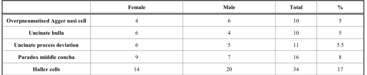
Table 6 demostrates the distribution of overpneumatised agger nasi cell, uncinate bulla, uncinate process deviation, paradox middle concha and Haller cells. None of these anomalies showed a significant relationship with age, gen-der, ethmoid infundibulum clarity and pathology presence (p>0.05).
DISCUSSION
The importance of paranasal sinus diseases for dentistry is a well known issue for years. Especially pathologic events in maxillary sinuses can mimic odontogenic diseases, also dental pathologies can effect maxillary sinuses or give the
symptoms of maxillary sinus diseases [14]. At the same time intraosseous implant procedures, which are getting more common lately, increases the interest on paranasal sinuses. Proximity of maxillary posterior area to maxillary sinuses can lead to some problems in implant surgery of patients with inadequate bone height, however sinus augmentation procedures overcome this problem [15]. Although complica-tions in sinus augmentation procedures are rare; problems like sinus membrane perforation and hemorrhage, problems in post-operative wound healing, sinusitis, infection of the graft may be seen. Many of these problems are related with sinus anatomy and pathologies present in sinuses [16]. Sinus diseases and anomalies have a great frequency in patients who will undergo sinus augmentation process. Presence of these cases cause increases in complications during surgery and post-operatively. Definition of these problems is of im-portance to reduce the risk of complications [17]. Radio-graphic evaluation is worthy in determination of sinus dis-eases. Anatomy of the osteomeatal area has a great impor-tance in evolution of maxillary sinus diseases and drainage of paranasal sinuses has a close relationship with this area [3].
Table 3. Relationship between age and pathologies for left and right maxillary sinuses.
Age Mann Whitney U Test
n Mean Min Max SD Mean Rank U p
No pathology 124 56,7 33,0 86,0 9,5 97,7
Pathology 76 57,5 32,0 85,0 9,4 105,1
Pathologies seen in CBCT for right sinus
Total 200 57,0 32,0 86,0 9,4
4359,5 0,375
No pathology 119 56,4 33,0 86,0 9,3 95,9
Pathology 81 57,9 32,0 85,0 9,6 107,2
Pathologies seen in CBCT for left sinus
Total 200 57,0 32,0 86,0 9,4
4275,5 0,175
Table 4. Relationship between right and left ethmoid infundibulum obstruction and right and left sinus pathologies.
Pathologies seen in CBCT for right sinus
No pathology Pathology
Chi-square Test
n % n % Chi-square p
Obstructed 10 8,1 30 39,5
Open 114 91,9 46 60,5
Right ethmoid infundibu-lum clarity
Total 124 100,0 76 100,0
27,124 0,000*
Pathologies seen in CBCT for left sinus
No pathology Pathology Chi-square test
n % n % Chi-square p
Obstructed 18 15,1 40 49,4
Open 101 84,9 41 50,6
Left ethmoid infundibulum clarity
Toplam 119 100,0 81 100,0
25,83 0,000*
Table 5. Relationship between concha bullosa presence and ethmoid infundibulum clarity.
Right ethmoid infundibulum clarity
Obstructed Open Total Statistical analysis
n % n % n % Chi-square p
No 28 70.00 94 58.75 122 61,00
Yes 12 30.00 66 41.25 78 39,00
Right concha bullosa presence
Total 40 100,00 160 100,00 200 100,00
4,226 0,238
Left ethmoid infundibulum clarity
Obstructed Open Total Statistical analysis
n % n % n % Chi-square p
No 40 68,97 96 67,61 136 68,00
Yes 18 22,41 46 20,42 64 32,00
Left concha bullosa presence
Total 58 100,00 142 100,00 200 100.00
0,576 0,902
Pathologies seen in CBCT for right sinus
No Yes Total Statistical analysis
n % n % n % Chi-square p
No 73 61,3 49 60,5 122 61,0
Yes 46 38.7 32 39.5 78 34.0
Right concha bullosa presence
Total 119 100,0 81 100,0 200 100,0
4,921 0,178
Pathologies seen in CBCT for left sinus
No Yes Total Statistical analysis
n % n % n % Chi-square p
No 85 68,5 51 67,1 136 68,0
Yes 39 31.5 25 32.9 64 32.0
Left concha bullosa presence
Total 124 100,0 76 100,0 200 100,0
2,081 0,556
Table 6. Distribution of overpneumatised agger nasi cell, uncinate bulla, uncinate process deviation, paradox middle concha and
Haller cells.
Female Male Total %
Overpneumatised Agger nasi cell 4 6 10 5
Uncinate bulla 6 4 10 5
Uncinate process deviation 6 5 11 5.5
Paradox middle concha 9 7 16 8
Haller cells 14 20 34 17
For this study we used cone beam computed tomography scans of maxillary posterior edentulous patients who had no sign of recent tooth extraction to distinguish maxillary sinus diseases of dental origin. Sinus pathologies and osteomeatal area anomalies are interpreted in cone beam computed to-mography images which is described as a useful method for
the diagnosis of maxillary sinus diseses and definition of sinonasal anatomy [9-13].
In dental and especially otorhinolaryngology literature there are various researches made with different radiographic and diagnostic methods for the definition of the prevalence
of maxillary sinus diseases and osteomeatal complex anoma-lies and relationships between these.
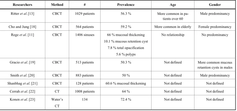
Table 7 demonstrates some studies made to define the prevalence and age and gender characteristics of maxillary sinus diseases.
In our study we found sinus pathologies in 39.3 % of the evaluated 400 sinuses and 50 % of the 200 patients. While our results are compatible with some of these studies [13, 19, 20], some studies showed higher prevalence values than ours’. In our opinion, differences in patient groups and inclu-sion criteria as a pathology are the main causes of the dis-cordances. For example, in Shanbhag et al. study [21] while 2 mm. of mucosal thickenings are considered as pathology; we counted minimum 3 mm. mucosal thickenings as patho-logic. Likewise, in Cho and Jung’s research [18] there is no definition of pathology inclusion criteria. Cerrah et al. [22] and Konen et al. [23] studies, which had higher frequencies, are researches performed in otolaryngology clinics including patients with sinonasal complaints. Mucosal thickening was the most common sinus pathology in our study and our re-sults are concordant with other works [11, 13, 21]. As we look in gender and age groups, while we found a male pre-dominancy, no relationship was seen between age and sinus pathologies. Of the above mentioned studies only one work concluded with female predominancy [18]. Besides, two studies indicated that pathologies are more common in eld-erly [13, 19]. This difference with our study may be the re-sult of definition of patient groups. Our workgroup was made of patients with maxillary posterior edentulous patients with higher ages.
CBCT study conducted by Smith et al. including 883 pa-tients to research prevalence and relationships between nasal septum deviation, concha bullosa and sinus diseases con-cluded with a 67.5 % septum deviation, 19.4 % concha bul-losa prevalence and no relationship was found between these
anomalies and sinus diseases [20]. Another CT study con-taining 172 patients consisting of 90 patients with paranasal sinus complaints and 82 asymptomatic patients defined hav-ing 28.8 % concha bullosa, 22.2 % septum deviation, 12.2 % paradox middle concha, 7.77 % agger nasi cells, 5.55 % Haller cells and 2.22 % uncinated process deviation in symp-tomatic group; having 12 % septum deviation, 7.31 % para-dox middle concha, 4.88 % agger nasi cells and 3.65 % Haller cells in asymptomatic group. No relation between these anomalies and age and gender was found in this study [24]. Fifty five percent concha bullosa and 44 % nasal sep-tum deviation was found in another study made using CT scans of 800 patients [25]. Vincent and Gendeh’s study with CT scans of 137 patients who will undergo or had undergone endoscopic sinus surgery resulted with a 25.5 % concha bul-losa prevalence and female predominancy. Nasal septum deviation was found in 46.7 % of patients and no statistically significant relationship was found between these two anoma-lies and sinus pathologies [26]. While concha bullosa and contralateral septum deviation was found to be related, no relation between these anomalies and sinus diseases was found in a CT study containing 998 scans, which resulted with a 65 % septum deviation and 44 % concha bullosa prevalence [27]. Results of previously mentioned Cerrah et
al. study with 1008 patients’ CT scans demonstrated 53.7 %
agger nasi cells, 41.6 % concha bullosa, 30.7 % septum de-viation, 18.2 % uncinated bulla, 17.1 % Haller cells, 8.4 % paradox middle concha and 4.9 % uncinate process devia-tion. Also these anomalies were related with sinus diseases in this study [22]. Case-control study of Dursun et al. which aimed to define the contractions between sinus pathologies and osteomeatal complex anomalies consisted of CT scans of 415 chronic rhinosinusitis and 60 asymptomatic patients. According to results of this study septum deviation was found 60.5 % of symptomatic and 16.6 % of asymptomatic group and statistically significant relationship was found between septum deviation and sinus pathologies. Concha
Table 7. Studies made to define the prevalence and age and gender characteristics of maxillary sinus diseases.
Researchers Method # Prevalence Age Gender
Ritter et al.[13] CBCT 1029 patients 56.3 % More common in pa-tients over 60
Male predominancy
Cho and Jung [18] CBCT 564 patients 59.2 % More common in elderly Female predominancy Rege et al. [11] CBCT 1406 sinuses 66 % mucosal thickening
10.1 % mucous retention cyst 7.8 % total opacification
5.6 % polyps
No relationship No predominancy
Gracio et al. [19] CBCT 513 patients 50.3 % Not defined More common mucous
retantion cysts in males
Smith et al. [20] CBCT 883 patients 50 % Not defined Male predominancy
Shanbhag et al. [21] CBCT 128 patients 60.6 % mucosal thickening Not defined Not defined
Cerrah et al. [22] CT 1008 patients 64 % Not defined Not defined
Konen et al. [23] Water’s CT
bullosa was found in 48.4% of symptomatic and 25 % of asymptomatic group and uncinated process deviation was seen in 29.6 % of symptomatic and 10 % of asymptomatic group. Besides, in terms of these two variables significant difference was found between symptomatic and asympto-matic patients. While no Haller cells and uncinate bulla was discovered in asymptomatic group, prevalence was 12.8 % and 2.4 % in symptomatic group respectively and with re-gard to Haller cells frequency relationship was found. No significant difference between two groups was determined in terms of paradox middle concha and agger nasi cells preva-lence [3]. Ünal et al. intended to define the interactions be-tween osteomeatal area apertures and sinus diseases and found 48.9 % Haller cells, 46.9 % paradox middle concha, 42.8 % concha bullosa, 40.8 % septum deviation, 16.3 % uncinate process deviation and 12.2 % uncinate bulla in 48 patients [28]. Another CT study including 120 symptomatic and 120 asymptomatic patients showed 40.8 % and 47.5 % concha bullosa, 12 % and 23 % paradox middle concha, 51 % and 62 % Haller cells, 83 % and 79 % agger nasi cells and 56 % and 60.8 % septum deviation, respectively [5].
We found nasal septum deviation in 64.5 % of the evalu-ated 200 patients. Concha bullosa was present in 47 % of the study group. Haller cells showed 17 % frequency and fol-lowed by 8 % paradox middle concha, 5.5 % uncinate proc-ess deviation and 5 % uncinate bulla and overpneumatized agger nasi cells. None of the evaluated anomalies showed a statistically significant relationship with age and gender, also with maxillary sinus diseases.
As mentioned before, there are various studies made to define the prevalence of osteomeatal complex anomalies and sinus diseases. Our results seem to be compatible with some of these studies. Main differences between our results and other studies may be the consequence of differences between evaluation criteria. For example, in our study we accepted a minimal deviation from the line that is drawn from crista galli to middle palatal suture as nasal septum deviation. While many studies did not define the criteria of inclusion as septum deviation, only one of the studies [20] reported the acception criteria as 4 mm. deviation from mid-sagittal plane as deviation and another [26] as deviation caused obstruction in osteomeatal area. We found none of the anomalies dis-cussed above to be related with sinus diseases. While this finding is in accordance with some of the studies, there is discrepancy between some. Situation here that must be ex-plained is the inclusion criteria as pathology and anomaly. We accepted 3 mm. or above changes in sinus mucosa as pathology, Cerrah et al. admitted Lund-Mackay classifica-tion which defines 2mm. or above changes in sinus mucosa as pathology [22]. Many of the other studies did not state any inclusion criteria as pathology. Another problem that must be clarified is the previously mentioned anomaly inclusion criteria. Haller cells are defined as infraorbital ethmoid air cells and agger nasi cells are the most anterior ethmoid air cells. We accepted cells not expanding beyond inferior wall of orbita as Haller cells and asymmetric overpneumatised cells as agger nasi cells. Any deviation of uncinate process’ parallelity from lamina papyracea, which defines the infer-omedial wall of orbita, as uncinate process deviation. Also, there may be controversies in the definition of concha bul-losa. Pneumatisation of middle concha may effect the
lamel-lar, bulbous or both parts of middle concha which is called as real concha bullosa [29].
Sinus diseases seem to sustain the interest on and many researches should be carried on to define the prevalence and cause of them. Sinus procedures and three dimensional im-aging techniques are getting more widespread in dentistry and definition of sinus diseases and anomalies is getting more important. In our study, we found a positive relation-ship between ethmoid infundibulum clarity and sinus health. Although we found no relationship between sinus patholo-gies and osteomeatal area anomalies, further researches in-cluding the dimensions of anomaly such as angular value of deviations or volume of the existing anomaly with standard-ized pathology inclusion criteria will be useful to define the interactions between osteomeatal area anomalies and sinus diseases.
CONFLICT OF INTEREST
The authors confirm that this article content has no con-flict of interest.
ACKNOWLEDGEMENTS
Declared none.
REFERENCES
[1] Laine FJ, Smoker WR. The osteomeatal unit and endoscopic sur-gery: anatomy, variations, and imaging findings in inflammatory diseases. AJR Am J Roentgenol. 1992; 159(4): 849–57.
[2] Kennedy DW. Prognostic factors, outcomes and staging in ethmoid sinüs surgery. Laryngoscope. 1992; 102(1): 1-18.
[3] Dursun E, Korkmaz H, Şafak M., et al. Paranazal sinüs infeksiyon-larında osteomeatal kompleksteki anatomik varyasyonlar. KBB Fo-rum. 1998; 6(3): 147-56.
[4] Bolger WE, Butzin CA, Parsons DS. Paranasal sinus bony anat-omic variations and mucosal abnormalities: CT analysis for endo-scopic sinus surgery. Laryngoscope. 1991; 101(1): 56-64. [5] Azila A, Irfan M, Rohaizan Y, et al. The prevalence of anatomical
variations in osteomeatal unit in patients with chronic rhinosinusi-tis. Med J Malaysia. 2011; 66(3): 191-94.
[6] Caversaccio M, Urs BU, Albert AM. Historical review of Haller’s cells. Ann Anat. 2011; 193(3): 185–90.
[7] Wormald PJ. The agger nasi cell: The key to understanding the anatomy of the frontal recess. Otolaryngol Head and Neck Surg. 2003; 129(5): 497-507.
[8] Karazincir S, Okuyucu Ş, Balcı A, et al. Konka bullosa septal deviasyon ilişkisi. KBB-Forum. 2007: 6(1), 19-21.
[9] Ilgüy D, Ilgüy M, Dölekoğlu S, et al. Evaluation of the posterior superior alveolar artery and the maxillary sinus with CBCT. Braz Oral Res. 2013; 27(5): 431-437.
[10] Donizeth-Rodrigues C, Fonseca-Da Silveira M, Gonçalves-De Alencar AH, et al. Three-dimensional images contribute to the di-agnosis of mucous retention cyst in maxillary sinus. Med Oral Pa-tol Oral Cir Bucal. 2013; 18(1): 151-57.
[11] Rege IC, Sousa TO, Leles CR, et al. Occurrence of maxillary sinus abnormalities detected by cone beam CT in asymptomatic patients. BMC Oral Health. 2012; 12(30): 1-7.
[12] Çakur B, Sümbüllü MA, Yılmaz AB. Alt konka hipertrofisi, nazal septum deviasyonu ve antral retansiyon kisti arasındaki ilişki. Atatürk Üniv Dis Hek Fak Der. 2011; 21(1): 5-9.
[13] Ritter L, Lutz J, Neugebauer J, et al. Prevalence of pathologic findings in the maxillary sinus in cone-beam computerized tomo-graphy. Oral Surg Oral Med Oral Path Oral Radiol Endod. 2011; 111(5): 634-40.
[14] Ruprecht A, Lam EWN. Paranasal Sinuses. In: White, SC., Pharoah, MJ. (Eds). Oral Radiology: Principles and Interpretation (6th edition). 2009; St. Louis Missouri: Mosby, Elsevier; pp. 506-25.
[15] Barone A, Santini S, Sbordone L, et al. A clinical study of the outcomes and complications sssociated with maxillary sinus aug-mentation. Int J Oral Maxillofac Implants. 2006; 21(1): 81-5. [16] Anavi Y, Allon DM, Avishai G, et al. Complications of maxillary
sinus augmentations in a selective series of patients. Oral Surg Oral Med Oral Path Oral Radiol Endod. 2008; 106(1): 34-8.
[17] Chan HS, Wang HL. Sinus pathology and anatomy in relation to complications in lateral window sinus augmentation. Implant Dent. 2011; 20(6): 406–12.
[18] Cho BH, Jung YH. Prevalence of incidental paranasal sinus opacification in an adult dental population. Korean J Oral Maxillo-fac Radiol. 2009; 39(2): 191-4.
[19] Gracco A, Parenti SI, Ioele C, et al. Prevalence of incidental maxil-lary sinus findings in Italian orthodontic patients: a retrospective cone- beam computed tomography study. Korean J Orthod. 2012; 42(6): 329-34.
[20] Kyle D. Smith, Paul C. Edwards, Tarnjit S. Saini, and Neil S. Nor-ton, “The Prevalence of Concha Bullosa and Nasal Septal Devia-tion and Their RelaDevia-tionship to Maxillary Sinusitis by Volumetric Tomography,” International Journal of Dentistry, vol. 2010, Article ID 404982, 5 pages, 2010. doi:10.1155/2010/404982
[21] Shanbhag S, Karnik P, Shirke P, et al. Cone-beam computed tomo-graphic analysis of sinus membrane thickness, ostium patency, and residual ridge heights in the posterior maxilla: implications for si-nus floor elevation. Clin Oral Implants Res. 2014; 25(6): 755-60.
[22] Cerrah YSS, Altuntaş EE, Uysal İÖ, et al. Bilgisayarlı tomografi ile saptanan paranazal sinüs anatomik varyasyonları. Cumhuriyet Tıp Derg; 2011; 33(1): 70-79.
[23] Konen E, Faibel M, Kleinbaum Y, et al. The value of the occipito-mental (Waters') view in diagnosis of sinusitis: a comparative study with computed tomography. Clin Radiol. 2000; 55(11): 856-60. [24] Kayalıoğlu G, Oyar O, Govsa F. Nasal cavity and paranasal sinus
bony variations: a computed tomographic study. Rhinology. 2000; 38(3):108–13.
[25] Earwaker J. Anatomic variants in sinonasal CT. Radiographics. 1993; 13(2): 381-415.
[26] Vincent TES, Gendeh BS. The association of concha bullosa and deviated nasal septum with chronic rhinosinusitis in functional en-doscopic sinus surgery patients. Med J Malaysia. 2010; 65(2): 108-11.
[27] Stallman JS, Lobo JN, Som PM. The incidence of concha bullosa and its relationship to nasal septal deviation and paranasal sinus disease. Am J Neuroradiol. 2004; 25(9): 1613-8.
[28] Ünal B, Arıkan OK, Bilgili Y, et al. Osteomeatal kompleks boşluklarının/mesafelerinin kemik ve mukozal genişliklerinin kronik sinüzit şiddeti ile ilişkisi – BT çalışması. KBB Forum. 2005;4(3): 110-4.
[29] Neto SAA, Martins PSL, Souza AS, et al. The role of osteomeatal complex anatomical variants in chronic rhinosinusitis. Radiol Bras. 2006; 39(3): 1-9.
View publication stats View publication stats