Acta Orthop Traumatol Turc 2013;47(1):65-67 doi:10.3944/AOTT.2013.2659
CASE REPORT
Correspondence: Eren Cansü, MD. Mehtap Sok. 34/7, Caddebostan, ‹stanbul, Turkey. Tel: +90 532 - 274 25 56 e-mail: [email protected]
Submitted: March 20, 2011 Accepted: December 22, 2011 ©2013 Turkish Association of Orthopaedics and Traumatology
Trigger finger at the carpal tunnel level:
three case reports
Fatih PARMAKSIZO⁄LU1, Eren CANSÜ2, Mehmet Bekir ÜNAL3
1
Department of Orthopedics and Traumatology, Yeni Yüzy›l University, ‹stanbul, Turkey;
2
Department of Orthopedics and Traumatology, Yüzy›l Hospital, Kocaeli, Turkey;
3
Department of Orthopedics and Traumatology, Medipol University, ‹stanbul, Turkey
Available online at www.aott.org.tr doi:10.3944/AOTT.2013.2659 QR (Quick Response) Code:
Although trigger finger occurs mostly due to a problem at the A1 pulley various other causes have also been reported. We present three patients with different tumors at the carpal tunnel as a cause of trig-gering. All patients were treated with local excision.
Key words: A1 pulley; carpal tunnel; stenosing tenosynovitis; trigger finger.
Trigger finger is a common hand disorder. Stenosing tenosynovitis of the flexor tendons leads to blocking of tendon movements at the A1 pulley, resulting in charac-teristic symptoms of the disorder. However, attention must be paid to diagnosis and treatment as various etio-logical factors may rarely cause similar manifestations. The surgical treatment method is the release of the A1 pulley. A soft tissue mass in the carpal tunnel level may also result in trigger finger.
We present 3 patients with a soft tissue tumor in the carpal tunnel. All patients were treated with surgical excision of the mass.
Case reports
Case 1
A 28-year-old female patient presented with triggering and pain in the fifth digit of the dominant right hand and pain in the wrist during flexion and extension of the fin-ger for two months. A mass of 1×0.6×0.3 cm was detect-ed over the fifth deep digital flexor tendon during explo-ration of the carpal tunnel (Fig. 1). The mass caused
locking of the fifth digit under the flexor retinaculum, in full flexion. The tumoral mass could pass under the reti-naculum by pushing. The locking was released by resec-tion of the mass and tendon movement was achieved with no problems. No recurrence was seen in the post-operative period. Histopathological examination revealed the mass to be a mixed type hemangioma.
Case 2
A 52-year-old female patient presented with locking on the third finger of the right hand for 7 months. Tenderness to palpation over the A1 pulley was noted. No additional pathology was detected at the clinical examination. Release of the A1 pulley was performed under local anesthesia. When the patient was asked to actively move her fingers during the surgery, it was observed that the pathology continued. After obtaining patient approval, the carpal tunnel was explored with a pneumatic tourniquet for hemostasis with axillary block. The locking disappeared following the excision of a mass of 2.5×2×1 cm, originating from the deep
tendon of the third digit (Fig. 2). Histological exami-nation of the mass was consistent with a ganglion cyst.
Case 3
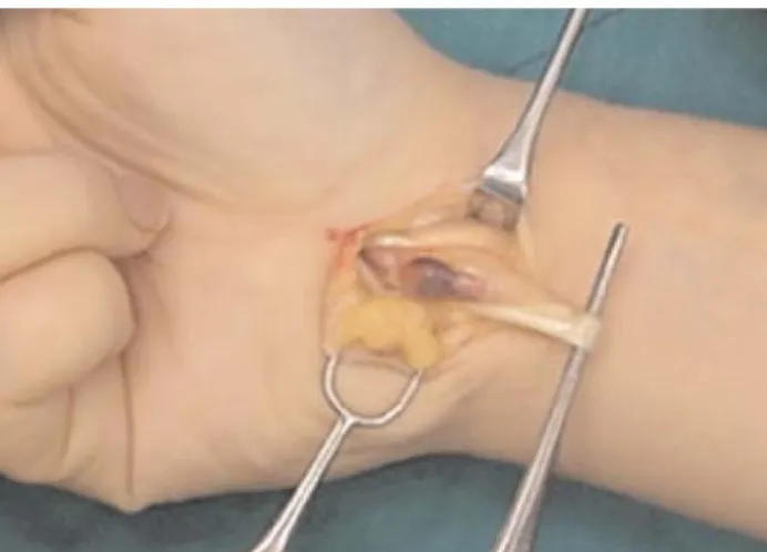
A 55-year-old male patient presented with a painful lock-ing on the fourth flock-inger of his non-dominant left hand for four months. His symptoms mimicked those of “trigger finger”. He also had a mass on the wrist which was mobile with the movements of the fingers. Magnetic res-onance imaging (MRI) findings showed that the mass originated from the deep flexor tendon of the fourth digit and the patient was offered surgery (Fig. 3). Symptoms disappeared following the excision of the mass of 4×1.5×1 cm, with the exploration of the carpal tunnel under axil-lary block and pneumatic tourniquet (Fig. 4). The mass was defined as schwannoma upon histopathological examination. No sensory or motor problems of the medi-an nerve were detected at follow-up.
In addition to above findings, no recurrence was observed in the patients’ follow-ups at the ninth, fourth and third years, respectively.
Discussion
Trigger finger is one of the most frequently seen disor-ders of the upper extremity.[1]
It presents itself with the trapping of the flexor tendons by the degenerated and thickened A1 pulley, usually as a result of stenosing tenosynovitis. The size mismatch between the flexor tendon and the pulley above is the most important and common cause.[2]
Inflammation of the tendon sheath secondary to thickening of the A1 pulley leads to swelling in the flexor tendon.[2]
Tenderness over the A1 pulley, a palpable nodule, triggering, and locking are common findings in physical examination. Despite its rather easy clinical diagnosis, probable issues other than
Acta Orthop Traumatol Turc
66
Fig. 1. The mass originating from the deep flexor tendon of the fifth digit is shown. [Color figure can be viewed in the online issue, which is available at www.aott.org.tr]
Fig. 2. Areas with tenosynovitis were also excised in addition to the mass shown in the circle. [Color figure can be viewed in the online issue, which is available at www.aott.org.tr]
Fig. 3. The space-occupying lesion originating from the flexor ten-dons and detected at the wrist level is seen in axial MRI sec-tion.
Fig. 4. The mass originating from the deep flexor tendon of the fourth digit is viewed following the exploration of the carpal tunnel. [Color figure can be viewed in the online issue, which is available at www.aott.org.tr]
Parmaks›zo¤lu et al. Trigger finger at the carpal tunnel level 67
the A1 pulley should also be considered among the rea-sons behind triggering. Sesamoid bone,[3]
finger exosto-sis,[2]
calcific tendonitis,[4]
abnormal insertion of the lum-brical muscles[5]
and post-traumatic lesions[6]
have been reported in the literature as causes of trigger finger. Other pathologies should be considered in the absence of pain and/or nodule over the A1 pulley in physical examination, requiring additional evidence by X-rays, computed tomography (CT) and MRI.
In a case reported by Lee and Pho,[2]
no signs of trig-ger fintrig-ger was present. However, radiography and CT findings revealed exostosis in the flexor tendon bed, per-formed due to local tenderness over the proximal pha-lanx. The complaints disappeared after the excision of the exostosis.
Some pathologies at the carpal tunnel level may also cause triggering by locking the flexor tendons under the flexor retinaculum during movement of the flexor ten-dons. This pathology in the wrist is defined as “trigger wrist” by some authors. However, according to Giannikas et al., this definition is controversial.[7] No
consensus has been reached as it is still debated whether triggering due to wrist movements or triggering of the finger should be defined as “trigger wrist” when the pathology is in the wrist. According to Desai et al., the term “trigger wrist” should be used only when a trigger-ing due to wrist movements is present.[8]
Lemon and Engber[9]
and Koob and Steffens[10]
reported “true” trig-ger wrist cases and stated that the term is generally mis-used to mean the triggering of the fingers at the wrist level. As the triggering is not initiated by wrist but fin-ger movements, a more accurate definition would be “trigger finger at the wrist”.[9]
Our cases were of trigger fingers not wrists, although the triggering was due to pathology at the wrist level.
Lemon and Engber[9]reported an example of “true”
trigger wrist. Triggering developed due to the displace-ment of the nodule at the extensor carpi radialis longus tendon into the second dorsal compartment by wrist movements and was treated with excision of the nodule. The authors pointed out that several intra-articular pathologies may cause restriction of movement of the wrist and painful click, and that extra-articular patholo-gies may present with similar symptoms. In addition to stenosing tenosynovitis, tumoral masses preventing the sliding of the tendons by forming a block is another cause of triggering at the fingers. Although several authors have reported tumors of different histopatho-logical character, these tumors rarely cause trigger fin-ger.[11-15]
As they may usually be located in the proximal of the A1 pulley (in the palm and the carpal tunnel), they are not always possible to detect at the physical exami-nation and be distinguish from typical trigger finger.
Other rare causes of trigger finger should also be considered in the preoperative period to avoid missing probable cases that do not present with classical symp-toms and findings, and further tests should be per-formed in case of uncertainty.
Even in patients with positive results of A1 pulley examination, more proximal regions, such as the palm and the carpal tunnel should also be examined. Despite the diagnosis of stenosing tenosynovitis at the A1 pulley, it should be kept in mind that an associated pathology may also be present in the more proximal region, e.g. the carpal tunnel, as seen in Case 2.
When A1 pulley release due to stenosing tenosynovi-tis is performed, we recommend surgery be done with local anesthesia. In this manner, it will be possible to determine if the locking still exists when the patient is asked to actively move his/her fingers during the sur-gery. Furthermore, the presence of other probable pathologies in the more proximal region can be observed and, thus, treated within the same session.
Conflicts of Interest: No conflicts declared.
References
1. Wolfe SW. Tenosynovitis. In: Green DP, Hotchkiss RN, Pederson WC, Wolfe SW, editors. Green’s operative hand surgery. Vol. 2, 5th ed. Philadelphia: Elsevier Churchill Livingstone; 2005. p. 2137-58.
2. Lee SJ, Pho RW. Report of an unusual case of trigger finger secondary to phalangeal exostosis. Hand Surg 2005;10:135-8. 3. Brown M, Manktelow RT. A new cause of trigger thumb. J
Hand Surg Am 1992;17:688-90.
4. Hansen U, Battista V. Pediatric trigger finger from calcific tendonitis. J Hand Surg Am 2007;32:1558-9.
5. Bartell TH, Shehadi SI. Trigger finger secondary to anom-alous lumbrical insertion: a case report and review of the lit-erature. Plast Reconstr Surg 1991;87:354-7.
6. Kalms SB, Højgaard AD. Trigger finger: report of an unusu-al case. J Trauma 1991;31:582-3.
7. Giannikas D, Karabasi A, Dimakopoulos P. Trigger wrist. J Hand Surg Eur Vol 2007;32:214-6.
8. Desai SS, Pearlman HS, Patel MR. Clicking at the wrist due to fibroma in an anomalous lumbrical muscle: a case report and review of literature. J Hand Surg Am 1986;11:512-4. 9. Lemon RA, Engber WD. Trigger wrist: a case report. J
Hand Surg Am 1985;10:61-3.
10. Koob E, Steffens K. True trigger wrist (a case report). [Article in German] Handchir Mikrochir Plast Chir 1988;20:288-90.
11. Oni OO. A tendon sheath tumour presenting as trigger fin-ger. J Hand Surg Br 1984;9:340.
12. Stockley I, Norris SH. Trigger finger secondary to soft tis-sue chondroma. J Hand Surg Br 1990;15:468-9.
13. Laing PW. A tendon tumour presenting as a trigger finger. J Hand Surg Br 1986;11:275.
14. Rankin EA, Reid B. An unusual etiology of trigger finger: a case report. J Hand Surg Am 1985;10:904-5.
15. Suematsu N, Hirayama T, Takemitsu Y. Trigger wrist caused by a giant cell tumour of tendon sheath. J Hand Surg Br 1985; 10:121-3.