O
h
r
c
i
r
g
a
in
e
a
s
l
R
e
Ramazan Atiç1, Celil Alemdar1, İbrahim Azboy2, Mehmet Bulut1, Mehmet Akif Çaçan2, Mehmet Gem1 1Department of Orthopaedics and Traumatology, Dicle University Medical Faculty, Diyarbakır, 2Department of Orthopaedics and Traumatology, İstanbul Medipol University Medicine Faculty, İstanbul, Turkey.
VAC in open fracture
Application of vac in type III open fractures
DOI: 10.4328/JCAM.5818 Received: 14.03.2018 Accepted: 03.04.2018 Published Online: 04.04.2018 Printed: 01.11.2018 J Clin Anal Med 2018;9(6): 498-503 Corresponding Author: Ramazan Atiç, Dicle University Medical Faculty, 21280, Diyarbakir, Turkey.
GSM: +905321728629 F.: +90 4122488440-4254 E-Mail: [email protected]
Abstract
Aim: Functional results of patients with Type III open long bone fractures treated with vacuum-assisted closure (VAC) and approximating sutures were evalu-ated. Material and Method: A total of 21 patients (4 female, 17 male with a mean age of 27 years, range: 3–64 years) with Gustilo-Andersen Type 3 open fracture were included in the study. The mean follow-up period was 25.67 months (range, 9–52 months). Thirteen patients had Type IIIB and eight patients had Type IIIC open fractures. Following a large debridement and irrigation in an operating room setting, wound dimensions were measured. Subsequently, VAC was applied. During the application, skin traction sutures were applied over the vacuum sponge. During the last dressing change in which a clean granulation tissue was obtained, the wound was closed following the measurement of its dimensions. Wound dimensions measured during the large debridement in the operating room were compared with the wound dimensions measured after the final VAC application. Results: Mean duration between trauma and operation was 7.57 hours (range, 2–23 hours). The wound was closed with delayed primary suturing in seven patients. In the remaining 14 patients, wound dimensions measured following the last VAC application were found to be decreased with a mean of 40.02% (range, 20-60%). Seven patients underwent a skin graft, two had a free flap application, four had a fasciocutaneous flap, and one had a fasciocutaneous flap + graft application. Two patients underwent revision opera-tions for graft failure. Five patients (23.8%) developed a deep wound infection. The infecopera-tions resulted in osteomyelitis in two patients (9.5%). Discussion: VAC application together with skin traction sutures in Type IIIB and C open fractures decreases wound size dimension of a graft or flap to be applied. Also it may reduce the requirement for secondary interventions.
Level of evidence: Level IV.
Keywords
Open Fracture; Vacuum-Assisted Closure; Skin Traction Suture; Wound Closure
| Journal of Clinical and Analytical Medicine 498
Introduction
Type III open fractures are high-energy injuries and may occur together with neurovascular defects, large soft tissue damage, and bone loss [1]. This in turn directly affects the treatment and clinical results. The risk of infection is higher in contaminated Gustilo-Anderson Type IIIB fractures, which cannot be closed primarily, and in Type IIIC fractures together with vascular in-jury [2]. Therefore, management of the infection is the main problem in these types of injuries [3]. Commonly, primary wound closure is not possible in these types of fractures. Early flap or graft applications might increase the complication rate includ-ing graft failure and infections [4].
Vacuum-assisted closure (VAC) applies negative pressure in open wounds. It was demonstrated that VAC significantly im-proves wound healing at the micro and macro level [5]. Vacuum-assisted closure contributes to wound healing by stimulating angiogenesis and the formation of granulation tissue in addi-tion to removing fluid and debris in the wound [6]. The VAC procedure is used mostly in chronic wounds. Recently, the use of VAC has been defined for temporary closure of traumatic wounds [7].
The present study evaluates clinical and radiological results of the cases that had undergone temporary closure with VAC dur-ing the early period of Type IIIB and C open fractures.
Material and Method
A total of 21 patients (17 males and 4 females with a mean age of 27 years, range 3–64 years) with Type IIIB and C open frac-tures of long bones who received VAC therapy during the early period of treatment between the years 2011 and 2014 were retrospectively evaluated. Thirteen patients were found to have Type IIIB and eight patients Type IIIC open fractures. Patients with diabetes mellitus, rheumatoid arthritis, and immune com-promised conditions were excluded. The mean duration of the follow-up and hospitalization were 25.67±14.36 months (range, 9–52 months) and 30.71±20.84 days (range, 8–75 days), re-spectively. Demographic data of patients is presented in Table 1.
Patients were evaluated at the emergency service in terms of soft tissue and bone defects, periosteal stripping, major vas-cular injury, compartment syndrome, and contamination. A computed tomography (CT) angiography was performed for patients who were considered to have vascular injury. Seven-teen patients had lower extremity and four patients had upper extremity injuries. Severe additional injuries were present in eight cases (additional injuries: fracture of the left femur neck, a bilateral cruris fracture and a left distal humerus fracture in one case; a multiple fracture of the left foot in one case; the fracture of right femur diaphysis in one case; a thoracic injury in one case (gunshot wound); a patellar tendon rupture and a defect in one case; a head trauma in one case; pneumothorax in one case; a tibia diaphysis fracture and bimalleolar fracture in the ankle joint in one case). Fractures were classified according to the Gustilo-Anderson classification.
Soft tissue and bone that had lost vitality were debrided and ir-rigated. Following percutaneous pinning or external fixation, the wound was closed using VAC (Confort NPWT C300, Konfort Özel Sağlık Hizmetleri Tıbbi Malzemeler Sanayi ve Ticaret LTD. ŞTI.,
Eskisehir, Turkey). Major neurovascular structures must not be in contact with the vacuum area so that they are not directly af-fected by the negative pressure. When necessary, muscle flaps were used to cover and protect these structures. When it was impossible to cover the bone structures, a vacuum sponge was applied directly over the bone. A sponge was cautiously placed into the wound to fill it completely, taking care not to go out of the wound to avoid squeezing the tissue. In order to decrease the size of the wound and facilitate delayed primary or second-ary closure, skin traction sutures were applied over the vacuum sponge (Figure 1). A VAC device was programmed to apply a continuous negative pressure of 100–125 mmHg and 130–160 mmHg in the upper and lower extremities, respectively. The VAC application was repeated as necessary at 48–72-hour intervals, along with debridement and irrigation.
For antibiotic prophylaxis, cefazolin 1 g was administered every eight hours, once a day after the wound was closed and genta-mycin 80 mg was applied for five days every 12 hours. Antibi-otic treatment was adjusted in cases that developed an infec-tion, according to the culture and antibiogram results. Delayed primary closure was applied when a clean granulation tissue was achieved and the wound could be primarily closed; second-ary closure with a graft and/or flap application was performed on the remainder of the cases.
Wound dimensions were evaluated using the wound dimensions after the first debridement and following the last VAC applica-tion. In addition, rates of infection, fracture union (at least three cortex continuity of bridging callus in the anteroposterior and lateral radiographs), duration of application of VAC, and the as-sociation of duration of VAC with the development of complica-tions were evaluated.
Statistical analysis was performed using SPSS for Windows 18.0 (SPSS Inc, Chicago, IL, USA) program. Student’s t test and Mann–Whitney U test were used in the evaluation of paramet-ric and nonparametparamet-ric variables, respectively. Categoparamet-rical data were compared using a chi-square test. P<0.05 was accepted as statistically significant.
Results
The mean time between the trauma and first debridement was 7.57 hours (range, 2–23). Osteosynthesis was obtained with ex-ternal fixator (n=13), percutaneous pinning (n=3), EF+
percuta-Figure 1. Appearance of the wound following initial debridement in a patient with an open fracture of the calcaneus due to a gunshot wound (A). VAC application together with skin traction sutures and shrinkage of the wound (B).
VAC in open fracture
neous pinning (n=3), and conservative treatment (n=3). For the second surgical intervention for fracture, plaque and screw, EF, and intramedullary nail fixation were used in eight, three, and two patients, respectively. Mean time to union was 4.34±1.47 months (range, 3–8 months).
The mean duration of VAC application was eight days (range, 3–16). A delayed wound closure was performed in seven pa-tients (Figure 2). In the remaining 14 papa-tients, mean decrease in the wound dimensions between pre and post-VAC applica-tion was found to be 40.02% (range, 20–60%). Those patients
underwent skin grafting (n=7), fasciocutaneous flap (n=4), free flaps (n=2), or fasciocutaneous flap + graft (n=1). Two patients who developed graft failure underwent revision surgery. No statistically significant difference was found in terms of du-ration of union between the patients who underwent delayed primary wound closure compared to those who underwent sec-ondary wound closure (Table 2). The dimensions of the wound following the first debridement was greater in patients who un-derwent secondary closure and the difference was statistically significant when compared with the dimensions of the wounds
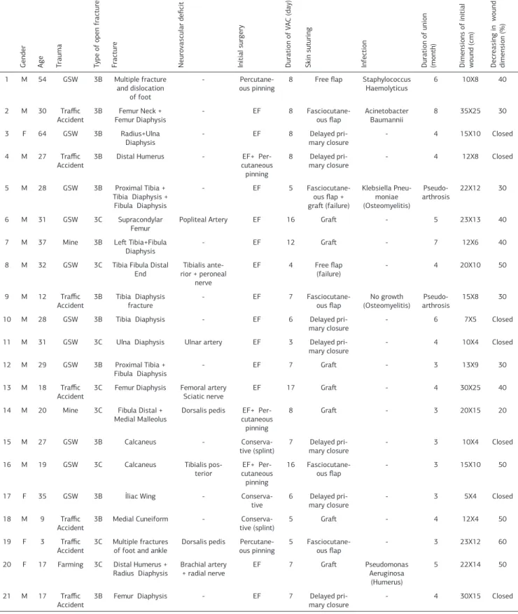
Table 1. Patient Information
Gender Age Trauma Type of open fracture Fracture Neuro
vascular deficit
Initial surgery Duration of V
AC (day)
Skin suturing Infection Duration of union (month) Dimensions of initial wound (cm) Decreasing in w
ound dimension (%) 1 M 54 GSW 3B Multiple fracture and dislocation of foot -
Percutane-ous pinning 8 Free flap Staphylococcus Haemolyticus 6 10X8 40 2 M 30 Traffic
Accident 3B Femur DiaphysisFemur Neck + - EF 8 Fasciocutane-ous flap Acinetobacter Baumannii 8 35X25 30 3 F 64 GSW 3B Radius+Ulna
Diaphysis - EF 8 mary closure Delayed pri- - 4 15X10 Closed
4 M 27 Traffic
Accident 3B Distal Humerus - cutaneous EF+ Per-pinning
8 Delayed
pri-mary closure - 4 12X8 Closed
5 M 28 GSW 3B Proximal Tibia + Tibia Diaphysis + Fibula Diaphysis - EF 5 Fasciocutane-ous flap + graft (failure) Klebsiella Pneu-moniae (Osteomyelitis) Pseudo-arthrosis 22X12 30 6 M 31 GSW 3C Supracondylar
Femur Popliteal Artery EF 16 Graft - 5 23X13 40
7 M 37 Mine 3B Left Tibia+Fibula
Diaphysis - EF 12 Graft - 7 12X6 40
8 M 32 GSW 3C Tibia Fibula Distal
End rior + peroneal Tibialis ante-nerve
EF 4 Free flap
(failure) - 4 20X10 50
9 M 12 Traffic
Accident 3B Tibia Diaphysis fracture - EF 7 Fasciocutane-ous flap (Osteomyelitis)No growth arthrosisPseudo- 15X8 30
10 M 28 GSW 3B Tibia Diaphysis - EF 6 Delayed
pri-mary closure - 6 7X5 Closed
11 M 31 GSW 3C Ulna Diaphysis Ulnar artery EF 3 Delayed
pri-mary closure - 4 10X4 Closed
12 M 29 GSW 3B Proximal Tibia +
Fibula Diaphysis - EF 7 Graft - 3 13X9 30
13 M 18 Traffic
Accident 3C Femur Diaphysis Femoral arterySciatic nerve EF 17 Graft - 4 30X25 40 14 M 20 Mine 3C Fibula Distal +
Medial Malleolus Dorsalis pedis cutaneous EF+ Per-pinning
8 Graft - 3 20X15 20
15 M 27 GSW 3B Calcaneus -
Conserva-tive (splint) 7 mary closure Delayed pri- - 3 10X4 Closed 16 M 19 GSW 3C Calcaneus Tibialis
pos-terior cutaneous EF+ Per-pinning
16
Fasciocutane-ous flap - 3 15X10 50
17 F 35 GSW 3B İliac Wing -
Conserva-tive 6 mary closure Delayed pri- - 3 5X4 Closed 18 M 9 Traffic
Accident 3B Medial Cuneiform - tive (splint)Conserva- 5 Graft - 4 12X4 50 19 F 3 Traffic
Accident 3C Multiple fractures of foot and ankle Dorsalis pedis ous pinningPercutane- 5 Fasciocutane-ous flap - 3 23X12 60 20 F 17 Farming 3C Distal Humerus +
Radius Diaphysis Brachial artery + radial nerve EF 7 Graft Pseudomonas Aeruginosa (Humerus)
5 22X14 50
21 M 17 Traffic
Accident 3B Femur Diaphysis - EF 7 mary closure Delayed pri- - 4 30X15 Closed EF: external fixator, GSW: Gunshot wound, VAC: Vacuum-assisted closure
of patients who underwent delayed primary closure (P=0.031). Delayed primary closure was applied to six out of thirteen pa-tients with Type IIIB open fractures (46.2%) and one out of eight patients with Type IIIC open fractures (12.5%). No signifi-cant difference was detected between Type IIIB and C cases in terms of necessity for delayed primary closure.
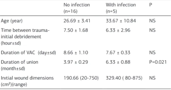
A deep infection developed in five (35.7%) out of fourteen pa-tients who underwent secondary closure, of whom two papa-tients (14.2%) developed osteomyelitis. None of the patients who un-derwent delayed primary closure (n=7) developed an infection (Table 3). Statistically significant difference was found between the primary and secondary closure in terms of deep infection (p=0.028). The microorganisms that caused infection were pseudomonas aeruginosa, acinetobacter baumannii, staphylo-coccus haemolyticus, and klebsiella pneumoniae. However, no microorganism could be isolated in one patient. The infection was treated through debridement and antibiotherapy in those patients. However one patient developed osteomyelitis refused further intervention. No statistically significant association was found between the dimensions of the first wound and develop-ment of infection (p=NS). No significant association was found between the development of infection and the duration of VAC application (p=NS). Trauma type and first treatment applied had no association with the development of an infection either. The duration of union was found to be significantly longer in patients who developed an infection (p=0.021). Pseudoarthrosis and osteomyelitis developed in two patients. In those patients debridement, antibiotherapy, and EF were performed. Union was achieved at the 19th month in one patient. However, in the other patient union could not be achieved. This patient refused further surgical intervention after a follow-up of 34 months. A G J K D F E B H C İ
Figure 2. AP direct X-ray obtained in the emergency department of a patient with Type IIIB open distal humerus fracture due to a traffic accident (A). Lateral direct X-ray obtained in the emergency department of the patient (B). Picture taken during the initial debridement (C). Postoperative AP radiograph of the patient who underwent external fixation + K wire application (D). Postoperative lateral radiograph of the patient (E). Shrinkage of the wound dimensions during VAC change (F). Appearance of the wound 1 month after the injury (G). Anteroposterior and lateral radiographs taken 4 months after the injury (H, I). Elbow flexion and extension 4 months after the injury (J,K).
Table 2. Comparison of closure techniques Delayed primary closure (n=7) Secondary closure (n=14) P Age (year±sd) 32.71 ± 5.60 24.92 ± 3.91 NS Time between trauma-initial
debridement (hour±sd)
3.71 ± 0.56 9.42 ± 2.09 P=0.007 Duration of VAC (day±sd) 6.43 ± 0.65 9.42 ± 1.34 NS Duration of union (month±±) 3.93 ± 0.38 4.58 ± 0.48 NS Inıtial wound dimensions
(cm²)(range)
118.71(20-450) 275.93 (48-275) P=0.031 Shrinkage (%) (range) 100 40.02 (20-60) p=0.001 NS: Non significant, VAC: Vacuum-assisted closure
Table 3. Comparison of patients by development of infection No infection
(n=16) With infection (n=5) P Age (year) 26.69 ± 3.41 33.67 ± 10.84 NS Time between
trauma-initial debridement (hour±sd)
7.50 ± 1.68 6.33 ± 2.96 NS
Duration of VAC (day±sd) 8.66 ± 1.10 7.67 ± 0.33 NS Duration of union
(month±sd) 3.97 ± 0.29 6.33 ± 0.88 P=0.021 Inıtial wound dimensions
(cm²)(range)
190.66 (20-750) 329.40 ( 80-875) NS NS: Non significant, VAC: Vacuum-assisted closure
VAC in open fracture
Discussion
The degree of soft tissue injury determines the treatment of Type III open fractures. If a wound cannot be closed primarily, a high risk of infection is the surgeon’s major concern [8]. Delayed primary closure or secondary closure with graft/flap may take a long time, possibly increasing the risk of infection [9].
The best way to decrease the risk of infection in open wounds is to remove tissues that are already infected or might provide a basis for infection with serial debridement. In this way the growth of bacteria in a medium of impaired blood flow could be prevented. VAC can be applied in those wounds to increase the efficacy of debridement. VAC is a safe technique for wound clo-sure that can be applied easily in such types of complex wounds [10]. It provides for the removal of interstitial fluid and debris through negative pressure. Thus, contaminated tissue fluid is removed from the fracture site during the period between the dressing changes and bacterial density in the tissue can be de-creased [11]. Also, local blood flow is inde-creased and oxygenation of the wound is provided [12]. VAC decreases the size of wounds that are difficult to close and provides an appropriate situation for secondary wound closure methods. In defects in the regions of weak soft tissue reserves such as the cruris, foot, and around the joints, good blood flow to the granulation tissue increases the success of grafts and flaps. Also, VAC increases cellular pro-liferation, angiogenesis, and microvascular blood flow and thus decreases edema and chronic inflammation, thereby increasing the success rate of grafts and contributing positively to wound healing [13]. VAC has been demonstrated to increase some mol-ecules and the expression of some cytokines with anabolic ac-tivity such as intracellular adhesion molecule-1 (MIF), vascular endothelial growth factor (VGEF), main fibroblast growth fac-tor, and type 1 collagen, thus decreasing the duration of wound healing and increasing the quality of healing [14]. In conclu-sion, VAC applications not only increase the efficacy of debride-ment but also prevent the contact of the open wound with the external environment. Moreover, the number of daily dressing changes, which are a misery for the patient, are decreased. Kilic et al. [15], applied VAC in the treatment of open wounds. They showed that VAC application decreases the duration of hospital stay and reduces the rate of infection and treatment costs. Stannard et al. [16] reported that the infection rate in their patients who received VAC was five times lower compared to the control group. In addition, with the closed circuit obsta-cle, bacteria that are present in the contaminated area are pre-vented from being transmitted to other patients and healthcare personnel in terms of hospital infections. Another advantage of VAC is that it prevents the open wound from being contaminat-ed by microorganisms of hospital origin. It is known that most of the pathogens that infect open wounds are not from the first trauma environment but through the hospital environment [17]. The VAC method, which was first applied to chronic wounds during the initial treatment, has also begun to be used safely in acute injuries. Govaert et al. [18] treated a pelvic fracture complicated by urogenital contamination. Hou et al. [19] re-ported that VAC decreased the duration of wound healing in 32 patients with Type III open tibial fracture and that application of local rotational flap was adequate in most patients and that VAC decreased the need for free flap procedures. However, they
also reported that negative effects of the procedure such as that rates of amputation and infection were increased when the duration of the VAC procedure exceeded a week. Concerns regarding long durations of VAC application were also reported by other researchers [20]. The mean duration of VAC in patients with and without development of infection in this present study was 7.67±0.33 days and 8.66±1.10 days, respectively. We found no significant association between the duration of VAC applica-tion and development of complicaapplica-tions.
In another study, Ulusal et al. [21] reported a significant de-crease in the wound dimensions with the use of VAC. In the present study, we also observed decreased wound dimensions in patients treated with VAC. In seven patients the wounds were closed with delayed primary repair. We performed efficient skin traction over the sponge following VAC application and sponge placement. We augmented the effect of the procedure to reduce the wound size. The dimensions of grafts or flaps required for the closure of the shrinking wounds are naturally decreased, which decreases the donor site morbidity. In addition, the dura-tion of surgical procedure is decreased and the procedure is made easier.
Conclusion
VAC application together with approximating sutures increases the comfort of the patient by decreasing the number of re-quired daily dressing changes, decreases the necessity of sec-ondary interventions by decreasing the wound dimensions, and decreases the donor site morbidity by decreasing the graft di-mensions when the wound is required to be closed secondarily. Ethical Statement
All procedures performed in studies involving human partici-pants were in accordance with the ethical standards of the institutional and/or national research committee and with the 1964 Helsinki declaration and its later amendments or compa-rable ethical standards.
Scientific Responsibility Statement
The authors declare that they are responsible for the article’s scientific content including study design, data collection, analy-sis and interpretation, writing, some of the main line, or all of the preparation and scientific review of the contents and ap-proval of the final version of the article.
Animal and human rights statement
All procedures performed in this study were in accordance with the ethical standards of the institutional and/or national re-search committee and with the 1964 Helsinki declaration and its later amendments or comparable ethical standards. No ani-mal or human studies were carried out by the authors for this article.
Funding: None Conflict of interest
None of the authors received any type of financial support that could be considered potential conflict of interest regarding the manuscript or its submission.
References
1. Alemdaroğlu KB, Tiftikçi U, Iltar S, Aydoğan NH, Kara T, Atlihan D. et al. Factors affecting the fracture healing in treatment of tibial shaft fractures with circular external fixator. Injury. 2009; 40(11):1151-6
2. Jordan DJ, Malahias M, Khan W, Hindocha S. The ortho-plastic approach to soft tissue management in trauma. Open Orthop J. 2014; 31:399-408.
3. Penn-Barwell JG, Rand BC, Brown KV, Wenke JC. A versatile model of open-fracture infection: a contaminated segmental rat femur defect. Bone Joint Res. 2014; 3:187-92.
4. Steiert AE, Gohritz A, Schreiber TC, Krettek C, Vogt PM. Delayed flap coverage of open extremity fractures after previous vacuum-assisted closure (VAC) therapy - worse or worth? J Plast Reconstr Aesthet Surg. 2009; 62:675-83.
5. Huang C, Leavitt T, Bayer LR, Orgill DP. Effect of negative pressure wound therapy on wound healing. Curr Probl Surg. 2014; 51:301-31.
6. Shin SJ, Han D, Song H, Jang YJ, Park DH, Park MC. Continuous high-pressure negative suction drain: new powerful tool for closed wound management: clinical experience. J Craniofac Surg. 2014; 25:1427-31.
7. Wei SJ, Cai XH, Wang HS, Qi BW, Yu AX. A comparison of primary and delayed wound closure in severe open tibial fractures initially treated with internal fixation and vacuum-assisted wound coverage: a case-controlled study. Int J Surg. 2014; 12:688-94.
8. Olesen UK, Juul R, Bonde CT, Moser C, McNally M, Jensen LT et al. A review of forty five open tibial fractures covered with free flaps. Analysis of complications, microbiology and prognostic factors. Int Orthop. 2015; 39:1159-66.
9. Lee JY, Jung H, Kwon H, Jung SN. Extended negative pressure wound therapy-assisted dermatotraction for the closure of large open fasciotomy wounds in nec-rotizing fasciitis patients. World J Emerg Surg. 2014; 15:29.
10. Gabriel A, Rauen B. Simplified negative pressure wound therapy with instilla-tion: advances and recommendations. Plast Surg Nurs. 2014; 34:88-92. 11- Payne C, Edwards D. Application of the Single Use Negative Pressure Wound Therapy Device (PICO) on a Heterogeneous Group of Surgical and Traumatic Wounds. Eplasty. 2014; 28:20.
12. Adogwa O, Fatemi P, Perez E, Moreno J, Gazcon GC, Gokaslan ZL et al. Nega-tive pressure wound therapy reduces incidence of postoperaNega-tive wound infection and dehiscence after long-segment thoracolumbar spinal fusion: a single institu-tional experience. Spine J 2014; 1:2911-7.
13. Zhang C, Liu D, Liang Z, Liu F, Lin H, Guo Z. Repair of refractory wounds through grafting of artificial dermis and autologous epidermis aided by vacuum-assisted closure. Aesthetic Plast Surg. 2014; 38:727-32.
14. Wang W, Pan Z, Hu X, Li Z, Zhao Y, Yu AX. Vacuum-assisted closure increases ICAM-1, MIF, VEGF and collagen I expression in wound therapy. Exp Ther Med. 2014; 7:1221-6.
15. Kiliç A, Ozkaya U, Sökücü S, Basilgan S, Kabukçuoğlu Y. Use of vacuum-as-sisted closure in the topical treatment of surgical site infections. Acta Orthop Traumatol Turc. 2009; 43:336-42.
16. Stannard JP, Volgas DA, Stewart R, McGwin G Jr, Alonso JE. Negative pressure wound therapy after severe open fractures: a prospective randomized study. J Orthop Trauma. 2009; 23:552–7.
17. Weitz-Marshall AD, Bosse MJ. Timing of closure of open fractures. J Am Acad Orthop Surg. 2002; 10:379-84.
18. Govaert G, Siriwardhane M, Hatzifotis M, Malisano L, Schuetz M. Prevention of pelvic sepsis in major open pelviperineal injury. Injury 2012; 43:533-6. 19. Hou Z, Irgit K, Strohecker KA, Matzko ME, Wingert NC, DeSantis JG, et al. De-layed flap reconstruction with vacuum-assisted closure management of the open IIIB tibial fracture. J Trauma. 2011; 71:1705-8.
20. Gopal S, Majumder S, Batchelor AG, Knight SL, De Boer P, Smith RM. Fix and flap: the radical orthopaedic and plastic treatment of severe open fractures of the tibia. J Bone Joint Surg Br. 2000; 82:959-66.
21. Ulusal AE, Sahin MS, Ulusal B, Cakmak G, Tuncay C. Negative pressure wound therapy in patients with diabetic foot. Acta Orthop Traumatol Turc. 2011; 45:254-60.
How to cite this article:
Atiç R, Alemdar C, Azboy İ, Bulut M, Çaçan MA, Gem M. Application of vac in type III open fractures. J Clin Anal Med 2018;9(6): 498-503.