Address for correspondence
Ezel Erşen, Department of Thoracic Surgery, Cerrahpaşa Medical Faculty, Istanbul University, 34098 Istanbul, Turkey, phone: +90 5057311567, e-mail: [email protected]
Introduction
Pectus excavatum (PE), also known as “funnel chest”, is the most common congenital chest wall anomaly associated with anterior chest wall defor-mity and depression, which affects approximately 1 : 400 live births [1].
Meyer was the first surgeon to try to repair the deformity, in 1911 [2]. He was followed by Sauer-bruch, who performed the first pectus repair using bilateral costal cartilage resection and sternal oste-otomy [3].
In 1949, Ravitch published his first paper, “The operative treatment of pectus excavatum,” and
de-Minimally invasive repair of pectus excavatum (MIRPE) in adults:
is it a proper choice?
Ezel Erşen1, Ahmet Demirkaya2, Burcu Kılıç1, Hasan Volkan Kara1, Osman Yakşi1, Nurlan Alizade1, Özkan Demirhan3,
Cem Sayılgan4, Akif Turna1, Kamil Kaynak1
1Department of Thoracic Surgery, Cerrahpaşa Medical Faculty, Istanbul University, Istanbul, Turkey 2Department of Thoracic Surgery, School of Medicine, Istanbul Acıbadem University, Istanbul, Turkey 3Department of Thoracic Surgery, Medical Faculty, Istanbul Bilim University, Istanbul, Turkey
4Department of Anesthesia and Reanimation, Cerrahpasa Medical Faculty, Istanbul University, Istanbul, Turkey
Videosurgery Miniinv 2016; 11 (2): 98–104 DOI: 10.5114/wiitm.2016.60456 A b s t r a c t
Introduction: The Nuss procedure is suitable for prepubertal and early pubertal patients but can also be used in adult patients.
Aim: To determine whether the minimally invasive technique (MIRPE) can also be performed successfully in adults. Material and methods: Between July 2006 and January 2016, 836 patients (744 male, 92 female) underwent correc-tion of pectus excavatum with the MIRPE technique at our institucorrec-tion. The mean age was 16.8 years (2–45 years). There were 236 adult patients (28.2%) (> 18 years) – 20 female, 216 male. The mean age among the adult patients was 23.2 years (18–45 years). The recorded data included length of hospital stay, postoperative complications, number of bars used, duration of the surgical procedure and signs of pneumothorax on the postoperative chest X-ray.
Results: The MIRPE was performed in 236 adult patients. The average operative time was 44.4 min (25–90 min). The median postoperative stay was 4.92 ±2.81 days (3–21 days) in adults and 4.64 ±1.58 (2–13) in younger patients. The difference was not statistically significant (p = 0.637). Two or more bars were used in 36 (15.8%) adult patients and in 44 (7.5%) younger patients. The difference was not statistically significant either (p = 0.068). Regarding the overall complications, complication rates among the adult patients and younger patients were 26.2% and 11.8% respectively. The difference was statistically significant (p = 0.007).
Conclusions: MIRPE is a feasible procedure that produces good long-term results in the treatment of pectus excava-tum in adults.
scribed 8 patients who were treated surgically with his technique [4].
Since then, several papers have been published regarding the modifications to Ravitch’s technique, although it maintained its prevalence as the stan-dard technique for the correction of PE. Yet, Ravitch’s technique also has some difficulties, as indicated by several reports which have highlighted its morbidi-ties and difficulmorbidi-ties [5].
In 1998, a new era began, when Nuss et al. re-ported their experience with 42 patients using a minimally invasive technique to correct the de-pression of PE [6].
Their technique was based on the use of a stain-less steel brace below the sternum, while there was no need for costal cartilage resection or sternal os-teotomy.
Since then, the Nuss technique has been used extensively by many surgeons, who focused on the surgical repair of the chest wall deformities. Several modifications in the Nuss technique have also been published. Some of them are currently in use (such as the modified Nuss technique described by Pile-gaard et al., which is applied routinely in our clinic for the correction of PE) [7].
However, there are also non-surgical interven-tions such as the vacuum bell and cosmetic inter-ventions such as the silicon implants and polyeth-ylene implants, which are manipulated in order to fix the deformity cosmetically. The vacuum bell can also be used intraoperatively to facilitate the retrosternal dissection and the insertion of the pectus bar [8, 9].
The Nuss technique is perfectly suitable for pre-pubertal and early pre-pubertal patients because of their chest wall compliance. While the technique can also be applied in adult patients due to the matured and rigid chest cavity, there are a number of problems including the prolonged operative time, increased rates of complications, bar displacement, higher pain rates and poor surgical outcome [10].
There is a tendency among surgeons not to oper-ate on adult patients unless they have severe defor-mity that causes pulmonary and cardiac problems.
Aim
The aim of our study was to determine wheth-er the minimally invasive technique (MIRPE) could be performed routinely and successfully not only in pediatric patients, but also in adults. In addition, we
evaluated asymptomatic adult patients with sym-metric and asymsym-metric deformities.
Material and methods
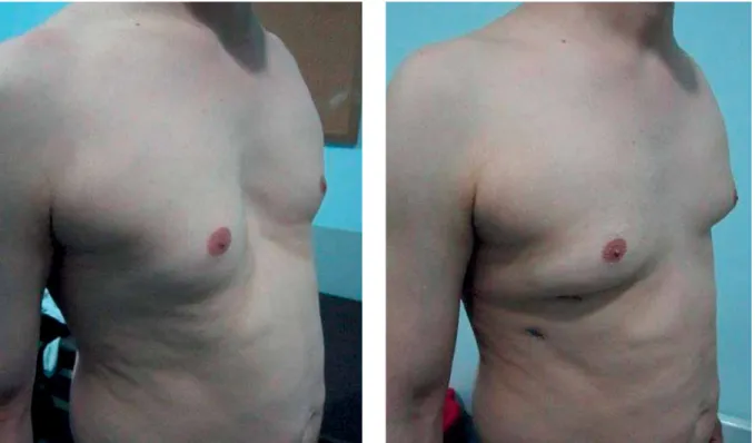
Between July 2006 and January 2016, 836 pa-tients (744 male and 92 female) underwent cor-rection of pectus excavatum with the MIRPE tech-nique at our institution. The mean patient age was 16.8 years (range: 2 to 45 years). All patients ob-tained an excellent cosmetic result (Photo 1). There were 236 adult patients (28.2%) (> 18 years) – 20 fe-male, 216 male. The mean age among the adult pa-tients was 23.2 years (range: 18–45 years). Patient demographics and preoperative characteristics are summarized in Table I. In all cases, a preoperative computed tomography (CT) scan was performed in order to evaluate the intrathoracic cavity for surgical planning. Echocardiography and pulmonary function tests were also performed in order to evaluate the cardiac and pulmonary performance. In addition, a preoperative nickel allergy test was performed. Indications were psychosocial complaints due to cosmetic appearance, reduced exercise tolerance, dyspnea on exertion and chest pain. The Haller in-dex (HI) was also used as an indication for surgery. HI greater than 3 was classified as severe deformity. The HI was calculated with CT. The HI of the patients ranged from 3.30 to 11 (mean: 4.4). All patients were operated on by the same surgeon. The data were retrospectively collected and analyzed. The recorded data included the length of hospital stay, postopera-tive complications, number of bars used, duration of the surgical procedure and signs of pneumothorax on the routine postoperative chest roentgenogram.
Surgical technique
All patients were positioned in the supine posi-tion with both arms abducted. An epidural catheter was placed for postoperative pain management be-fore the general anesthesia. Double-lumen intuba-tion was used routinely except for patients younger than 10 years of age. In this group of patients, the operation was carried out using apnea intervals.
The deepest point of the deformity, xiphoid and entry and exit points of the bar were marked on the skin. The incision for the scope was made from the right lateral side at the mid-axillary line and just below the level of the nipple to visualize the chest cavity cranially and caudally. A 5-mm trocar was
in-troduced into the thorax and the deepest point of the deformity was defined using a 30° scope.
A template (Zimmer Biomet Inc., Warsaw, Indi-ana, USA) was formed in order to make a model of
how the chest was intended to look after the cor-rection. Following this, a Pectus Support Bar (Zim-mer Biomet Inc., Warsaw, Indiana, USA) was bent to match this template. Having employed the modified Nuss technique as described by Pilegaard et al. [7, 11], we used bars that were shorter than the bars described by Nuss. Using a shorter bar, the stabilizer could be placed closer to the exit of the pectus bar from the thoracic cavity. This modification is believed to decrease the occurrence of bar displacement.
Lateral incisions for introducing the bar were ap-proximately 2 cm on the right side and 3 cm on the left side. We stabilized all bars on the left side.
With lateral incisions, a subcutaneous tunnel was created with blunt dissection, through the entry and exit points of the bar. A steel introducer (Zimmer Biomet Inc., Warsaw, Indiana, USA) was inserted into the thoracic cavity at the level of the entry point. It was pushed below the sternum and just above the pericardium securely using videothoracoscopy. An umbilical tape was secured to the tip of the intro-ducer, while the introducer was withdrawn with its convex side facing down. This resulted in the tunnel-ing of the umbilical tape from left to right. The tape was tied to the tip of the bar. With the guidance of the umbilical tape, the bar was passed through the
Photo 1. Pre- and postoperative images of a 45-year-old patient who underwent MIRPE
Table I. Demographic variables and preoperative characteristics of adults patients (n = 236)
un-dergoing MIRPE
Characteristic Results
Age, mean [years] 23.2 (18–45)
Gender, n (%):
Male 216 (91.5)
Female 20 (8.4)
Depth of defect (Haller index) 4.4 (3.3–11) Preoperative symptoms (patients), n (%):
Cosmetic concern 126 (53)
Dyspnea on exertion 16 (6)
Shortness of breath at rest 0
Cardiac arrhythmia, palpitations 0
Chest pain 38 (16)
Fatigue, decreased energy 27 (11)
Electrocardiogram changes 8 (3)
thorax from the right to the left incision in a con-cave-up position. The bar was flipped 180° in order to buttress the sternum and correct the deformity.
A stabilizer was placed on the left side of the bar as closely as possible to the entry into the thorac-ic cavity to avoid rotation. A no. 5 sternal wire was used to fix the stabilizer to the bar on the left side. The bar was also secured on the right side using one no. 1 polydioxanone (PDS) (No. 1 Pedesente Doğsan, Trabzon, Turkey) suture around the ribs. Using ad-ditional absorbable 2-0 vicryl sutures, the bar was fixed to the adjacent tissue.
Additional bars were implanted as a single bar did not provide satisfactory cosmetic correction and the HI was greater than 5. We also placed the bars asymmetrically in patients with asymmetric defor-mity.
Later, a slim 14 Fr silicone tube was inserted into the pleural cavity through the trocar site. The prox-imal end of the silicone tube was placed in a small cup of saline solution in order to form a modified underwater seal device. The lung was reinflated, intrathoracic air was evacuated and lung re-expan-sion was controlled with the videothoracoscope at the end of the procedure. The tube was withdrawn, while the anesthesiologist applied positive end-expi-ratory pressure.
A chest X-ray was taken on the same day of the surgery in order to evaluate the presence of pneu-mothorax. All patients took antibiotics intravenously for 3 days.
Pain management was the most important issue following the surgery. It was managed with an epi-dural catheter for the first 2 days. On the third day, the catheter was removed and nonsteroidal anti-in-flammatory drugs (NSAID) and myorelaxant drugs were administered orally for 5 weeks postoperative-ly. The patient was monitored in the outpatient clinic 1 week after surgery for clinical evaluation with an X-ray and 1 month after surgery for general evalua-tion. After 2.5–3 years, we called the patients back for removal of the implanted system.
For the first 6 weeks, we did not allow the patient to carry a heavy weight (more than 2 kg in front of the body or more than 5 kg on the shoulders). Cy-cling and rotation of the upper body of more than 15° were also prohibited. The patient was also re-quested to sleep in the supine position without turn-ing to either side. In addition, heavy contact sports (e.g. boxing, hockey, and self-defense sports such as
karate and judo) were all forbidden until the removal of the bar.
Statistical analysis
Statistical analysis was performed using Pear-son’s χ2 testfor bivariate analysis. All statistical
anal-yses were performed using IBM SPSS Statistics, ver-sion 20.0 (IBM Corp., Armonk, N.Y.). Values of p < 0.05 were considered statistically significant.
Results
The modified Nuss operation was performed in all 236 adult patients. The median length of the bars was 11 inches (range: 9–14 inches) for adults and 10 inches (range: 7–14 inches) for younger patients. The average operative time was 44.4 min (range: 25–90 min).
The median postoperative stay was 4.92 ±2.81 days (range: 3–21 days) in adults and 4.64 ±1.58 (2–13) in younger patients. The difference was not statistically significant (p = 0.637). Two or more bars were used in 36 (15.8%) adult patients and 44 (7.5%) younger patients. These figures were not statistically significant either (p = 0.068). Intraopera-tive variables and characteristics of hospital stay are summarized in Table II.
There were no perioperative deaths. Cardiac inju-ry developed in one case where a small ventricular defect was repaired rapidly with anterior thoracot-omy. One patient developed aspiration pneumonia which was treated with antibiotics. Seven (2%) pa-tients had asymptomatic residual pneumothorax
Table II. Intraoperative variables and character-istics of hospital stay (adult patients, n = 236)
Characteristic Result
Operative time [min] 44.2 (25–90)
Estimated blood loss [ml] 25 (5–500)
Bars placed, n (%): 1 200 (84) 2 or more 36 (15) Mortality, n (%) Intraoperative death 0 (0.0) 30-day mortality 0 (0.0)
which was resolved without chest tube placement. Postoperative pleural effusion was detected in 2 pa-tients. The effusions were minimal and they were resolved spontaneously. Wound infection developed in 4 patients. Wound care and antibiotics were suf-ficient for recovery in this group. One patient devel-oped thoracic outlet syndrome after the correction of the deformity. The first rib caused severe obstruc-tion of the right subclavian artery. The resecobstruc-tion of the first rib and the division of the anterior scalene muscle and fibrous bands provided complete relief in this case. Bar displacement developed in 12 (5%) adult patients, while it was observed in 5 (0.8%) younger patients. The difference between the two groups was statistically significant (p < 0.05). All complications related to the surgical procedure are provided in Table III.
Regarding the overall complications, the compli-cation rates among the adult patients and younger patients were 26.2% and 11.8% respectively. The dif-ference was statistically significant (p = 0.007).
Discussion
The surgical treatment of pectus excavatum has changed in the last few decades. Although the Ravitch procedure is still a widely used approach for adult patients with pectus excavatum, there is an increasing trend for using the minimally invasive technique also known as the Nuss procedure. There are sufficient data regarding the use of the Ravitch repair in adult patients [12–16], although the treat-ment of pectus excavatum with the minimally inva-sive technique in adults is still controversial. Many surgeons still recommend the use of the Ravitch approach, since there are problems such as the fre-quent need for multiple bars and higher rate of com-plications that accompany the minimally invasive approach.
The minimally invasive technique has been used for pectus excavatum repair since 1998 [6], although only a limited number of studies have been pub-lished on this topic.
In 2002, Coln et al. reported their early expe-rience with the minimally invasive technique in adults. Eight patients (5 male and 3 female), aged between 19 and 32 years, underwent the minimal-ly invasive Nuss repair [17]. They used one bar with one or two stabilizers. The mean operative time was 1.32 h, while the mean hospital stay was 4 days. There were no early complications, although in one case the separation of a stabilizer required reoper-ation, while in another case bar displacement was reported as a late complication. Although the short time results were encouraging, this was a small se-ries with a short follow-up period.
Kim et al. [18] reported their experience with the minimally invasive pectus repair with 51 patients. They assigned the patients to three different age groups. Twelve patients were older than 20 years, and 12 patients were aged between 12 and 20 years. Two bars and lateral stabilizers were used routinely in the adult patients, while two bars were used only in 9 adolescents. The mean operative times were longer in the adult group (127.3 ±44.9 min). The mean hospital stay was 10 ±8.5 days for adult pa-tients. These values are very high compared with our results. In our study, the operative time was 44.4 min and the mean hospital stay was 4.92 ±2.81 days.
The postoperative complication rate in the ado-lescent and adult groups was 58.3%, and bar rota-tion was reported in 8.3% of adolescents and 33.3% of adults.
Table III. Complications
Complications N (%)
Bar displacement 12 (5)
Cardiac injury 1 (0.4)
Thoracic outlet syndrome 1 (0.4)
Wound infection 4 (1)
Pneumonia 1 (0.4)
Removal of stabilizer pain 0
Sternotomy/thoracotomy 1 (0.4)
One more bar 2 (0.8)
Removal before time due to pain 0
Pneumothorax 7 (2)
Pneumothorax which needed drainage 0
Pleural effusion 2 (0.8)
Pleural effusion which needed drainage 0
Prolonged pain 4 (1)
Readmission for pain 0
Recurrence 0
Respiratory distress 0
Bleeding requiring transfusion or reoper-ation
In our study, bar displacement developed in 5% of the adult patients, whereas the complication rate in the same group was 26.2%.
Recent reports have demonstrated more promis-ing outcomes in adults [19–21]. Pilegaard published his experience with 52 adult patients who were over 30 years of age [21]. The median age of the patients was 37 (range: 30–53). One bar was used in 15 (29%) patients, while two bars were used in 35 patients, and three bars were used in 2 patients. Two stabilizers were used in 10% of the bars. The median duration of surgery was 60 min and medi-an postoperative hospital stay was 4 days. Although 25 patients had pneumothorax, only 1 of them re-quired a chest tube. There was no bar rotation, while 1 patient with lateral migration of the bar was erated. The bars were removed early because reop-eration caused infection and the patient refused to take antibiotics for a long time.
In another study, Teh et al. evaluated the results of 19 patients aged 17 years or above [22]. The mean operative time was 2.1 ±0.2 h. Twelve patients required two bars, while one bar was used in 7 tients. We used two or more bars in 36 (15.8%) pa-tients in adults. The mean hospital stay was 5.8 days, which was similar to our result. One patient de-veloped pneumonia and 6 patients had residual pneumothorax which resolved without chest tube placement. Pneumothorax rates were quite high in comparison with our study. We believe this is mainly due to our evacuation technique, where we used an underwater seal mechanism.
In 2008, Pilegaard and Licht published a larger study including 180 adult patients [11]. The patients were aged 18 years or above (mean: 22 years). One hyndred and sixty of them were male patients. They used two bars in 57 patients and three bars in 2 pa- tients. They found that more than one bar was used in adults compared with younger patients and re-ported it as statistically significant. In our study, we also found that more than one bar was used fre-quently in adults, but we found that this was not statistically significant.
The median duration of the procedure was 41 min, which was very similar to our operative time, since we performed the same modified technique as described by Pilegaard. The median hospital stay – 5 days (3 to 29 days) – was also similar. The hospital stay was not significantly longer compared with the younger patients. The mean hospital stay was 4.92
±2.81 days in our study, which was not significantly different in comparison with the younger patients, who had a mean hospital stay of 4.64 ±1.58 days.
They detected pneumothorax in 86 (48%) cases, while 4 patients required tube drainage. In our study, there were only 7 (5%) patients with pneumothorax, which resolved spontaneously. Other complications included pneumonia in 4 patients, pleural effusion in 4 patients, empyema in 1 patient, seroma in 1 pa- tient and deep infection in 5 patients. Three (2%) patients underwent reoperation because of the dis-location of the bar.
In 13 (7%) patients, the stabilizer was removed early due to intolerable pain. In our study, we did not remove any bars or stabilizers due to pain. Yet, 12 (5%) patients underwent reoperation for bar dis-placement.
Absorbable stabilizers were used in 8 patients. We did not experience any displacement or rotation in the last 3 years. The mean duration of bar appli-cation was 36 ±4.6 months (30–48 months). One hundred and thirty-six (57%) patients had their bars removed, while there was no recurrence.
Pain appears to be the major problem following the minimally invasive pectus repair. It is believed that there is greater stress in all ribs in adults com-pared to children after the Nuss procedure [23].
On the other hand, force distribution in adults is more diffuse and often frequently located in the posterior part of the chest wall. Moreover, there are papers which report the increasing use of analgesics and narcotics in older patients [24].
We managed the pain in adult patients with the same amount of analgesics that we used in younger patients. As also reported in the literature [25], we found that patients with more than one bar had less pain in the adult group.
We also experienced a very rare problem in one of the patients. The patient developed vascular tho-racic outlet syndrome after the correction of the de-formity. The first rib caused severe obstruction of the right subclavian artery. The patient was treated with resection of the first rib and division of the anterior scalene muscle and fibrous bands [26].
Conclusions
Data from our present clinical experience indicate that many adults with pectus deformities can be op-erated on using the minimally invasive technique.
We can achieve the same good results as the younger patients with the same operative time as well as the same number of bars. Although complica-tions are quite high in comparison with the younger patients, patients do not have to stay longer after the operation compared with the younger patients.
In conclusion, minimally invasive pectus repair for the treatment of pectus excavatum is feasible and provides good long-term results in adult pa-tients. As surgeons become more experienced, com-plications can be minimized and improved outcomes can be achieved.
Conflicts of interest
The authors declare no conflict of interest.
References
1. Johnson WR, Fedor D, Singhal S. Systematic review of surgical treatment techniques for adult and pediatric patients with pectus excavatum. J Cardiothor Surg 2014; 9: 25.
2. Meyer L. Für chirurgischen behandlung der angeborenen trich-terbrust. Klin Wochenschr 1922; 1: 647.
3. Sauerbruch F. Operative beseitigung der angeborenen trichter-brust. Deutsche Zeitschr f Chir 1931; 234: 760.
4. Ravitch MM. The operative treatment of pectus excavatum. Ann Surg 1949; 129: 429-44.
5. Kelly RE Jr. Pectus excavatum: historical background, clinical picture, preoperative evaluation and criteria for operation. Semin Pediatr Surg 2008; 17: 181-93.
6. Nuss D, Kelly RE Jr, Croitoru DP, et al. A 10 year review of a min-imally invasive technique to the correction of pectus excava-tum. J Pediatr Surg 1998; 33: 545-52.
7. Pilegaard HK, Licht PB. Early results following the Nuss opera-tion for pectus excavatum: a single-instituopera-tion experience of 383 patients. Interact Cardiovasc Thorac Surg 2008; 7: 54-7. 8. Snel BJ, Spronk CA, Werker PM, et al. Pectus excavatum
recon-struction with silicone implants: long-term results and a review of the English-language literature. Ann Plast Surg 2009; 62: 205-9. 9. Haecker FM, Sesia SB. Intraoperative use of the vacuum bell for
elevating the sternum during the Nuss procedure. J Laparoen-dosc Adv Surg Tech A 2012; 22: 934-6.
10. Del Frari B, Schwabegger AH. Clinical results and patient sat-isfaction after pectus excavatum repair using the MIRPE and MOVARPE technique in adults: 10-year experience. Plast Re-constr Surg 2013; 132: 1591-602.
11. Pilegaard HK, Licht PB. Routine use of minimally invasive sur-gery for pectus excavatum in adults. Ann Thorac Surg 2008; 86: 952-7.
12. Fonkalsrud EW, Dunn JC, Atkinson JB. Repair of pectus excava-tum deformities: 30 years of experience with 375 patients. Ann Surg 2000; 231: 443-8.
13. Fonkalsrud EW, Bustorff-Silva J. Repair of pectus excavatum and carinatum in adults. Am J Surg 1999; 177: 121-4.
14. Mansour KA, Thourani VH, Odessey EA, et al. Thirty-year expe-rience with repair of pectus deformities in adults. Ann Thorac Surg 2003; 76: 391-5.
15. Genc O, Gurkok S, Gozubuyuk A, et al. Repair of pectus defor-mities: experience and outcome in 317 cases. Ann Saudi Med 2006; 26: 370-4.
16. Fonkalsrud EW, De Ugarte D, Choi E. Repair of pectus excava-tum and carinaexcava-tum deformities in 116 adults. Ann Surg 2002; 236: 304-14.
17. Coln D, Gunning T, Ramsay M, et al. Early experience with the Nuss minimally invasive correction of pectus excavatum in adults. World J Surg 2002; 26: 1217-21.
18. Kim DH, Hwang JJ, Lee MK, et al. Analysis of the Nuss procedure for pectus excavatum in different age groups. Ann Thorac Surg 2005; 80: 1073-7.
19. Olbrecht VA, Arnold MA, Nabaweesi R, et al. Lorenz bar repair of pectus excavatum in the adult population: should it be done? Ann Thorac Surg 2008; 86: 402-8; discussion 408-9.
20. Hanna WC, Ko MA, Blitz M, et al. Thoracoscopic Nuss procedure for young adults with pectus excavatum: excellent midterm results and patient satisfaction. Ann Thorac Surg 2013; 96: 1033-6; discussion 1037-8.
21. Pilegaard HK. Extending the use of Nuss procedure in patients older than 30 years. Eur J Cardiothorac Surg 2011; 40: 334-7. 22. Teh SH, Hanna AM, Pham TH, et al. Minimally invasive repair for
pectus excavatum in adults. Ann Thorac Surg 2008; 85: 1914-8. 23. Nagasao T, Miyamoto J, Tamaki T, et al. Stress distribution on
the thorax after the Nuss procedure for pectus excavatum re-sults in different patterns between adult and child patients. J Thorac Cardiovasc Surg 2007; 134: 1502-7.
24. Grosen K, Pfeiffer-Jensen M, Pilegaard HK. Postoperative con-sumption of opioid analgesics following correction of pectus excavatum is influenced by pectus severity: a single-centre study of 236 patients undergoing minimally invasive correction of pectus excavatum. Eur J Cardiothorac Surg 2010; 37: 833-9. 25. Nagaso T, Miyamoto J, Kokaji K, et al. Double bar application
de-creases postoperative pain after the Nuss procedure. J Thorac Cardiovasc Surg 2010; 140: 39-44, 44.e1-2.
26. Kılıç B, Demirkaya A, Turna A, et al. Vascular thoracic outlet syndrome developed after minimally invasive repair of pectus excavatum. Eur J Cardiothorac Surg 2013; 44: 567-9.