doi:10.3944/AOTT.2012.2592
Correspondence: Neslihan Aksu, MD. ‹stanbul Bilim Üniversitesi T›p Fakültesi, Florence Nightingale Hastanesi, Mehmetçik Cad. Cahit Yalç›n Sok. No: 1, Mecidiyeköy, ‹stanbul, Turkey.
Tel: +90 212 - 212 88 11 e-mail: [email protected] Submitted: December 28, 2010 Accepted: October 30, 2011 ©2012 Turkish Association of Orthopaedics and Traumatology
Available online at www.aott.org.tr doi:10.3944/AOTT.2012.2592 QR (Quick Response) Code:
Objective: Our aim was to evaluate the results of minimally invasive plate osteosynthesis (MIPO)
using locking plates in diaphyseal humerus and proximal humerus fractures.
Methods: Nine patients who underwent open reduction and MIPO for the treatment of diaphyseal
and proximal humerus fractures between June 2006 and October 2009 were included in this study. One S3®
and 8 PHILOS®
plates were used. Mean age was 75.2 (range: 32 to 86) years and all patients were females. Mean follow-up was 33.9 (range: 14.8 to 54.8) months. According to AO/ASIF classifi-cation, four patients had 12C1, two patients 12A1, one patient 12A2, and two patients 11A2 fractures. Axillary and radial nerves were explored and protected in all patients. Patients were evaluated radi-ographically for union and functionally using the Constant-Murley score.
Results: None of the patients had nonunion, avascular necrosis, axillary or radial nerve paralysis or
implant failure. Mean Constant-Murley score was 86.8±2.2 (range: 70 to 100). Mean union time was 3.2 (range: 2.5 to 5) months.
Conclusion: MIPO of humerus diaphysis and proximal fractures allows for preservation of blood
sup-ply in fracture fragments, owing to less soft tissue and periosteal injury. When the procedure is per-formed with the lateral double incision, exposure and preservation of the axillary and radial nerves are necessary. Early return of function in the shoulder and elbow joints and favorable healing time are the major advantages of this method in this rare subset of humerus fractures.
Key words: Fracture; humerus; minimally invasive; MIPO; osteosynthesis; PHILOS® .
Treatments performed to preserve the blood supply of the bone using internal splinting, bridging and new plate designs providing limited contact with the bone are referred to as biological flexible fixation methods. These methods emphasize the importance of soft tissue on fracture healing and do not compromise circulation by using anatomic reduction to achieve a rigid fixation. In the indirect approach, acceptable reduction of the fracture is advised to provide movement of the frac-tured tips with a relatively stable fixation. Alignment of
the fracture by bridging is favored instead of absolute rigid fixation with compression. The use of indirect reduction for alignment of the fractures minimizes soft tissue stripping.[1]
In addition, indirect reduction reduces surgical trauma and promotes formation of the callus by “biological flexible fixation”. Biological flexi-ble fixation is achieved with minimal bone-implant con-tact, long-distance bridging and locked internal fixators with fewer screws. Implants prevent micro movements during fixation with plates, which provides absolute
sta-Minimally invasive plate osteosynthesis (MIPO) in
diaphyseal humerus and proximal humerus fractures
Neslihan AKSU, Sinan KARACA, Ayhan Nedim KARA, Zekeriya U¤ur IfiIKLAR
bility. Biological flexible fixation tolerates the micro movements between the fracture fragments, and in fact, requires some motion for union. This micro movement is preserved by using locking screws between the bone and implant.[2]
By doing so, the method does not com-promise an early and full recovery.[3]
Nevertheless, a live bone is required to achieve successful results.[2]
In their experimental study on sheep femurs, Baumgaertel et al. investigated the radiological, biomechanical and microangiographic differences between anatomic rigid, fixation and biological (bridging) fixation, and proved that indirect reduction and bridge plating was superior to rigid anatomic fixation employed by direct reduc-tion.[4] Mineralization in fracture callus occurs faster
and more effectively in indirect reduction, compared to anatomic reduction.[4]
In a study on cadavers, Farouk et al. demonstrated that conventional plating techniques impaired the blood flow to the bone more than the minimally invasive approach that utilized percutaneous plates.[5]
Preserved blood flow in biological plating tech-niques may lead to clinical results, such as delayed unions, recurrence of the fracture, decrease in infection incidences, as well as increase in union rates and decrease in graft utilization.
In plate applications, incisions are made on the distal end of the fracture line. In diaphyseal fractures, the plate is moved above the periosteum and placed percuta-neously to bypass the fracture line, and screws are placed through the proximal and distal incisions (bridge-plat-ing). Today, the minimally invasive plate osteosynthesis (MIPO) approach, as defined by Krettek et al. is favored, whereas wide incisions without exposing the fracture line had been earlier preferred in biological fixations.[6-9]
In the MIPO method, minimal incisions are performed to place and screw the plate away from the fracture line. After fracture reduction and extremity alignment are confirmed by fluoroscopy, the plate is moved above the periosteum through these incisions. The plate is then screwed through the proximal and distal incisions. It is not essential to shape the plate to exactly fit the contours of the bone in plate fixations. In this method, implants providing minimal surface contact with the bone are preferred.[6-9]
Although conservative treatment returns successful results of 90% in diaphyseal fractures of the humerus, there are absolute and relative surgical treatment indica-tions in cases, such as multiple traumas, open fracture, bilateral fracture, floating elbow, and obesity. Soft tissue interposition and circulatory disorders are more com-monly seen in segmental fractures of the humerus when
compared to other fractures. The incidence of nonunion increases if these types of fractures are treated conserva-tively.[10]
In treatment of diaphyseal fractures of the humerus with open reduction internal fixation, a nonunion rate of 5.1% and radial nerve palsy complication rate of 5.8% to 17.6% can be expected.[11-13]Today, MIPO is a
popular treatment method in long bone surgeries, returning lower complication rates.[13-16]
In our study, we evaluated the results of open reduction and locked plate fixation using the minimal-ly invasive double incision in diaphyseal fractures of the humerus and proximal diaphyseal fractures.
Patients and methods
Nine patients who underwent MIPO between June 2006 and October 2009 were retrospectively evaluated. The mean age of the patients was 75.2 (range: 32 to 86) years. One patient had a proximal fracture, one had a diaphyseal fracture and the remaining 7 had proximal diaphyseal fractures. According to AO/ASIF classifica-tion, 4 patients were classified as Type 12C1 (of which 3 had accompanying fractures of Type 11B3, 11A2 and 11A1), 2 were classified as Type 12A1, another 2 as Type 11A2, and one as Type 12A2.
Patients were seated in the beach chair position at an angle of 60° and the C-arm was placed across the patient. The first incision was performed longitudinally, starting from the anterolateral edge of the acromion to 6-8 cm downwards. The deltoid muscle was freed from the avas-cular fibrosis raphe between the anterior and medial del-toid head. The axillary nerve was suspended and pre-served. Then, the skin and subcutaneous tissue were laid open with a distal lateral incision of 6-8 cm, depending on the level the fracture distally reached. The radial nerve was found between the brachialis and brachioradi-alis muscles with blunt dissection, suspended and pre-served (Fig. 1). To prevent axillary and radial palsy caused by compression and stretching, Parabeuf retrac-tors were used instead of retracretrac-tors with lever arms, e.g. Hohmann (Fig. 1). Rotational movements were execut-ed with the forearm in supination, elbow in 70° flexion, and shoulder in 60° abduction. Thus, the axis was fixed with traction applied from the elbow as the biceps mus-cle was aligned perpendicular to the bicondylar axis. The reduction was verified on anteroposterior and lateral flu-oroscopic images. A submuscular tunnel, starting over the periosteum toward the distal, was prepared with an elevator applied through the proximal incision. A 4.5 mm locking screw guide applied at the locked plate was used as a handle to distally move the plate from the
prox-imal incision and place it in the tunnel opened over the periosteum. In 2 patients S3®(DePuy, Kirkel-Limbach,
Germany) plates (Fig. 2) and in 7 patients PHILOS®
(Proximal Humerus Internal Locked System; Synthes-Stratec Medical, Oberdorf, Switzerland) plates were used for fixation. First the proximal, then the distal frag-ments were fixed to the plate using 4.5 mm locking screws. A minimum of 3 to 4 screws were used for each fracture fragment fixation. Mitek anchor sutures (DePuy, Johnson and Johnson, Boston, MA, USA) were additionally used in 2 patients for rotator cuff repair (Fig. 2). A drain was placed through the distal incision in all patients and removed one day after surgery. The average drainage was 60 (range: 40 to 120) cc. All patients wore a shoulder sling for 3 weeks. Wrist, active and passive elbow movements and shoulder pendulum exercises
were begun the first week as a part of standard fracture rehabilitation course. Radiological and functional evalu-ations of the patients took place on the postoperative 3rd and 6th weeks and 3rd and 6th months. Anteroposterior and lateral follow-up radiographs were taken. When a callus was observed in three of four (medial and lateral, anterior and posterior) cortexes, it was considered as a union. The mean follow-up time was 33.9 (range: 14.8 to 54.8) months. The 3rd, 6th and 12th month Constant scores were statistically evaluated according to Dunn's (multiple comparison) test.
Results
Nonunion, avascular necrosis, axillary or radial nerve palsy was not observed in any patient. Average surgery time was noted as 72.6 (range: 66 to 150) minutes. The
Fig. 1. (a) Axillary nerve is preserved in proximal and radial nerve in distal incisions; (b) plate application through proximal incision; (c) both inci-sions are closed. [Color figure can be viewed in the online issue, which is available at www.aott.org.tr]
(a) (b) (c)
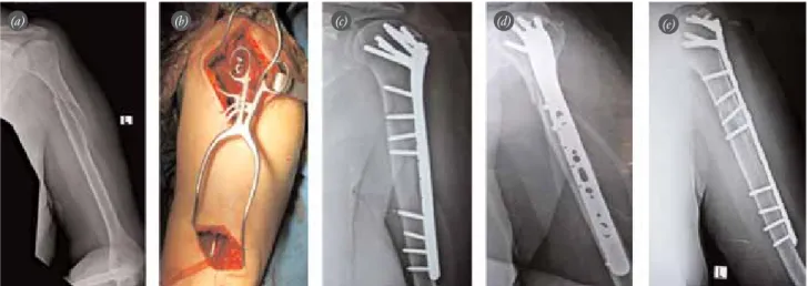
Fig. 2. Views of 82-year-old female patient. (a) Type 11A1 and 12C12 fractures are seen. (b) Surgical intervention. (c, d) Early postoperative radiographs showing fixation with S3®humerus plate and rotate cuff repair with anchor suture. (e) Union is seen on the 3rd month radi-ograph. [Color figure can be viewed in the online issue, which is available at www.aott.org.tr]
mean Constant-Murley score was 86.8±2.2 (range: 70 to 100). The average time to union was 3.2 (range: 2.5 to 5) months (Figs. 3 and 4). All patients had an abduction above 90° at the end of the 3rd and above 150° at the 6th month (Fig. 3). The mean Constant-Murley score was 64.8±1.7 at the 3rd, 73.5±1.3 at the 6th and 86.8±2.2 at the end of the 12th month. According to Dunn’s test, there was a significant difference between the Constant scores taken at the above intervals (p=0.0001). The Constant score at the end of first year was statistically higher than those measured at the 3rd and 6th months (p=0.007), and the Constant score at the 6th month was higher than the score the 3rd month (p=0.007). Mayo
wrist (performance) score was excellent in all patients at the end of the 3rd month (mean: 92.5).
Discussion
Fixations that allow for identifiable movements of frac-ture fragments under functional loading are considered to be relatively flexible fixations.[17] Short plates are
exposed to long bending forces; however, the bending stress disperses in long plates, making the fixation flex-ible.[18]
In the minimally invasive intervention per-formed with lateral double incision, full reduction and compression of the fractured fragments is not accom-plished. Thus, this fixation is relatively flexible. In
Fig. 3. (a-c) Range of movement at the 3rd postoperative month. [Color figure can be viewed in the online issue, which is available at www.aott.org.tr]
(a) (b) (c)
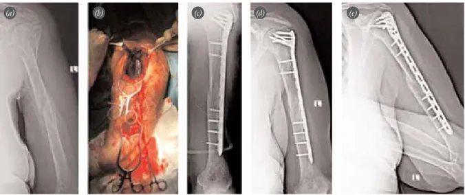
Fig. 4. Views of 70-year-old female patient. (a) AO Type 11B2 and 12C22 fractures. (b) Surgical intervention. (c) Early postoperative radiograph showing fixation with PHILOS® plate. Union is seen on the 3rd month on (d) anteroposterior and (e) lateral radi-ographs. [Color figure can be viewed in the online issue, which is available at www.aott.org.tr]
these types of fixations, the fracture line heals with the formation of a callus that fast bridges the fractured tips. Healing by means of callus is faster and more effective than direct healings in absolute hard fixations with no callus. The soft tissue and periosteum damage is limit-ed as the fracture site is not openlimit-ed, and this explimit-edites union by preventing the impairment of blood flow at the fracture ends.[17]
In contrast to those advantages, nonunion, malalignment, problems with implants, and skin issues regarding these methods have also been reported in the literature.[19-23]
Interposition of the soft tissue between fractured pieces and circulatory disorder among the segmental pieces are more commonly noticed in segmental humerus fractures than other fractures. Open surgical treatment of the segmental fractures may result in nonunion as the already impaired circulation of the seg-mented piece can worsen due to soft tissue stripping.[10]
In MIPO, soft tissues interposed between the segmental fractured pieces can be removed with an elevator to achieve reduction. However, it is important to not strip the soft tissue off the fractured fragments.
As there is no need to lay out the radial nerve in MIPO applications, the plates for humerus diaphyseal fractures have been applied on the anterior face of the humerus in both cadaveric and clinical studies in the literature.[13,16,18,24,25] In diaphyseal fractures extending
toward the proximal end, it would be more appropriate to place the plate laterally in MIPO. This enables the reduction of the proximal piece abducted by the del-toid muscle.[14,15]
The axillary and radial nerve can be injured during the surgical fixation of the humeral fracture. Recognizing the layout of these nerves is the key to avoiding compli-cation. The axillary nerve runs around the neck of the humerus from posterior to anterior, at 6.1±0.7 (range: 4.5 to 6.9) cm distal of the upper tip of the humeral head.[26]
In our study, the proximal intervention was per-formed in the form of incision from the anterolateral edge of the acromion to 6-8 cm toward the distal and the axillary nerve was suspended and preserved.
The radial nerve lies in the posteromedial of the proximal of the humerus object, in the posterior in the middle of the object and in the lateral in the distal of the object. Anterior intervention to the humerus in many MIPO applications is preferred because it does not need to expose the radial nerve. However, although there is no necessity for a radial nerve explo-ration, screws can still injure the nerve in the posterior during anterior interventions.[18,23]
The radial nerve lies
with the deep brachial artery in the spiral groove in the posterior compartment between the medial and lateral attachment points of the triceps. In plate applications on the anterior of the humerus, the screws applied through the anterior to posterior can injure the radial nerve on the posterior; this region is at the level of the deltoid insertion.[23,27]
The radial nerve enters the later-al intermuscular septum (LIS) at 11.8±2.1 (range: 8.1 to 19.0) cm proximal to the lateral epicondyle, going from the posterior compartment to the anterior com-partment, and continues obliquely in regard to the humerus. The nerve is merely mobile at this point.[28]
The radial nerve is at risk in 2 regions during fracture and fixation. The first is the distal part of the deltoid tuberosity of 6.3±1.7 cm, in the middle part of the pos-terior body of the humerus. The second region is 10.9±1.5 cm proximal to the lateral epicondyle; the metaphyseal width level on the 1/3 distal lateral humerus side.[27]
The radial nerve is in direct contact with the humerus in a small part of the supracondylar ridge. This is the section after the LIS but before the brachioradialis.[29,30]
If the radial nerve is not to be exposed, the bottom tip of the plate should not reach the LIS and below it. As the plates reached the LIS and bottom level in all patients in our study, we exposed the radial nerve and preserved it.
The posterior radial collateral artery and lateral antebrachial cutaneous nerve injuries should be watched out for in percutaneous screwing. The poste-rior antebrachial cutaneous nerve lies with the radial nerve in the spiral groove as the radial nerve penetrates the LIS, enters the anterior compartment at 5 cm prox-imal of the lateral epicondyle, shows up under the skin, and passes through the anterior of the lateral epi-condyle.[31]
In our study, no injury was observed in the nerve as none of the plates extended toward the distal. In order to protect the axillary and radial nerves, Parabeuf retractors were used instead of Hohmann retractors with leverage arms. Traction was not applied to suspended nerves. Attention was paid to avoid drill bit and screw injuries. The size of the plate was chosen to ensure a fixation on the distal fracture segment with 4 locking screws. In segmental diaphyseal fractures with proximal metaphysis, a minimum of 2 and maxi-mum of 4 locking screws were applied on the segmen-tal piece. Percutaneous interfragmentary screws were used in 3 patients with long spiral fractures.
Lau et al.[15]
applied the MIPO technique in their series of 17 patients with proximal diaphyseal humerus fracture, using PHILOS®
interven-tion. The axillary nerve was not exposed in the proxi-mal. To avoid exposing the radial nerve, plate size was chosen to ensure that the plate distal was more than 8 cm above the lateral epicondyle. Yet, neuropraxia was seen in the radial nerve in 3 patients and full recovery was achieved in the 3rd, 6th and 8th month follow-ups. One patient had a delayed union due to implant loos-ening, 4 cases experienced impingement syndrome due to implementation of the plate at an elevated level and 2 of these patients gained full range of motion follow-ing the removal of the plates.[15]
In Rancan et al.’s[14]
study, MIPO was performed with lateral intervention using PHILOS®
plates on 29 metadiaphyseal proximal humerus fractures. The axillary nerve was not exposed proximally but the radial nerve was laid out distally. In the preoperative period follow-ups, one patient had subacromial impingement syndrome and another had to be reoperated using the same technique, due to the disintegration of the distal fracture segment from the plate, following incorrect insertion of the screws and subsequent screw breaking.[14]
Fracture fixation using minimally invasive interven-tion reduces the soft tissue damage and periosteal strip-ping, minimizing the damage to the biological envi-ronment of the fracture. Due to less soft tissue strip-ping off the fractured pieces, blood circulation of the fracture fragments is preserved. In conclusion, surgical intervention with lateral minimally invasive double incision is a safe and effective method for fixation of diaphyseal and proximal fractures of the humerus, pro-viding satisfactory healing time and excellent function-al results and is an function-alternative to open surgery.
Conflicts of Interest: No conflicts declared.
References
1. Miclau T, Martin RE. The evolution of modern plate osteosynthesis. Injury 1997;28 Suppl 1:A3-6.
2. Perren SM. Evolution of the internal fixation of long bone fractures. The scientific basis of biological internal fixation: choosing a new balance between stability and biology. J Bone Joint Surg Br 2002;84:1093-110.
3. Rozbruch SR, Müller U, Gautier E, Ganz R. The evolution of femoral shaft plating technique. Clin Orthop Relat Res 1998;(354):195-208.
4. Baumgaertel F, Buhl M, Rahn BA. Fracture healing in bio-logical plate osteosynthesis. Injury 1998;29 Suppl 3:C3-6. 5. Farouk O, Krettek C, Miclau T, Schandelmaier P, Guy P,
Tscherne H. Minimally invasive plate osteosynthesis and vas-cularity: preliminary results of a cadaver injection study. Injury 1997;28 Suppl 1:A7-12.
6. Krettek C, Müller M, Miclau T. Evolution of minimally invasive plate osteosynthesis (MIPO) in the femur. Injury 2001;32 Suppl 3:SC 14-23.
7. Wenda K, Runkel M, Degreif J, Rudig L. Minimally invasive plate fixation in femoral shaft fractures. Injury 1997;28 Suppl 1:A13-9.
8. A¤ufl H, Reiso¤lu A, Zincircio¤lu G, Eryanilmaz G. Treatment of intraarticular comminuted supracondylar femoral fractures by indirect plate osteosynthesis. [Article in Turkish] Acta Orthop Traumatol Turc 2002;36:384-9. 9. A¤ufl H, K›ranyaz Y, Sezen H, Eryan›lmaz G. Treatment of
distal tibia fractures by biological plating. [Article in Turkish] Acta Orthop Traumatol Turc 1999;33:173-8. 10. Charles A, Rockwood Jr, David PG, Robert WB, James DH.
Rockwood and Green’s fractures in adults. Lippincott-Raven; 1996. p. 197-201.
11. Ring D, Jupiter JB. Diaphyseal fractures of the humerus. In: Norris TR, editor. Orthopaedic knowledge update: shoulder and elbow. 2nd ed. Rosemont, IL: American Academy of Orthopaedic Surgeons; 2002. p. 237-47.
12. Liporace FA. Fractures of the humeral shaft. In: Lieberman JR, editor. AAOS comprehensive orthopaedic review 2009. p. 563-68.
13. Zhiquan A, Bingfang Z, Yeming W, Chi Z, Peiyan H. Minimally invasive plating osteosynthesis (MIPO) of middle and distal third humeral shaft fractures. J Orthop Trauma 2007;21:628-33.
14. Rancan M, Dietrich M, Lamdark T, Can U, Platz A. Minimal invasive long PHILOS®-plate osteosynthesis in metadiaphyseal fractures of the proximal humerus. Injury 2010;41:1277-83.
15. Lau TW, Leung F, Chan CF, Chow SP. Minimally invasive plate osteosynthesis in the treatment of proximal humeral fracture. Int Orthop 2007;31:657-64.
16. Kobayashi M, Watanabe Y, Matsushita T. Early full range of shoulder and elbow motion is possible after minimally inva-sive plate osteosynthesis for humeral shaft fractures. J Orthop Trauma 2010;24:212-6.
17. Perren SM, Claes L. Biology and biomechanics in fracture management. In: Rüedi TP, Murphy VM (eds). [AO principle in fracture management]. Translated by: A¤ufl H. [Chapter in Turkish] Istanbul: Nobel T›p Kitabevi; 2006. p. 7-29. 18. Apivatthakakul T, Arpornchayanon O, Bavornratanavech S.
Minimally invasive plate osteosynthesis (MIPO) of the humeral shaft fracture. Is it possible? A cadaveric study and preliminary report. Injury 2005;36:530-8.
19. Lujan TJ, Henderson CE, Madey SM, Fitzpatrick DC, Marsh JL, Bottlang M. Locked plating of distal femur frac-tures leads to inconsistent and asymmetric callus formation. J Orthop Trauma 2010;24:156-62.
20. Henderson CE, Kuhl LL, Fitzpatrick DC, Marsh JL. Locking plates for distal femur fractures: is there a problem with fracture healing? J Orthop Trauma 2011;25 Suppl 1:S8-14.
21. Clavert P, Adam P, Bevort A, Bonnomet F, Kempf JF. Pitfalls and complications with locking plate for proximal humerus fracture. J Shoulder Elbow Surg 2010;19:489-94. 22. Schepers T, van Lieshout EM, De Vries MR, Van der Elst
M. Increased rates of wound complications with locking plates in distal fibular fractures. Injury 2011;42:11125-9. 23. López-Arévalo R, de Llano-Temboury AQ, Serrano-Montilla
of diaphyseal humeral fractures with the minimally invasive percutaneous plate (MIPPO) technique: a cadaveric study and clinical results. J Orthop Trauma 2011;25:294-9.
24. Apivatthakakul T, Patiyasikan S, Luevitoonvechkit S. Danger zone for locking screw placement in minimally inva-sive plate osteosynthesis (MIPO) of humeral shaft fractures: a cadaveric study. Injury 2010;41:169-72.
25. Concha JM, Sandoval A, Streubel PN. Minimally invasive plate osteosynthesis for humeral shaft fractures: are results reproducible? Int Orthop 2010;34:1297-305.
26. Bono CM, Grossman MG, Hochwald N, Tornetta P 3rd. Radial and axillary nerves. Anatomic considerations for humeral fixation. Clin Orthop Relat Res 2000;(373):259-64. 27. Carlan D, Pratt J, Patterson JM, Weiland AJ, Boyer MI, Gelberman RH. The radial nerve in the brachium: an
anatomic study in human cadavers. J Hand Surg Am 2007;32: 1177-82.
28. Cox CL, Riherd D, Tubbs RS, Bradley E, Lee DH. Predicting radial nerve location using palpable landmarks. Clin Anat 2010;23:420-6.
29. Holstein A, Lewis GM. Fractures of the humerus with radi-al-nerve paralysis. J Bone Joint Surg Am 1963;45:1382-8. 30. Whitson RO. Relation of the radial nerve to the shaft of the
humerus. J Bone Joint Surg Am 1954;36:85-8.
31. Tubbs RS, Apaydin N, Uz A, Sullivan A, Loukas M, Shoja MM, et al. Anatomy of the lateral intermuscular septum of the arm and its relationships to the radial nerve and its prox-imal branches. Laboratory investigation. J Neurosurg 2009; 111:336-9.