N-[4-(3-Methyl-3-phenyl-cyclobutyl)-thiazol-2-yl]-N'-pyridin-2-ylmethylene-chloro-acetic Acid Hydrazide:
Synthesis and Configurational Assignment Based on X-ray,
1H, and
13C NMR and Theoretical Calculations
1Sibel Demir
a,*, Muharrem Dinçer
b, Alaaddin Çukurovali
c, and Ibrahim Yılmaz
d aTechnical Science Vocational High School, Gaziantep University, Gaziantep, 27310 TurkeybDepartment of Physics, Faculty of Arts and Sciences, Ondokuz Mayıs University, Kurupelit, Samsun, 55139 Turkey cDepartment of Chemistry, Faculty of Science, Firat University, Elazığ, 23119 Turkey
dDepartment of Chemistry, Faculty of Science, University of Karamanoğlu Mehmet Bey, Karaman, 70200 Turkey
*e-mail: [email protected] Received January 13, 2016
Abstract—In this study, quantum chemical calculations based on the density functional theory have been
car-ried out to examine the effects of N-[4-(3-methyl-3-phenyl-cyclobutyl)-thiazol-2-yl]-N'-pyridin-2-ylmeth-ylene-chloro-acetic acid hydrazide. The calculated values are compared with the experimental data available for these molecules as a mean of validation of our proposed chemistry model. Aided by normal coordinate analysis and potential energy distributions, a confident vibrational assignment of all fundamentals is pro-posed herein. Additional support is given by 1H and 13C NMR spectra recorded with the sample dissolved in CDCl3 and by predicted chemical shifts at the B3LYP/6-31G(d)/6-311G+(d) levels obtained using the gauge-invariant atomic orbital method. The calculated HOMO and LUMO energies also confirm that the charge transfer occurs within the molecule. Thiazole-based compounds are potential storehouse for exploit-ing CH···O and CH···N hydrogen bondexploit-ing interactions for molecular self-assembly.
DOI: 10.1134/S1063774517060086
INTRODUCTION
Molecular modeling is a method which combines computational chemistry techniques with graphics visualization for simulating and predicting the three-dimensional structure, chemical processes and physi-cochemical properties of molecules and solids [1]. We understand for model chemistry (or theoretical model chemistry), the implementation of a theoretical model which should be uniformly applicable to molecular systems of any size and type up to a maximum size determined only by the practical availability of com-puter resources. This can be accomplished by linking a density functional for exchange and correlation with a particular basis set [2].
Thiazoles represent a very interesting class of com-pounds due to their wide applications in pharmaceuti-cal, phytosanitary, analytical and industrial aspects, e.g., as antibacterial [3], fungicide [4, 5], anti-inf lam-matory [6–8], anthelmintics, antitubercular [9, 10], anti-HIV [11], antidegenerative [12] and hypothermic
[13] activities, and herbicides [14] and have biological activities [15–20]. In recent years, thiazole-based chemоsensors have also been investigated and shown to be successfully applicable in biological systems [21–25].
It is known that 2-aminothiazole is a biologically active compound with a broad range of activity and also it is an intermediate in the synthesis of antibiotics and dyes. Numerous thiazole derivative Schiff bases and their transition metal complexes have been inves-tigated by various techniques [26–33].
The objective of this work is to perform a detailed calculation of the molecular structure of the N-[4-(3- methyl-3-phenyl-cyclobutyl)-thiazol-2-yl]-N'-pyri-din-2-ylmethylene-chloro-acetic acid hydrazide (NNP2CH), as well as to predict their infrared (IR), and nuclear magnetic resonance (NMR), by using a new model chemistry within density functional theory (DFT) [34] and Hartree-Fock (HF), and to validate the calculated results by comparison with the experi-mental data available for these molecules as well as with the results of other theoretical models. Beginning
1The article is published in the original.
STRUCTURE OF ORGANIC
COMPOUNDS
model of the molecular structure is indicated in syn-thesis schema (Fig. 1).
EXPERIMENTAL
General Method
1H and 13C NMR spectra were recorded on a
Bruker Avance III 400 spectrometer. Chemical shifts are reported on δ scale relative to TMS. Fourier-trans-form infrared (FT-IR) spectra were measured by an ATI Unicam-Mattson 1000 FT-IR spectrometer in the frequency range of 4000–400 cm−1 using KBr
discs. CCDC 775740 contains the supplementary crystallographic data. The data can be obtained free of charge via http://www.ccdc.cam.ac.uk/conts/retriev-ing.html or from the Cambridge Crystallographic Data Centre, 12 Union Road, Cambridge CB2 1EZ, UK; fax: (+44) 1223-336-033; or e-mail: [email protected].
Crystal Structure Determination and Refinements
X-ray data collection was carried out using a X-AREA diffractometer with a graphite monochromatized MoKα radiation. Cell refinement: X-AREA; Data reduction: X-RED32 [35]; Program used to solve structure: SHELXS-97; Program(s) used to refine structure: SHELXL-97 [36]; Molecular graphics: ORTEP-3 for Windows [37].
X-ray diffraction study of NNP2CH (C23H22ClN3OS)
has been carried out and the data obtained are pre-sented in Table 1. Diffraction intensities for NNP2CH were collected at 296 K on a Bruker Smart Apex (CCD) diffractometer (MoKα, λ = 0.71073 Å). The organic hydrogen atoms were generated in ideal posi-tions. Anisotropic thermal parameters were applied to all non-hydrogen atoms. A summary of key crystallo-graphic information is given in Table 1.
Fig. 1. Reaction sequence of synthesis of the title compound.
H3C N N S NH N=СH + С СH2 Сl Сl 1.4-Dioxare r.t. O H3C СH2 O Сl N N S С N N=СH
Table 1. Crystallographic characteristics and the X-ray
data collection and structure-refinement parameters for the title compound aR 1 =∑||Fo| − |Fc||/∑|Fo|; bwR2 = [∑w( )2/∑w( )2]1/2. Formula C23 H22 Cl N3 O S Formula weight 424.94 T, K 296 Wavelength, λ, Å 0.71073
Crystal system Monoclinic
Sp. gr., Z P21/c, 4 a, Å 8.1194(7) b, Å 25.4054(12) c, Å 11.1315(8) β, deg 108.568(6) V, Å3 2176.6 (3) Dcalc, g cm–3 1.260 F(000) 888 h, k, l ranges –10 ≤ h ≤ 8 –32 ≤ k ≤ 32 –14 ≤ l ≤ 14 Reflections measured 12950 Reflections unique 4608 Rint 0.029
Reflections with [I > 2σ(I)] 2503
R1 [I > 2σ(I)]a 0.037
wR2 [I > 2σ(I)]b 0.094
S 0.83
Structure determination SHELXS-97
Refinement Full matrix
∆ρmax/∆ρmin, e/Å3 0.14/−0.22
−
2 2
o c
Synthesis
The synthesis of the title compound was simply carried out in the following reaction scheme. A solu-tion of 0.3485 g (1 mmol) of N-pyridin-2-ylmethy- lene-N'-[4-(3-methyl-3-phenyl-cyclobutyl)-thi-azol-2-yl]-hydrazine was dissolved in 20 mL of diox-ane containing triethylamine (1 mmol). To this solution, 90 μL (1 mmol) of chloroacetyl chloride solution in 20 mL 1,4-dioxane was added dropwise for two hours with stirring at room temperature. The mix-ture was stirred for two hours more and then neutral-ized with 5% aqueous ammonia (if necessary, but gen-erally it is necessary). The compound precipitated was filtered, washed with copious water and crystallized from ethanol.
Pale yellow crystals: yield: 71%; m.p.: 111°C (EtOH); IR (KBr, ν cm−1): 3102 (–NH–), 2986–
2865 (aliphatics), 1716 (C=O), 1580 (C=N thiazole), 738 (>C–Cl), 628 (C–S); 1H NMR (CDCl
3, TMS,
δppm): 1.57 (s, 3H, –CH3), 2.54 – 2.63 (m, 4H,
–CH2– in cyclobutane ring), 3.84 (quint, j = 8.8 Hz,
1H, >CH– in cyclobutane ring), 4.84 (s, 2H, –CH2– Cl), 7.04 (s, 1H, = CH–S in thiazole ring), 7.12–7.19 (m, 3H, aromatics), 7.26 – 7.34 (m, 3H, aromatics), 7.74 – 7.78 (m, 1H, aromatics), 7.94 (d, j = 7.7 Hz, 1H, aromatics), 8.35 (s, 1H, –N=CH–), 8.60 – 8.62 (m, 1H, aromatics); 13C NMR (CDCl 3, TMS, δ ppm): 167.89, 158.89, 154.13, 152.92, 152.15, 149.91, 148.13, 136.89, 128.45, 125.58, 124.95, 120.97, 114.53, 43.41, 41.00, 39.16, 31.17, 30.16; anal. calc. for C22H21ClN4OS (424.95): C, 62.18; H, 4.98; N, 13.18; S, 7.55; found: C, 62.21; H, 5.03; N, 13.34; S, 7.89.
Computational Details
The molecular structure of this compound in the ground state was optimized by Becke 3–Lee–Yang– Parr (B3LYP) functional and by combining the results of the GaussView program [38]. Finally, the calculated normal mode vibrational frequencies and NMR were also calculated with these methods.
In order to obtain stable structures, the geometrical parameters in the ground state (in vacuo) were fully optimized at B3LYP levels of theory using the 6-31G(d) and 6-311G+(d) basis sets. The optimized structural parameters were used in the vibrational fre-quency calculations at B3LYP levels to characterize all stationary points as minima. Then vibrationally aver-aged nuclear positions of this compound were used for harmonic vibrational frequency calculations resulting in IR frequency together with intensities. Vibrational frequencies for these species were calculated using these methods and then scaled by 0.9613 [38, 39] and 0.9680 [39] for B3LYP/6-31G(d) and 6-311G+(d), respectively. The vibrational band’s assignments have been made by using both the animation option of GaussView 3.0 graphical interface for Gaussian pro-gram [40].
RESULTS AND DISCUSSION
X-Ray Studies and Optimized Molecular Geometry
The single crystal X-ray analysis reveals that the molecule was located on a two-fold axis (Fig. 2a). It crystallizes in the monoclinic system (sp. gr. P21/c)
with one molecule per the unit cell. Hydrogen-bond geometry is given in Table 2.
Each macrocycle use thiazole nitrogen (N1) and oxygen from the chloroacetic acid (O1) as hydrogen-bond acceptors, whereas the aromatic proton (H19) and hydrazide group (H15) act as donors [41]. The lat-tice stabilization is obtained through C–H···X (X = O, N) hydrogen bonds and π-ring stacking interactions. The X-ray crystal structure analysis revealed a
string-Fig. 2. ORTEP-3 drawing of the title compound with the
atom-numbering scheme-(the thermal ellipsoids are drawn at the 30% probability level and H atoms are shown as small spheres of arbitrary radii) (a) and the theoretical geometric structure of the title compound (b).
(а) (b) O1 Cl1 C22 C21 N2 N3 N1 H15 C1 C2 C4 C3 C5 C7 C6 C8 C10 C11 C12 C13 C15 C16 C17 C18 C19 C20 N4 C14 C9 S1
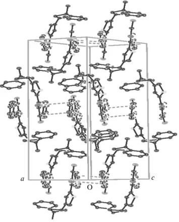
like arrangement of macrocyclic connected by inter-molecular C–H···O hydrogen bonding (H19···O1 = 2.53 Å, C19–H19···O1 = 137°). This pattern was build up the lattice as shown in Fig. 3, this arrangement was held in place by C–H···O interactions. The intramo-lecular hydrogen bonding was between the thiazole nitrogen (N1) and the CH (H15) of the molecule (N1···H15 = 2.22 Å).
The optimized structure parameters of the NNP2CH calculated by DFT method and listed in
Table 3 are in accordance with the atom numbering scheme given in Fig. 2a. As can be seen from Table 3, slight variations in the bond lengths and angles are observed because the molecular states are different during the experimental and theoretical processes. The agreement between the theoretical and experi-mental results (bond lenght and bond angles) has been expressed by obtained linear function formulas: y = 1.0421x– 0.0459R2 = 0.9946 (6-31G(d)), y = 1.0445x –
0.0502, R2 = 0.9947 (6-311G+(d)) for bond lengths;
y = 1.0445x – 0.0502R2 = 0.9947 (6-31G(d)), y =
0.9737x + 2.5398R2 = 0.9708 (6-31G(d)), y = 0.9751x +
2.3621R2 = 0.9688 (6-311G+(d)) for bond angles. The
geometrical parameters for the title molecule are nor-mal and consistent with those recently reported for thiazole derivative [42].
A logical method for globally comparing the struc-tures obtained with the theoretical calculations is by superimposing the molecular skeleton with that obtained from X-ray diffraction, giving a root mean squared error (RMSE) of 0.830 Å for B3LYP/6-31G(d) and 0.857 Å for B3LYP/6-311G+(d). Figure 4 shows the atom-by-atom superimposition of the structures calculated (magenta) on the X-ray structure (mahogany) of the title compound.
IR Spectroscopy
Vibrational spectral measurements were made for
N-[4-(3-methyl-3-phenyl-cyclobutyl)-thiazol-2-yl]-N'-pyridin-2-ylmethylene-сhloro-acetic acid
hydra-zide. Optimized geometrical structure and harmonic vibrational frequencies were computed by DFT (B3LYP) methods using the same basis sets. Complete assignments of the observed spectra were suggested. The calculated vibrational frequencies (scaled) and approximate description of normal modes obtained using B3-base DFT methods are listed in Table 4. Gaussview program [43] was used to assign the
calcu-Fig. 3. Packing diagram of the title compound.
a c
O
Fig. 4. Atom-by-atom superimposition of the structures
calculated (magenta) and X-ray structure (mahogany) of the title compound (hydrogen atoms have been omitted for clarity) for B3LYP/6-31G(d) level (a) and B3LYP/6-311G+(d) level (b).
(а) (b)
Table 2. Hydrogen-bond geometry (Å, deg) for the title
compound i Symmetry code: –1 + x, y, –1 + z. ii Cg4 is a phenyl ring. D–H···A, Å D–H, Å H···A, Å D···A, Å D–H···A, deg C15–H15···N1 0.93 2.22 2.816(2) 121 C19–H19···O1(i) 0.93 2.53 3.275(3) 137 C1–H1···Cg4(ii) 0.93 2.86 3.755(2) 162
lated harmonic frequencies. The FT-IR spectrum of the title compound is shown in Fig. 5.
The FT-IR spectra have some characteristic bands of the stretching vibrations of the C–H, C–H2, C–H3,
C–C, C–N, C=N, C–S, and C–Cl groups. The aro-matic structure shows the presence of C‒H stretching vibrations in the region 2900–3150 cm–1, which is the
characteristic region for the identification of the C–H stretching vibrations. In this region, the bands are not appreciably affected by the nature of the substituent [44]. The C–H aromatic stretching mode was observed at 3162 and 3100 cm–1 experimentally, and
calculated at 3104–3080 cm–1 for 6-31G(d) and at
(3092/3072)–3083 cm–1 for 6-311G+(d) basis sets. In
the higher frequency region, almost all vibrations belong to symmetric and asymmetric νCH2 and νCH3
(in cyclobutane ring and chloroacetic acid group) stretching vibrations, respectively. The ranges of fre-quencies obtained by B3LYP method in this region are (2950/2947)–3009 cm–1 and (3011/3008)–3061 cm–1
for 6-31G(d) (νsCH2(B) and νasCH2(E)) and 2948–
3015 cm–1 and 3012–3068 cm–1 for 6-311G+(d) levels
(νsCH2(B) and νasCH2(E)), respectively. Bands at
2993–2986 cm–1 for 6-31G(d) and 2991 cm–1 for
6-311G+(d), and 2921 for 6-31G(d) and 2924 cm–1
for 6-311G+(d) correspond to the asymmetric and symmetric C–H3 stretching modes, respectively.
Vibrations characteristic to the thiazole ring were observed at 1580, 1534, and 628 cm–1 due to νC=N,
νC–C, and νC–S, while that have been calculated at 1475, 1523, and 820 cm–1 for B3LYP/6-31G(d), and at
1464, 1571/1526, and 823 cm–1 for B3LYP/6-311+G(d)
basis set. This bands were observed at 1580, 1534, and 738 cm–1 experimentally, and calculated at 1476, 1526,
and 726 cm–1 (6-31G(d) basis set) for
N'-benzylidene-
N-[4-(3-methyl-3-phenylcyclobutyl)-thiazol-2-yl]-chloro-acetic acid hydrazide [42]. On the other hand,
νC=O stretching and νC–Cl stretching bands were observed at 1716 and 781 cm–1, that have been
calcu-lated using B3LYP method at 1733, 771 and 1722, 774 cm–1 for 6-31G(d) and 6-311G+(d) basis sets,
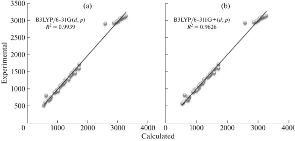
respectively. These assignments are also supported by Demir et al. [42]. Other calculated vibrational fre-quencies can be seen in Table 4. The correlations between the experimental and calculated vibrational parameters are given in Fig. 6. According to our calcu-lations, B3LYP method correlates well for the vibra-tional parameters.
Chemical Shifts of Nuclei of Carbon and Hydrogen Atoms
The chemical shifts of the relevant protons were given in Table 5 together with the values calculated theoretically (i) by our method of examining only the ring current effects [45] of the aromatic moieties in two series of compounds and (ii) by exclusive gauge
Table 3. Selected optimized and experimental geometry
parameters of the title compound in ground state
Parameter Experi-mental Calculated DFT/B3LYP 6-31G(d) DFT/B3LYP 6-311G+(d) Bond lengths, Å S1–C1 1.706(2) 1.737 1.735 S1–C3 1.727(2) 1.768 1.766 N1–C3 1.293(2) 1.302 1.300 N1–C2 1.377(2) 1.382 1.382 C2–C1 1.341(2) 1.364 1.362 C11–C22 1.765(2) 1.794 1.795 N3–C15 1.253(2) 1.285 1.282 N3–N2 1.393(3) 1.382 1.380 C3–N2 1.408(2) 1.414 1.416 N2–C21 1.382(2) 1.405 1.408 C6–C8 1.532(4) 1.540 1.539 C6–C5 1.540(3) 1.562 1.563 C6–C7 1.550(2) 1.563 1.563 C5–C4 1.544(2) 1.558 1.557 C16–N4 1.331(2) 1.346 1.343 N4–C20 1.341(3) 1.335 1.333
Bond angles, deg
C1–S1–C3 88.11(8) 87.677 87.686 C3–N1–C2 110.44(14) 111.942 112.007 C15–N3–N2 121.94 (14) 123.485 124.177 N1–C3–S1 115.32 (13) 114.553 114.523 N2–C3–S1 122.36 (12) 122.490 122.392 C21–N2–N3 120.83(13) 112.971 112.909 C6–C5–C4 89.53(14) 89.411 89.181 C1–C2–N1 114.59(16) 114.211 114.113 C1–C2–C4 127.02(16) 126.590 125.870 C4–C7–C6 89.08(14) 89.375 89.182 O1–C21–C22 124.41(17) 123.683 123.779 N2–C21–C22 114.27(15) 114.939 114.868 N4–C16–C15 114.63(17) 113.496 113.997 N3–C15–C16 120.26(17) 119.734 119.311 C21–C22–Cl1 110.98(14) 111.162 111.183 C16–N4–C20 116.29(19) 117.938 118.138
Torsiyon angles, deg
C11–C22–C21–O1 −3.7 0.141 –0.003
O1–C21–N2–C3 –4.8 –0.114 0.002
S1–C3–N2–N3 –156.8 –179.302 –179.992
C22–C21–N2–N3 –1.4 –0.220 0.000
Table 4. Comparison of the observed and calculated vibrational spectra of the title compound
Vibrational modes: ν, stretching; s, symmetric; as, asymmetric; α, scissoring; γ, rocking; ω, wagging; δ, twisting; θ, ring breathing;
β, in-plane bending. Abbreviations: A, thiazole ring; B, cyclobutane ring; C, phenyl ring; D, pyridin ring; E, chloro acetic acid group; F, hydrazit group.
Assignments Experimental Calculated
IR with KBr, cm–1 B3LYP 6-31G(d) B3LYP 6-311G+(d)
νC–H(A) 3264 3141 3134 νC15–H15 3215 3121 3115 νsC–H(D) 3162 3104 3092/3072 νasC–H(D) 3132 3091/3072 3091/3072 νsC–H(C) 3100 3080 3083 νasC–H(C) 3080 3070/3062/3049 3072 νasC–H2(E) 3053 3061 3068 νasC–H2(B) 3020 3011/3008 3012 νsC–H2(E) 3010 3009 3015 νasC–H3 2954 2993–2986 2991 νC–H(B) 2928 2961 2966 νsC–H2(B) 2862 2950–2947 2948 νsC–H3 2577 2921 2924 νC=O 1716 1733 1722 νC15–N3 1714 1615 1610 νC=C(C) 1608 1600–1576 1594 νC–C(D) 1566 1574–1563 1560/1571 νC–C(A) 1534 1523 1571/1526 γC–H(C) 1493 1487/1434 1485 νC3=N1(A) 1580 1475 1464 αC–H3 1479 1462 1468 γC–H(D) 1464 1456/1422 1422 αC–H2(B) 1444 1442 1439 αC–H2(E) 1435 1413 1410 ωC–H3 1397 1375 1370 ωC–H2(E) + νC3=N1 + γC–H(D + F) 1368 1292 1341 νC9–C6 1251 1282 1284/1282 νC3–N2–N3 + γC–H(F) + ωC–H2(E) 1240 1229–1205/1180–1175 1226–1202 ωC–H2(B) 1222 1203 1199 αC–H(C) 1203 1166 1166 δC–H2(E) 1148 1152 1163 αC–H(D) – 1136 1137 γC–H (A) 1125 1132 1136 δC–H2 + γC–H(B) 1093 1111/1029 1029 αC–H(D) 1078 1079 1081 Θ(A) 1021 1006/875 1009 Θ(C) 970 978 985 δ(D) 947 971/947 980 Θ(D) 990 970 978–976 δ(C) 1021 950/926 959 Θ(B) 923 932 936 β deformation (E) 897 909 910 νC–S (A) 628 820 823 νC–Cl 781 771 774 β deformation (C) 768 745 748/687 β deformation (A) 747 727/676 676 β(E) 738 710 773 ω(C) 704 687 748/687 β(D) 590 609–574 616 β deformation (E + F) 545 566/531 578
invariant atomic orbitals (GIAO) [46, 47] calculations. Comparison of the experimental and theoretically cal-culated chemical shift differences between the relevant protons (Table 5) demonstrates the excellent agree-ment in both direction (high- or low-field) and amount; thus, the 1H and 13C NMR spectra of
corre-sponding compounds in two series are correctly described by the HF and DFT calculations. The NMR spectral data for NNP2CH were compiled in Table 5. These chemical shifts were calculated with the GIAO method [48] and 6-31G(d) and 6-311G+(d) basis sets.
1H chemical shift values (with respect to TMS) were
calculated and found to be 6.26–0.29 ppm at 6-31G(d) level and 6.79–0.02 ppm at 6-311G+(d),
whereas the experimental results were observed to be 8.35–1.57 ppm. The 1H NMR spectrum is indicated
as evident from the appearance of a singlet for methyl protons at δ = 1.57 ppm. The singlet is assigned to H8' (C8) atoms that have been calculated at 3.24 ppm (6-31G(d)) and 3.44 ppm (6-311G+(d,p)) for the B3LYP level. All carbon atoms give peaks at the range of 148.37–5.82 ppm for 6-31G(d) and 167.56–1.87 ppm for 6-311G+(d,p) level. The result shows that the range 13C NMR chemical shift of the typical organic
molecule is usually greater than 100 ppm [49, 50]; the accuracy ensures reliable interpretation of spectro-scopic parameters. The chlor, oxygen and sulfur atoms that are present at three different positions in the mol-ecule shows electronegative property, so that the chemical shift of C22, C21, C1, and C3 seems to be 43.15, 158.65, 114.27, and 167.57 ppm, respectively. As can be seen from Table 5, the theoretical 1H and 13C
chemical shift values for the title compound are gener-ally closer to the experimental 1H and 13C chemical
shift data.
Molecular Electrostatic Potential
Molecular electrostatic potential (MEP) has proved itself as an effective tool for quantitatively assessing various non-covalent interactions, such as hydrogen-bonding, halogen bonding, and cation–π interactions [51–54]. Electrostatic properties of mole-cules can be computed approximately using discrete point charges located on the atomic sites at the van der Waals surface or at surfaces farther away from the mol-ecules [55–59].
The quantitative analysis of V(r) (electrostatic potential) initially consisted mainly of locating and identifying the most negative potentials, Vmin, which
Fig. 5. The FT-IR spectrum of the title compound. 1500 1000 1500 2000 2500 3000 3500 4000 10 20 30 40 50 60 70 Wavenumbers Transmittance, %
Fig. 6. Correlation graphics of calculated and experimental frequencies of the title compound for B3LYP/6-31G(d,p) level (a) and
B3LYP/6-311G+(d,p) level (b). (а) 500 1000 1500 2000 2500 3000 3500 Calculated 1000 2000 3000 4000 Exp e rim e n ta l B3LYP/6-31G(d, p) R2 = 0.9939 0 (b) 1000 2000 3000 4000 B3LYP/6-311G+(d, p) R2 = 0.9626 0
were usually associated with: (a) the lone pairs of the more electronegative atoms, such as N, O, F, Cl, S and Br; and (b) unsaturated, aromatic and strained car-bon-carbon bonds. These Vmin could is often related to reactive properties, for instance, the pKα values of azine nitrogens [60], and epoxide carcinogenicity [61]. More recently, attention has been focused on the elec-trostatic potential computed on the molecular “sur-face”, and both positive and negative extrema, VS,max
and VS,min, have been used to establish quantitative
relationships. For example, we have shown that, for a large variety of molecules, the VS,max and VS,min
cor-relate with hydrogen bond acidity and basicity, respec-tively [62]. This overall approach has been useful, but it has been limited in scope; the Vmin, VS,min, and VS,max
values are certainly key features of the molecular elec-trostatic potential, but they do not convey all the infor-mation that is contained in V(r).
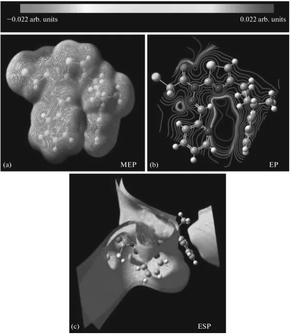
The potential surface scan with the B3LYP/6-311G+(d) level of theoretical approximations has been performed for the title molecule (Fig. 7). The negative (red and yellow) regions of MEP were related to elec-trophilic reactivity and the positive (blue) ones to nuc-leophilic reactivity shown in Fig. 7a. As can be seen, this molecule has several possible sites for electro-philic attack. Negative regions were found in the stud-ied molecule around the O1 atom of the carbonyl group, the S1 atom of thiazole ring, the N4 nitrogen atom of the pyridine ring. The negative V(r) values are –0.0562 a.u. for O1, which is the most negative region; –0.0562 a.u. for O1; –0.0264 a.u. for S1; –0.0333 a.u. for N4 atom, which is the least negative region. Thus, it would be predicted that an electrophile would pref-erentially attack the title molecule at the O1 and N4 positions. Furthermore, we found a maximum value of +0.032 a.u. on the C19–H19 bond on the positive regions of V(r), indicating that this site is probably involved in nucleophilic processes.
The lines are drawn in the contour map clearly show the f low of the electron density in title molecule (Fig. 7b). The red color dominating area, where the nitrogen and oxygen atoms were located, is found to be highly negative, and other colored parts signify the positive region of the molecule. Additionally, the total electron density is mapped to the electrostatic poten-tial surface; the isosurface representation of the total electrostatic potential of the title compound is shown in Fig. 7c.
Mulliken Population Analysis
The calculation of effective atomic charges plays an important role in the application of quantum mechan-ical calculations to molecular systems. The charge dis-tribution on different atoms (C, N, O, S, and Cl) for NNP2CH from Mulliken population analysis (MPA) procedures using B3LYP method is listed in Table 6.
Table 5. Theoretical and experimental 13C and 1H isotropic chemical shifts (with respect to TMS, all values in ppm) for the title compound
* Average. Atom Experimental (CDCl3) Calculated B3LYP/6-31G(d) B3LYP/6-311G+(d) C1 114.27 100.01 96.26 C2 152.71 144.85 149.11 C3 167.57 148.37 163.71 C4 30.94 17.79 5.59 C5 40.78 21.44 10.81 C6 38.93 29.78 15.44 C7 40.78 24.76 13.57 C8 29.93 5.82 1.87 C9 152.70 140.51 147.28 C10 124.72 107.97 113.26 C11 125.35 109.7 115.39 C12 128.21 104.85 108.70 C13 125.35 110.14 115.97 C14 124.72 108.25 113.47 C15 149.67 137.19 147.99 C16 151.92 142.12 148.22 C17 114.27 101.27 103.01 C18 136.66 117.24 127.45 C19 120.76 103.28 105.24 C20 147.92 135.31 143.31 C21 158.65 155.9 167.56 C22 43.15 36.63 27.45 H1 7.04 3.16 3.23 H4 3.84 0.29 0.02 H5* 2.61 0.76 1.51 H7* 2.65 1.09 1.51 H8* 1.57 3.24 3.44 H10 7.10 4.36 4.41 H11 7.20 4.48 4.67 H12 7.24 4.26 4.35 H13 7.30 4.57 4.75 H14 7.31 4.52 4.73 H15 8.35 6.26 6.79 H17 7.34 4.98 5.06 H18 7.38 4.9 5.33 H19 7.41 4.42 4.51 H20 7.78 6.17 6.42 H22* 4.84 1.38 1.00
Graphical representations of atomic charges and nat-ural charges on the atom are shown in Fig. 8.
Molecular Orbitals
The frontier orbitals of a chemical species, highest occupied molecular orbital (HOMO) and lowest unoccupied molecular orbital (LUMO), are quite important for the determination of their reactivity. Fukui et al. [63] were first who recognized this. The
EHOMO is often associated with the electron donating
ability of the molecules. The EHOMO indicates the molecular ability in donating electrons to appropriate acceptor molecules with low energy empty molecular orbital. ELUMO indicates the ability of the molecule to
accept electrons. The lower value of ELUMO, the more
probable it is that the molecule would accept elec-trons. Consequently, concerning the value of the energy gap ΔE = ELUMO – EHOMO, higher values of ΔE
will provide lower reactivity to a chemical species.
Fig. 7. Molecular electrostatic potential map calculated at B3LYP/6-311G+(d) level (a), the contour map of molecular
electro-static potential surface of the title compound (b), and the isosurface representation of total electroelectro-static potential of the title com-pound (c).
MEP EP
ESP
(a) (b)
(c)
Table 6. Mulliken charges of
N-[4-(3-methyl-3-phenyl-cyclobutyl)-thiazol-2-yl]-N'-pyridin-2-ylmethylene-chloro-ace-tic acid hydrazide with 6-31G(d) and 6-311+G(d)
Atoms Mulliken charge
6-31G(d) Atoms Mulliken charge 6-311G+(d) Atoms Mulliken charge 6-31G(d) Atoms Mulliken charge 6-311G+(d) Cl1 –0.039 Cl1 0.344 H1 0.174 H1 0.247 O1 –0.442 O1 –0.131 H4 0.130 H4 0.212 S1 0.320 S1 0.054 H5A 0.137 H5A 0.245 N1 –0.524 N1 0.010 H5B 0.162 H5B 0.260 N2 –0.363 N2 –0.016 H7A 0.137 H7A 0.246 N3 –0.315 N3 0.343 H7B 0.161 H7B 0.262 N4 –0.469 N4 0.132 H8A 0.140 H8A 0.247 C1 –0.431 C1 –0.328 H8B 0.149 H8B 0.253 C2 0.366 C2 0.481 H8C 0.149 H8C 0.253 C3 0.306 C3 0.235 H10 0.125 H10 0.216 C4 –0.174 C4 –0.079 H11 0.125 H11 0.205 C5 –0.291 C5 –1.351 H12 0.122 H12 0.200 C6 0.016 C6 1.459 H13 0.125 H13 0.204 C7 –0.289 C7 –1.355 H14 0.125 H14 0.217 C8 –0.452 C8 –1.236 H15 0.232 H15 0.339 C9 0.147 C9 1.647 H17 0.152 H17 0.233 C10 –0.177 C10 –0.694 H18 0.149 H18 0.234 C11 –0.127 C11 –0.460 H19 0.144 H19 0.227 C12 –0.133 C12 –0.423 H20 0.151 H20 0.222 C13 –0.127 C13 –0.458 H22A 0.228 H22A 0.305 C14 –0.177 C14 –0.677 H22B 0.228 H22B 0.306 C15 0.045 C15 –0.597 C16 0.276 C16 –0.322 C17 –0.151 C17 0.947 C18 –0.105 C18 –1.409 C19 –0.138 C19 –0.089 C20 0.041 C20 –0.169 C21 0.616 C21 0.093 C22 –0.454 C22 –1.084
Fig. 8. Comparison of different basis sets for calculated atomic charges of the title compound. 1.5 1.0 0.5 0 0.5 1.0 1.5 2.0 2.0 Atoms Cl1 S1 N2 N4 C2 C4 C6 C8 Cl0 Cl2 Cl4 Cl6 Cl8 C20 C22 H4 H5B H7B H8B H10 H12 H14 H17 H19 H22A At o mic c har ges B3LYP/6-31G(d) B3LYP/6-311G+(d)
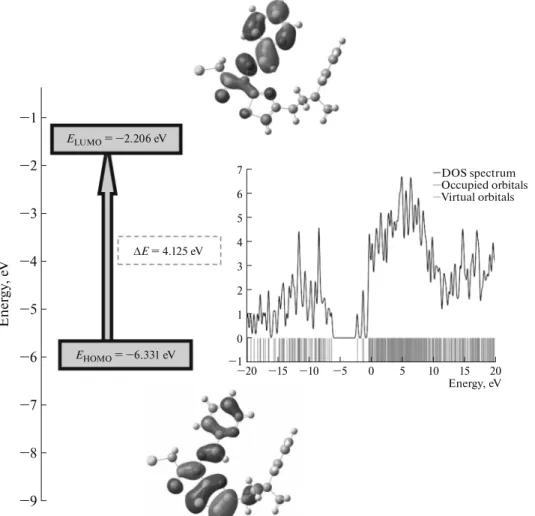
Fig. 9. Molecular orbital surfaces and energy levels given in boxes for the HOMO, LUMO and calculated the total electronic
den-sity of states diagram of the title compound computed at B3LYP/6-311G+(d) level.
0 1 2 3 4 5 6 7 1 15 10 5 0 5 10 15 20 20 Energy, eV DOS spectrum Occupied orbitals Virtual orbitals 9 8 7 6 5 4 3 2 1 En e rgy , e V ELUMO = 2.206 eV EHOMO = 6.331 eV ΔE = 4.125 eV
Lower values of the energy difference will indicate the higher reactivity of the molecules because the energy to remove an electron from the last occupied orbital to the first unoccupied orbital will be low [64, 65].
A visual inspection of the molecular orbitals, as shown in Fig. 9 for the title compound, explains this observation. Thus, while the LUMO orbitals reside exclusively in the thiazole, furan, cyclobutane ring and hydrazide group, we have localization of the HOMO on the furan, thiazole ring and hydrazide group. HOMO and LUMO energies, calculated by B3LYP/6-31G(d) and 6-311G+(d), are listed in Table 7. Besides, Gauss-Sum 3.0 Program [66] was used to calculate group contributions to the molecular orbitals (HOMO and LUMO) and prepare the density of the state (DOS), as shown in Fig. 9. The DOS plot provides a diagrammatic view of the molecular orbital contribu-tions, computed by the GaussSum 3.0 program [65] at B3LYP/6-311G+(d).
CONCLUSION
In the present work, a complete X-ray analysis has been made for proper diffraction results of N-[4-(3- methyl-3-phenyl-cyclobutyl)-thiazol-2-yl]-N'-pyri-din-2-ylmethylene-сhloro-acetic acid hydrazide. A complete vibrational and molecular structure anal-ysis has been performed based on the quantum mechanical approach to DFT calculation using B3LYP/6-31G(d) and B3LYP/6-311G+(d) basis sets. Chemical shifts of nuclei of carbon and hydrogen atoms, and the vibrational frequencies of the funda-mental modes of the title compound have been pre-cisely assigned and analyzed, and the theoretical results have been compared with the experimental vibrations. The intermolecular charge transfer is evi-denced by Mulliken charge population analysis. HOMO and LUMO energy gap reveal that the energy gap ref lects the chemical activity of the molecule. To predict the reactive sites for electrophilic and nucleo-philic attack for the NNP2CH molecule, the MEP at
the B3LYP/6-311G+(d) optimized geometry was cal-culated.
REFERENCES
1. J. R. Hill, L. Subramanian, and A. Maiti, Molecular
Modeling Techniques in Materials Science (Taylor and
Francis/CRC, Boca Raton, FL, 2005).
2. J. B. Foresman and A. E. Frisch, Exploring Chemistry
with Electronic Structure Methods (Gaussian,
Pitts-burgh, PA, 1996).
3. N. Agarwal, S. Kumar, A. K. Srivastava, and K. P. C. Sarkar, Ind. J. Het. Chem. 6, 291 (1997).
4. R. C. Sup, R. Y. Sukp, and C. W. Bang, Korean J. Med. Chem. 5, 72 (1995).
5. S. I. Sumistov and Z. A. Bofoshko, USSR Patent No. 154861 (1964).
6. J. D. Hadjipavlou-Litina and A. Geronikaki, Arz. Forsch. Drug Res. 46, 805 (1996).
7. L. D. Hadjipavlou, A. Geronikaki, and E. Sotiropou-lou, Res. Commun. Chem. Pathol. 79, 355 (1993). 8. A. Geronikaki, D. Hadjipavlou-Litina, and M.
Amourgia-nou, Il Farmaco 58, 489 (2003).
9. M. Tsuruoka and I. Seikutsugaka, Med. Biol. 10, 296 (1947).
10. J. R. Merchant, G. Martysen, and N. S. Venkatesh, Indian J. Chem. B 20, 493 (1981).
11. G. Maass, U. Immendoerfer, B. Koenig, et al., Antimi-crob. Agents Chemother. 37, 2612 (1993).
12. M. P. Anna, G. Athina, M. Remi, et al., Bioorg. Med. Chem. 11, 2983 (2003).
13. R. P. Kapoor, M. K. Rastogi, R. Khanna, et al., Indian J. Chem. B 23, 390 (1984).
14. J. V. Metzger, in Comprehensive Heterocyclic Chemistry, (Pergamon, Oxford, 1984), p. 235.
15. B. Dash, M. Patra, and S. Praharaj, Indian J. Chem. B
19, 1378 (1980).
16. J. Michel Grivy, F. Tellez, S. Bernes, et al., Inorg. Chim. Acta 339, 532 (2002).
17. A. Benalte-Garcia, F. J. Garcia-Barros, F. J. Higes-Rolando, et al., Polyhedron 18, 2907 (1999).
18. K. Lemma, J. Berglund, N. Farrell, et al., J. Biol. Inorg. Chem. 5, 300 (2000).
19. M. J. M. Campbell, Coord. Chem. Rev. 15, 279 (1975). 20. S. Padhye and G. B. Kauffman, Coord. Chem. Rev. 63,
127 (1985).
21. S. Bhattcharya and M. Thomas, Tetrahedron Lett. 41, 10313 (2000).
22. B. Imperiali, J. Org. Chem. 63, 6727 (1988).
23. D. Parker, S. P. White, and M. Brookhart, Chem. Commun. 1, 47 (2000).
24. A. Torrado, G. K. Walkup, and B. Imperiali, J. Am. Chem. Soc. 120, 609 (1988).
25. R. B. Thompson, Z. Ge, M. Patchan, et al., Bioelec-tronics 11, 557 (1996).
26. J. Sitkowski, L. Stefaniak, T. Dziembowska, et al., J. Mol. Struct. 381, 177 (1996).
27. R. Castro, J. A. Garcia-Vazquez, J. Romero, et al., Polyhedron 12, 2241 (1993).
Table 7. HOMO–LUMO energy calculated by B3LYP
with 6-31G(d) and 6-311G+(d) methods Parameters B3LYP/6-31G(d) B3LYP/6-311G+(d) HOMO, eV –6.036 –6.331 LUMO, eV –1.858 –2.206 ΔE, eV 4.178 4.125 HOMO–1, eV –6.361 –6.657 HOMO–2, eV –6.603 –6.910 LUMO+1, eV –0.941 –1.276 LUMO+2, eV –0.782 –1.238
28. S. Saydam, Synth. React. Inorg. Met. Org. Chem. 32, 437 (2002).
29. H. Ieda, H. Fujiwara, and Y. Fuchita, Inorg. Chim. Acta 319, 203 (2001).
30. E. Borras, G. Alzuet, J. Borras, et al., Polyhedron 19, 1859 (2000).
31. S. Saydam and C. Alkan, Pol. J. Chem. 75, 29 (2001). 32. M. James, H. Kawaguchi, and K. Tatsumi, Polyhedron
16, 1873 (1996).
33. G. Mohamed, Spectrochim. Acta, Part A 57, 411 (2001).
34. R. G. Parr and W. Yang, Density Functional Theory of
Atoms and Molecules (Oxford University Press, New
York, 1989).
35. Stoe and Cie, X-AREA Version 1.18 and X-RED32
Ver-sion 1.04 (Stoe and Cie, Darmstadt, Germany, 2002).
36. G. M. Sheldrick, SHLEXS-97. Program for Refinement
of Crystal Structures (University of Göttingen,
Ger-many, 1997).
37. L. J. Farrugia, J. Appl. Crystllogr. 30, 565 (1997). 38. J. B. Foresman and A. Frisch, Exploring Chemistry with
Electronic Structure Methods (Gaussian, Pittsburg,
1996).
39. J. P. Merrick, D. Moran, and L. Radom, J. Phys. Chem. A 111, 11683 (2007).
40. R. Dennington, T. Keith, J. Millam, et al., GaussView
Version 3.07 (Semichem, Shawnee Mission, KS, 2003).
41. V. Haridas, S. Sahu, and P. Venugopalan, Tetrahedron
67, 727 (2011).
42. S. Demir, M. Dincer, A. Cukurovali, et al., Int. J. Quantum Chem. 112, 1016 (2012).
43. M. J. Frisch, G. W. Trucks, and H. B. Schlegel,
Gauss-ian 03 Revision C.02 (GaussGauss-ian, Pittsburgh, PA, 2003).
44. A. Teimouri, M. Emami, A.N. Chermahini, et al., Spectrochim. Acta Part A 71, 1749 (2009).
45. S. Klod and E. Kleinpeter, J. Chem. Soc. Perkin Trans.
2, 1893 (2002).
46. J. R. Ditchfield, Mol. Phys. 27, 789 (1974).
47. J. P. Cheeseman, G. W.Trucks, T. A. Keith, et al., J. Chem. Phys. 104, 5497 (1996).
48. K. Wolinski, J. F. Hilton, and P. Pulay, J. Am. Chem. Soc. 112, 8251 (1990).
49. H. O. Kalinowski, S. Berger, and S. Braun, Carbon-13
NMR Spectroscopy (Wiley, Chichester, 1988).
50. K. Pihlaja, E. Kleinpeter (Eds.), Carbon-13 Chemical
Shifts in Structural and Sterochemical Analysis, Ed. by
K. Pihlaja and E. Kleinpeter (VCH, Deerfield Beach, 1994).
51. J. W. Zou, Y. J. Jiang, M. Guo, et al., Chem. Eur. J. 11, 740 (2005).
52. H. Hagelin, T. Brinck, M. Berthelot, et al., Can. J. Chem. 73, 483 (1995).
53. O. Lukin and J. Leszczynski, J. Phys. Chem. A 106, 6775 (2002).
54. L. Joubert and P. L. A. Popelier, Phys. Chem. Chem. Phys. 4, 4353 (2002).
55. P. C. Mishra and A. Kumar, Top. Curr. Chem. 174, 27 (1995).
56. C. A. Reynolds, J. W. Essex, and W. G. Richards, J. Am. Chem. Soc. 114, 9075 (1992).
57. J. Meister and W. H. E. Schwartz, J. Phys. Chem. 98, 8245 (1994).
58. C. G. Mohan, A. Kumar, and P. C. Mishra, J. Mol. Struct. Theochem. 332, 171 (1995).
59. R. Bonaccorsi, A. Pullman, E. Scrocco, et al., Theor. Chim. Acta (Berlin) 24, 51 (1972).
60. P. Politzer and J. S. Murray, Reviews in Computational
Chemistry, Ed. by K. B. Lipkowitz and D. B. Boyd
(VCH, New York, 1991).
61. P. Politzer, P. R. Laurence, and K. Jayasuriya, Env. Health Persp. 61, 191 (1985).
62. H. Hagelin. T. Brinck, M. Berthelot, et al., Can. J. Chem. 73, 483 (1995).
63. K. Fukui, T. Yonezawa, and H. Shingu, J. Chem. Phys.
20, 722 (1952).
64. N. Khalil, Electrochim. Acta 48, 2635 (2003).
65. I. Lukovits, K. Palfi, I. Bako, et al., Corrosion 53, 915 (1997).
66. N. M. O’Boyle, A. L. Tenderholt, and K. M. Langer, J. Comput. Chem. 29, 839 (2008).
![Table 1. Crystallographic characteristics and the X-ray data collection and structure-refinement parameters for the title compound a R 1 =∑||F o | − |F c ||/∑|F o |; b wR 2 = [∑w( ) 2 /∑w( ) 2 ] 1/2 .FormulaC23 H22 Cl N3 O SFormula weight424.94T, K296Wa](https://thumb-eu.123doks.com/thumbv2/9libnet/4554365.82934/2.918.75.437.404.1055/crystallographic-characteristics-collection-structure-refinement-parameters-formulac-sformula.webp)


![Table 6. Mulliken charges of N-[4-(3-methyl-3-phenyl-cyclobutyl)-thiazol-2-yl]-N'-pyridin-2-ylmethylene-chloro-ace- N-[4-(3-methyl-3-phenyl-cyclobutyl)-thiazol-2-yl]-N'-pyridin-2-ylmethylene-chloro-ace-tic acid hydrazide with 6-31G(d) and 6-311+G(d)](https://thumb-eu.123doks.com/thumbv2/9libnet/4554365.82934/10.918.77.823.117.1091/mulliken-charges-cyclobutyl-ylmethylene-cyclobutyl-pyridin-ylmethylene-hydrazide.webp)