Elif Damla Ar›san
1*, Serdar Ar›san
2,
Murat Can Kiremit
2, Narçin
Palavan-Ünsal
1and Erbil Ergenekon
21‹stanbul Kültür University, Science and Letters Faculty, Ataköy Campus, 34156 Bak›rköy, ‹stanbul-Turkey
2fiiflli Etfal Research and Training Hospital, 1stUrology Clinics, fiiflli, Istanbul-Turkey
Abstract
Chronic pelvic pain syndrome (CPPS) is a common and serious health problem affecting the quality of life in men. According to National Institute of Health classification system abacterial chronic pelvic pain has two sub category which are defined as 3a and 3b. The major difference between these two groups inflammation status. In this study we aim to investigate the antioxidant enzyme levels such as Glutathion peroxidase (Gsh-Px), superoxide dismutase (SOD), catalase (CAT) and lipid peroxidation products in CPPS patients. All enzyme levels were determined in patients and disease free control group blood samples by spectrophotometrically. In our study, the enzyme levels were not a good indicator for comparison of 3a or 3b type CPPS patients. However, Gsh-Px, SOD and lipid peroxidation products were significantly different from disease free control samples. Although high generation of H202due to
SOD activity, we did not detect any change in CAT activity in patients blood samples compare to control group.
Key words: Prostatitis, superoxide dismutase, catalase,
glutathion peroxidase, lipid peroxidase.
Introduction
Chronic prostatitis is a major healthcare issue, and management of chronic prostatitis symptoms is still poor (Potts and Pasqualotto, 2003). Considerable confusion exists about the clinical findings associated with prostatitis and the effect of these conditions on male reproductive physiology. Chronic prostatitis is defined by lower urinary tract symptoms, predominantly with pain. This diagnosis is made when neurologic, infectious, and urologic disease (e.g. benign prostatic hyperplasia or kidney stones) has been excluded. Symptoms in patients with chronic prostatitis may last several weeks or more and may recur periodically for many years. The National Institutes of Health (NIH) classification and definition of the four categories of prostatitis defines category 3a as an inflammatory chronic pelvic pain syndrome with white blood cells (WBCs) in semen, expressed prostatic secretions (EPSs), or voided bladder 3 (VB3) (1995). Category 3b is defined as non inflammatory chronic pelvic pain syndrome without WBCs in semen, EPSs, or VB3 (Orsilles and Piante-Depaoli, 1998). However, the presence of inflammatory cells in EPSs correlates poorly with the presence of inflammatory cells in semen.
Predominantly neutrophils were found as a response to generation of reactive oxygen species (ROS) in semen (Krieger et al., 1996). Increased levels of free radicals may cause defect spermatozoa and impair sperm function in infertile men (Roberts and Jacobsen, 2000). There is still controversy concerning the gold standard treatment for this chronic disease, thus a proper diagnostic technique for inflammatory and non inflammatory CPPS still remains to be defined (Shahed and Shoskes, 2000). It may be caused by auto-immune response disorders or another factor (Orhan et al., 2001). In recent studies, ROS levels of prostatitis in patients’ semen samples do not verify the illness. So it is the cause reason of DNA damages and other signaling pathway inducers performing activity. Previous data established that the total antioxidant
Antioxidant enzyme profile in chronic pelvic pain syndrome
patients
*Correspondence author:
Istanbul Kültür University, Science and Letters Faculty, Ataköy Campus, 34156 Bak›rköy
‹stanbul-Turkey
E-mail: [email protected]
status of prostatitis patients is not in the normal range. Accelerated ROS amount cause the over expression of antioxidant enzymes. Although many studies, the exact mechanism of antioxidant status of prostatitis is still under debate (Potts and Pasqualotto, 2003).
In this study we aim to answer the roles of antioxidant enzyme levels whether may have a role in inflammatory type of CPPS.
Material and methods
Patients, definitions and selection criteria
Out of a study group comprising 71 patients who met the diagnostic criteria for chronic prostatitis; 40 met the criteria for NIH category 3a and 31 met that NIH criteria category 3b, classified clinically on the basis of four-glass test results (Table1). Apart from these 71 patients, a further 12 were excluded from the study due to their incompatibility with the set criteria. The following criteria were approved for research studies on chronic nonbacterial prostatitis/CPPS (Chronic prostatitis workshop, 1995). The Helsinki Declaration was strictly observed regarding the use of human samples. Also, all studies were undertaken with the approval and institutional oversight of the Institutional Review Board for Ethics of Human Subjects. All 71 patients were later compared on the basis of Endtz test results. 36 healthy donors with normal semen characteristics as proven by various reports wererecruited as controls. Donors with a history of genitourinary infection, symptoms, or instrumentation were excluded from the study. 46 of 71 patients were (65 %) smoker and 7 of 71 (10 %) gave up smoking in 5 years and the rest never used. The case and control group were not used to take antioxidant pills regularly and they were eating vegetables and meat products regularly.
Semen analysis
Semen samples were collected by masturbation into sterile containers after at least 72 hours of sexual abstinence. After liquefaction, semen specimens were evaluated for semen volume, appearance, and viscosity. Semen characteristics (concentration, motility, and morphology) were examined in accordance with WHO criteria (WHO laboratory manual, 1999). Computer assisted semen analyzer were loaded for 5µl for each semen sample and motile cells were examined (Table 2).
White blood cells
The presence of white blood cells (WBCs) in semen specimens were assessed by the myeloperoxidase (Endtz) test (Shekarriz et al., 1995). A 20µl volume of liqufied semen was placed in a 1,5ml cryogenic vial, followed by 20 µl of phosphate buffer saline (pH 7.0) and 40µl of benzidine solution. The sample was mixed, allowed to sit for 5 min and examined for cells that had Type 3a (n=40) Type 3b (n=31) Control (n=36) Total (n=107) Age (Mean, range) 33.7 (22-54) 36.35 (24-63) 32.44 (21-58) 34.04 (21-63)
Smoking
Yes 28/40 (70 %) 18/31 (58.1 %) 20/36 (55.5 %) 66/107 (61.6 %)
No 12/40 (30 %) 13/31 (41.9 %) 16/36 (44.5 %) 41/107 (38.4 %)
PSA levels ng/dl
(Mean- range) 1.043 (0.458-2.84) 1.048 (0.54- 2.94) 0.995 (0.324-2,58) 1.028 (0.324-2.94)
Other medical problems* 3 (7.5 %) 4 (12.9 %) 2 (5.5 %) 9(8.4 %)
*Hepatitis, HIV, hypertension, cardiovascular disease.
stained brown, indicating cells positive for peroxidase. Leukocytospermia was defined as 1 x 10⁶ or more
WBC/ml of semen (Table 2).
Antioxidant enzyme activities in blood
Blood samples were centrifuged for 2 min at 1500 rpm at room temperature and plasma was separated into a clean tube. 200µl blood plasma samples were analyzed each enzyme activity spectrophotometrically (Shimadzu, Japan). Glutathione peroxidase (Gsh-Px) and superoxide dismutase (SOD) enzyme activity were determined according to manufacturer instructions using specific kit (Ransel and Ransod, RANDOX, England). Catalase activity (CAT) was measured in hemolysates at 25°C by the method of Aebi (1984). The decomposition rate of the substrate H2O2 was monitored spectrophotometrically at 240 nm for 30 s. The activity is expressed as mU/L. 1 U is equal to 1 µmol of H2O2 decomposed/min. Lipid peroxidation was estimated by measurement of thiobarbituric acid
reactive substances (TBARS) in erythrocyte lysates by the method previously described by Aydin et al. (2001). After the reaction of MDA with thiobarbituric acid, the reaction product was followed spectrophotometrically at 532 nm, using malondialdehyde as a standard. The results are expressed as nmol/mL (Table 3).
Statistical analysis
The enzyme activity results were expressed as mean ± standard error (SEM). All experiments were replicated in triplicate. Student t-test was used for comparison between NIH classified. CPPS patients and control subjects. The 0.05 level was selected as point of the minimal statistical significance
Results
We determined lipid peroxidation by TBARS method, SOD and related Gsh-Px enzyme activity and CAT activity in serum samples. Antioxidant defense levels in serum of control, type 3a and 3b CPPS patients are
Variable Controls (n= 36) CPPS (Endtz negative) CPPS (Endtz positive)
(n= 60) (n= 11)
Concentration (x 106/ml) 59.9 ± 1.2 37.6 ± 0.9 40.1 ± 2.3
Motility ( %) 60.2 ± 4.3 51.0 ± 6.7 47.2 ± 5.0
WHO morphology ( %) 63.9 ± 2.4 31.3 ± 2.7 29.6 ± 1.3
Data presented as mean ± SE CPPS= Chronic pelvic pain syndrome
Table 2. Comparison of semen characteristics between controls and patients with chronic pelvic pain prostatitis with or without
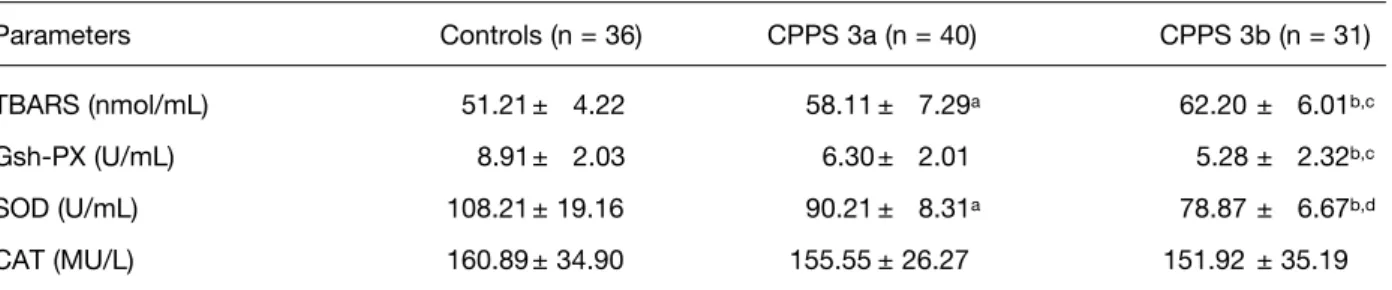
leukocytospermia. Parameters Controls (n = 36) CPPS 3a (n = 40) CPPS 3b (n = 31) TBARS (nmol/mL) 51.21 ± 4.22 58.11 ± 7.29a 62.20 ± 6.01b,c Gsh-PX (U/mL) 8.91± 2.03 6.30 ± 2.01 5.28 ± 2.32b,c SOD (U/mL) 108.21 ± 19.16 90.21 ± 8.31a 78.87 ± 6.67b,d CAT (MU/L) 160.89 ± 34.90 155.55 ± 26.27 151.92 ± 35.19
Values are indicated by mean ± SD.
a As compared with controls, P < 0.05. bAs compared with controls, P < 0.001.
c As compared with the CPPS 3a group, P < 0.05 dAs compared with the CPPS 3a group, P < 0.001 e As compared with controls, P < 0.01.
summarized in Table 3. Lipid peroxidation was 51.21 nmol/ml in control patients. This enzyme activity was increased in CPPS patients. In type 3a lipid peroxidation was 58.11 nmol/ml and in type 3b, this level was estimated as 62.20 nmol/ml. There was slightly significant difference between type 3a and type 3b (p=0.045). When we compared the activity of lipid peroxidation between control and CPPS patients, the significant difference was calculated (p=0.034). In general, antioxidant levels of control patients had always the highest enzyme activity for Gsh-Px and SOD. SOD enzyme activity was decreased approxiamately 17 % in type 3a and 27 % in type 3b patients. When we investigated the Gsh-Px activity, we estimated concomitant results for this enzyme with SOD activity. The Gsh-Px enzyme activity was lower in type 3a patients by 30 % compare to control group
This enzyme activity was decreased by 40 % in type 3b patients. When all results were calculated for SOD and Gsh-Px enzyme analysis, a significant (p<0.05) reduction of levels for each case was obtained significantly correlated (p<0.001). CAT activity determined to understand the how diminished SOD activity induce H2O2 production in patients samples. However CAT activity was found stable and there was no significant difference between all groups (p=0.078).
Discussion
A wide variety of reactive oxygen and nitrogen species can attack DNA directly and form mutagenic lesions. Radical oxygen species may also cause the formation of DNA adducts indirectly by initiating autocatalytic lipid peroxidation, which generates a large variety of potential genotoxic breakdown products, including alkoxyl radicals (LO), peroxyl radicals (LOO), and aldehydes, such as MDA (Dotan et al., 2004; Meagher and Fitzgerald, 2000). Antioxidant enzymes such as SOD, peroxidases and CAT act as sensitive regulators to protect cells from damage by oxygen free radicals (Potts and Pasqualotto, 2003; Kinnula et al., 2004; Stoechlmacher, 2002). Recent studies on colorectal, lung, breast, bladder and gastrointestinal cancers, asthma, Diabetus mellitus, schizophrenia, parkinson revealed a decreased antioxidant activity in these patient samples compared to disease free controls (Pociot et al., 1993; Tikly et al., 2004).
In this study, we determined the antioxidant profile of patients who have CPPS 3a or 3b compare to control group. We checked the Gsh-Px results which were upregulated in disease free conditions. It is well
known that, Gsh-Px enzyme activity is related to SOD enzyme production. Substrate of Gsh-Px is H2O2 mainly produced as end product of SOD activity. We have found elevated lipid peroxidation or TBARS activity in CPPS 3a and 3b groups versus controls. Our results supported previous reports (Pasqualotto et
al., 2000). The protection ability of high antioxidant
enzyme levels in patient profile has diminished but these results do not provide for a satisfactory classification reason between type 3a and 3b because among these groups the differences in enzyme activity has no significance (p>0.05). Nevertheless control samples were remarkably different from patient enzyme activity (p<0.05). SOD enzyme levels were concomitantly changed with Gsh-Px levels in both groups. There was no significant difference for CAT activity between type 3a and 3b CPPS.
We could speculate that the circulating antioxidant enzymes might be used up in the attempt to counteract the enhanced lipid peroxidation in the CPPS 3a and 3b. Another speculation is that the enhanced lipid peroxidation occurs as a consequence of the insufficient power of a depleted antioxidant defense system for a prolonged time. Furthermore, as Gsh-Px and SOD are themselves susceptible to oxidation by the oxidative reactive molecules and lipid peroxides, they could be inactivated by their own substrates (Piogelet et al., 1990). Blum and Fridovich (1985) have found that Gsh-Px activity may be inactivated in oxidative stress conditions by superoxide anion and toxic ligands such as MDA could partially inhibit Gsh-Px activity. Another reason might be occurred by non-regular diet which has depicted trace elements such as Cu, Mn, Zn, and Se could lead to the inactivation of the antioxidant enzymes (Gate et.
al., 1990). Arisan et al., (2006) suggested that low
antioxidant profile in CPPS might be a reason genetic allelic variation which was occurred on transcription site of key enzymes such as Manganase-SOD. Diminished Gsh-Px activity in CPPS patients correlated with SOD activity decreament might be a reason of calculated high radical oxygen species in CPPS. It has been obviously understood that alone normal CAT activity was probably unable to detoxify H2O2in to H2O completely. An accumulation of H2O2 might occur, resulting in higher production of ·OH radical. This highly reactive oxidant molecule binds and oxidizes DNA, lipid, and proteins, and it reacts with structures from its close neighborhood. Any oxidative lesion that is not repaired can lead to mutations, increasing the risk for many chronic illnesses (Arisan et al., 2006). Additionally, lowered
SOD activity could result in accumulation of highly diffusible and potent superoxide anion, which causes deleterious effects at sites far from the tissue.
However, this stability might not be very important to prevent high radical oxygen species attack because of other ezymes disability. On the other hand, our results are compatible with previous reports which prove that patients with Tardive dyskinasea (TD) had a lower SOD activity than those without TD (Zhang et al., 2002). However, these reports have not shown the relationship between specific MnSOD activity and the occurrence of TD, and not all studies did a clear identification of the relationship between polymorphism and enzyme activity.
In conclusion, we assessed the relationship between CPPS and control group focusing on oxidant and antioxidant profile balance. We assumed that antioxidant injury caused by the impaired antioxidant enzyme production, might contribute to adverse developments in the pathogenic cascade of CPSS patients. Our findings lead to the suggestion that oxidative disorder-linked medical health problems can be associated with genetic risk factors such as mutation on gene expression profile.
References
Aebi H. Catalase in vitro, Methods Enzymol.
105:121–126, 1984.
Arisan ED, Arisan S, Kiremit MC, Tigli H, Caskurlu T, Palavan-Unsal N, Ergenekon E. Manganase superoxide dismutase polymorphism in chronic pelvic pain syndrome patients. Prostate Cancer
Prostatic Dis. 9(4):426-431, 2006.
Aydin A, Hilmi O, Sayal A, Ozata M, Sahin G and Isimer A. Oxidative stress and nitric oxide related parameters in type II Diabetes mellitus: effects of glycemic control. Clin Biochem. 34: 65–70, 2001. Blum J and Fridovich I. Inactivation of glutathione
peroxidase by superoxide radical. Arch Biochem
Biophys. 240: 500–508, 1985.
Chronic Prostatitis Workshop. December 7- 8, 1995 National Institute of Diabetes and Digestive and Kidney Diseases National Institutes of Health Bethesda, Maryland
Dotan Y, Lichtenberg D and Pinchuk I. Lipid peroxidation cannot be used as a universal criterion of oxidative stres. Prog Lipid Res. 3: 1–28, 2004.
Gate L, Paul J, G. Nguyen Ba, Tew KD and Tapiero H. Oxidative stress induced in pathologies: the role of antioxidants. Biomed Pharmacother. 53 169–180, 1990.
Kinnula VL, Lehtonen S, Koistinen P, Kakko S, Savolainen M and Kere J. Two functional variants of the superoxide dismutase genes in Finnish families with asthma. Thorax. 59: 116-119, 2004. Krieger JN, Berger RE, Ross SO, Rothman I and Muller
CH. Seminal fluid findings in menwith nonbacterial prostatitis and prostatodynia. J Androl. 17: 310-318, 1996.
Meagher E.A and Fitzgerald G.A. Indices of lipid peroxidation in vivo: strengths and limitation. Free
Radic Biol Med. 28: 1745–1750, 2000.
Orhan I, Onur R, Ilhan N and Ardicoglu A. Seminal plasma cytokine levels in the diagnosis of chronic pelvic pain syndrome. Int J of Urol. 8: 495-499, 2001.
Orsilles MA and Piante-Depaoli M. Oxidative stress-related parameters in prostate of rats with experimental autoimmune prostatitis. Prostate. 34: 270-274, 1988.
Potts JM and Pasqualotto FF. Seminal oxidative stress in patients with chronic prostatitis. Andrologia, 35: 304-308, 2003.
Pigeolet E, Corbisier P, Houbion A, Lambert D, Michiels C, Raes M, Zachary MD and Remacle J. Glutathione peroxidase, superoxide dismutase, and catalase inactivation by peroxide and oxygen derived free radical. Mech Ageing Dev. 51: 283–297, 1990.
Roberts RO and Jacobsen SJ. Epidemiology of prostatitis. Curr Urol Rep. 1: 135-141, 2000. Shahed AR and Shoskes DA. Oxidative stress in
prostatic fluid of patients with chronic pelvic pain syndrome: correlation with gram positive bacterial growth and treatment response. J Androl. 21: 669-675, 2000.
Shekarriz M, Sharma RK, Thomas AJ and Agarwal A. Positive myeloperoxidase staining (Endtz test) as a indicator of excessive reactive oxygen species formation in semen. J Assist Reprod Genet. 12: 70–74, 1974.
Stoehlmacher J, Ingles SA, Park DJ, Zhang W and Lenz HJ. The -9Ala/-9Val polymorphism in the mitochondrial targeting sequence of the
manganese superoxide dismutase gene (MnSOD) is associated with age among Hispanics with colorectal carcinoma. Oncol Rep. 9: 235-238, 2002.
World Health Organization, WHO laboratory manual for the examination of human semen and sperm-cervical mucus interaction (4th ed), Cambridge University Press, Cambridge, 1999.
Pociot F, Lorenzen T and Nerup J. A manganese superoxide dismutase (SOD2) gene polymorphism in insulin-dependent diabetes mellitus. Dis
Markers, 11: 267-274, 1993.
Tikly M, Marshall SE, Haldar NA, Gulumian M, Wordsworth P and Welsh KI. Oxygen free radical scavenger enzyme polymorphisms in systemic sclerosis. Free Radic Biol Med. 36: 1403-1407, 2004