REGULAR ARTICLES
Pharmacokinetics of pentoxifylline and its 5-hydroxyhexyl metabolite
following intravenous administration in cattle
Kamil Uney1&Bunyamin Tras1&Orhan Corum2&Ramazan Yildiz3&Mehmet Maden4
Received: 29 June 2018 / Accepted: 10 September 2018 / Published online: 15 September 2018 # Springer Nature B.V. 2018
Abstract
This study investigated the pharmacokinetics of pentoxifylline (PTX) and its 5-hydroxyhexyl metabolite (M-I) after single-dose intravenous (IV) administration (10 mg/kg) of PTX in six healthy cattle. The safety of PTX was evaluated by clinical observation and biochemical analysis. Plasma concentrations of PTX and M-I were simultaneously determined by reverse-phase high performance liquid chromatography. Pharmacokinetic parameters were calculated using non-compartmental methods. Salivation and discomfort were observed for 2 h following the drug administration. Serum direct bilirubin, total bilirubin, and phosphorus levels at 24 h following the drug administration were significantly different from the control values (0 h) (P < 0.05). Pharmacokinetic variables of PTX were characterized by a short terminal elimination half-life (1.05 ± 0.19 h), a large volume of distribution (6.30 ± 1.76 L/kg), and high total body clearance (5.31 ± 1.27 L/h/kg). The mean ratio between the area under the concentration-time curves of M-I and PTX was 1.34. These results indicate that single-dose administration of PTX at 10 mg/kg IV in cattle resulted in therapeutic concentrations similar to those observed in humans and horse. However, further studies are necessary to determine the safety and pharmacokinetics following repeated administrations of PTX.
Keywords Pentoxifylline . M-I metabolite . Pharmacokinetics . Intravenous . Cattle
Introduction
Pentoxifylline (PTX), 1-(5-oxohexyl)-3,7-dimethylxanthine, is a non-selective phosphodiesterase inhibitor synthesized f r o m t h e o b r o m i n e . P T X i s c o m m o n l y u s e d f o r hemorheological conditions in humans such as peripheral vas-cular disease and microcirculation. Hemorheological effects of PTX originate from increased flexibility of erythrocytes, decreased blood viscosity by reducing thromboxane and fi-brinogen concentration and increasing fibronolytic activity,
and inhibition of platelet adhesion(Aviado and Porter1984;
Harris et al.2010). PTX increases the level of intracellular
cyclic adenosine monophosphate (cAMP) via inhibition of phosphodiesterase, calcium-dependent phosphoprotein phos-phatase and transglutaminase, and magnesium-dependent ac-tivation of protein kinase(Aviado and Porter1984; Bessler
et al.1986). This effect decreases the release of tumor necrosis
factor (TNF)-α, interleukin (IL)-1β, IL-6, and IL-8 (Cakmak et al.2012). PTX was first used in humans for the treatment of
intermittent claudication and then vasculitis, endotoxemia, sepsis, diabetic ulcers, seizure disorders, cancer, and collagen disorders due to its hemorheological and anti-inflammatory properties(Samlaska and Winfield1994).
PTX is highly metabolized in humans and animals and is converted into its many metabolites. In humans, PTX is metabolized into at least seven metabolites, including ac-tive 5-hydroxyhexyl metabolite (M-I, lisofylline) and M-V, which have hemorheological effect (Hinze1972; Ambrus
et al.1995). However, the inhibitory effect of M-I on plate-let aggregation and TNF-α is more potent than that of M-V (Harris et al.2010). The M-I is reported to be effective in diseases such as sepsis, cancer, and type 1 diabetes (Yang et al.2005).
* Kamil Uney [email protected] 1
Department of Pharmacology and Toxicology, Faculty of Veterinary Medicine, University of Selcuk, 42031 Konya, Turkey
2 Department of Pharmacology and Toxicology, Faculty of Veterinary Medicine, University of Kastamonu, 37200 Kastamonu, Turkey 3
Department of Internal Medicine, Faculty of Veterinary Medicine, University of Mehmet Akif Ersoy, 15030 Burdur, Turkey 4
Department of Internal Medicine, Faculty of Veterinary Medicine, University of Selcuk, 42031 Konya, Turkey
PTX is also used in veterinary medicine as an extra-label drug. PTX is recommended for vasculitis, atopy, contact aller-gy, dermatomyositis, and systemic lupus erythematosus in dogs (Sykes and Papich2014) and for cutaneous vasculitis, laminitis, endometritis-placentitis, and foal septicemia in hors-es (Liska et al.2006). It may also have potential therapeutic use as a hemorheological agent for some diseases (e.g., vas-culitis, laminitis, endometritis-placentitis, and septicemia) in cattle. The pharmacokinetics of PTX has been determined in horse (Crisman et al.1993; Liska et al.2006), dog (Marsella et al.2000), broiler chicken (De Boever et al.2005), rabbit (Adcock et al.2007), rat (Italiya et al. 2017), and mice (Honess et al.1993; Wyska2010). However, there is no in-formation about PTX pharmacokinetics in cattle. The purpose of the present study was to determine the pharmacokinetics of PTX and M-I in cattle following intravenous administration of PTX at a dose of 10 mg/kg and determine its safety through clinical observation and biochemical analysis.
Materials and methods
Chemicals
PTX (≥ 99%) and M-I (≥ 99%) analytic standard was obtained from Sigma–Aldrich (St. Louis, Mo., USA). Methanol, sodi-um acetate buffer, and acetic acid (Merck, Darmstadt, Germany) were used at a purity that was suitable for high-performance liquid chromatography (HPLC). For drug ad-ministration to the animals, an aqueous solution of PTX was prepared by dissolving the standard powder with 100 mL of sterile water to prepare the infusion solution of PTX for each cattle at the dose of 10 mg/kg.
Animals
This study used six 28 ± 4-month-old Holstein cattle weighing 370 ± 50 kg body weight, which had not received any drug administration in the past 2 months. Drug administration was conducted in a special farm (Nigde/Turkey). Animals that had no disease history and were determined to be healthy after examination were included in the study. These animals were kept in a section separate from the other animals at the farm, and individual ear tag numbers were used for differentiating these animals. Before the study, an acclimation period of 1 week was applied for the animals. Cattle were fed with commercially available feed two times a day (07:00 in the morning, 19:00 in the evening) in accordance with their weight and age. In addition, alfalfa hay and water was provid-ed ad libitum. Animals were kept at restraint stations during the 12-h study period. All study protocols were approved by the Ethics Committee of the Faculty of Veterinary Medicine (University of Selcuk, Konya, Turkey).
Drug administration and sample collection
Before administration, catheters were placed into the right and left jugular veins for drug administration and blood sample collection. Animals received PTX at a dose of 10 mg/kg by a slow intravenous (IV) injection over 5-min period using a sy-ringe infusion pump set to 20 mL/min. Blood samples for PTX and M-I analyses (3 mL) were collected into heparinized tubes before drug administration (0 h) and at 0.05, 0.1, 0.17, 0.25, 0.33, 0.5, 0.75, 1, 1.5, 2, 3, 4, 6, 8, 10, and 12 h following drug administration. Blood samples for biochemical analyses (3 mL) were collected into gel-containing tubes at 0 h (control) and at 6 and 24 h after PTX administration. Collected blood samples were centrifuged at 4000×g for 10 min. Plasma and serum samples were stored at− 70 °C until the time of analysis.
Biochemical analyses
Serum albumin (ALB), alkaline phosphatase (ALP), alanine aminotransferase (ALT), aspartate aminotransferase (AST), cholesterol (CHOL), creatine kinase (CK), creatinine (CREA), direct bilirubin (DBIL), gamma glutamyltransferase (GGT), glucose (GLU), lactate dehydrogenase (LDH), total bilirubin (TBIL), total protein (TP), urea, calcium (Ca), chlo-rine (Cl), potassium (K), sodium (Na), and phosphorus (P) levels were measured in an automatic analyzer using commer-cially available kits (Ilab300, Biomerieux, Italy).
Analytical method
Plasma concentrations of PTX and M-I were determined in a HPLC (Shimadzu, Tokyo, Japan) system according to the method introduced by Marsella et al. (2000) with modifica-tions. The HPLC system consisted of a pump (LC-20 AT with CBM-20A system controller), autosampler (SIL 20 A), degasser (DGU-14A), column oven (CTO 10A), and a SPD-10A UV–VIS detector. The HP PC-controlled LC solution software program (Shimadzu, Japan) was used for data analy-sis. A total of 300μL methanol was added to 200 μL plasma and vortexed for 30 s. Then, the mixture was centrifuged at 10000×g for 10 min. The resulting clean supernatant was transferred to autosampler vials, and 10μL was injected into the HPLC-UV system. PTX and M-I separation was per-formed using Gemini™ C18 column (250 × 4.6 mm; internal diameter, 5 μm; Phenomenex, Torrance, CA). The column temperature was maintained at 40 °C, and the autosampler temperature was maintained at room temperature. The wave-length was set to 275 nm. The mobile phase containing meth-anol and 0.025 M sodium acetate buffer (40:60 v/v) was trans-ferred to the HPLC with a flow rate of 1 mL/min using a pump. Next, PTX and M-I stock solutions at the concentration of 1 mg/mL were prepared with deionized water. Calibration standards of 0.04–40 μg/mL were prepared by adding PTX
and M-I stock solutions into blank cattle plasma. Low (0.4μg/ mL), moderate (4μg/mL), and high (40 μg/mL) concentra-tions of PTX and M-I quality control samples were used to determine recovery, accuracy, and precision values of the HPLC method. Recovery was calculated by comparing the peak areas of the substance, which would be quantified on plasma samples that were evaluated like any sample, with the peak areas of the standards. Recovery for PTX and M-I was 96–103%. The lowest standard solutions of PTX and M-I (0.01–0.1 μg/mL) were loaded into blank plasma samples to determine the limit of detection (LOD) and limit of quantita-tion (LOQ). On the chromatogram, a concentraquantita-tion with S/G ratio of 3 was determined as the LOD and a concentration with S/G ratio of 6 was determined as the LOQ. The LOD and LOQ values for PTX and M-I were determined as 0.02μg/ mL and 0.04μg/mL, respectively. Intra-assay and inter-assay repeatabilities were used as a measure of precision. Six repeat-ed analyses were performrepeat-ed for each level of quality control samples at low (0.4μg/mL), moderate (4 μg/mL), and high (40μg/mL) concentrations for the detection of intra-assay and inter-assay differences. These levels were repeated in 6 differ-ent days. The concdiffer-entration was quantified in all samples, and the percentage coefficients of variation (CV) were calculated using the concentration in enriched plasma samples. The intra-assay and inter-intra-assay CV for PTX were≤ 4.90% and ≤ 6.35%, whereas those for M-I were≤ 3.63% and ≤ 4.02%, respectively.
Pharmacokinetic calculations
For each animal, the concentration-time curves of PTX and M-I were plotted using WinNonlin 6.1.0.173 (Pharsight Corporation, Scientific Consulting Inc., NC, USA) software. Pharmacokinetic variables were quantified using a non-compartment model analysis. In the study, the following phar-macokinetic parameters were determined: terminal elimina-tion half-life (t1/2 z), mean residence time (MRT), area under the concentration-versus time curve (AUC), volume of distri-bution at steady state (Vdss), and total clearance (ClT). The area under the curve was calculated using a linear/log method. t1/2 z and MRT0–∞variables were determined using the following equalities:
t1=2λZ ¼0:693
λ
λ is the elimination rate constant. MRT ¼AUMCAUC
AUMC is the area under the first moment curve; AUC is the area under the plasma concentration-time curve.
For PTX and M-I, peak plasma concentration (Cmax), and time to reach Cmax(Tmax) were quantified by direct observa-tion of the plasma concentraobserva-tion-time curve of each animal. The Cmaxof PTX was the highest concentration observed at the end of the infusion.
Statistical analyses
All values were presented as mean ± SD. The harmonic mean was calculated for t1/2 zand MRT values. Biochemical param-eters were analyzed using a paired t test. SPSS 22.0 software (IBM Corp, Armonk, NY) was used for statistical analysis. P < 0.05 was accepted as the limit of significance.
Results
Safety
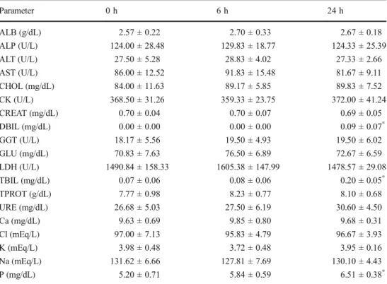
The effect of PTX administration at a dose of 10 mg/kg IV in cattle on biochemical parameters are presented in Table 1. Following PTX administration in cattle, DBIL, TBIL, and P levels were significantly higher at 24 h in comparison to the control values (0 h) (P < 0.05). No significant difference was found in other biochemical parameters (P > 0.05). Food and water intake of the animals was normal throughout the study. Except for clinical symptoms, such as salivation and discom-fort, which lasted approximately 2 h, no adverse drug reac-tions were observed in the animals.
Pharmacokinetic parameters
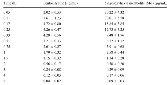
Semilogarithmic plasma concentration-time curves of PTX and M-I following 10 mg/kg IV PTX administration in cattle are presented in Fig.1, and plasma concentrations at sampling times are presented in Table2. After IV administration of PTX at 10 mg/kg in cattle, PTX and M-I were detected at up to 6 h following administration.
Pharmacokinetic parameters of PTX and M-I are presented in Table3. t1/2 z, MRT0-∞, AUC0-∞, Vdss, and ClTvalues of PTX were 1.05 h, 1.19 h, 1.97 h*μg/mL, 6.30 L/kg, and 5.31 L/h/kg, respectively. t1/2 z, MRT0-∞, AUC0-∞, Cmax, and Tmaxvalues of M-I were 1.51 h, 1.54 h, 2.58 h*μg/mL, 30.01 μg/mL, and 0.10 h, respectively. AUC0-∞(M-I)/AUC0-∞(PTX)ratio was 1.34.
Discussion
The efficacy of PTX is dose-dependent, and 10 mg/kg of dose is recommended in horses for hemorheological activity(Liska et al.2006). In the present study, PTX was administered to
cattle at recommended dose for hemorheological effects in microcirculation diseases.
Except for approximately 2-h long transient salivation and discomfort symptoms after PTX administration, no local or systemic adverse effect was observed in the animals. Lethargy, eye closing, and deep respiration were reported in chickens following PTX administration (100 mg/kg, IV and oral) (De Boever et al.2005), and transient side effects, such as sweating, increased heart rate, and fasciculations in mus-cles, were reported in horses (8.5 mg/kg, IV) (Liska et al.
2006). In our study, PTX caused the increase in DBIL,
TBIL, and phosphorus levels in cattle. However, this increase was within the reference range reported for cattle (Radostitis et al.2007). PTX caused no significant change in serum bio-chemical parameters of horses (8.5 mg/kg, IV) and dogs (8– 30 mg/kg, IV and oral) (Rees et al.2003; Liska et al.2006).
The t1/2 zof PTX in cattle was 1.05 h, which is similar to that reported in chicken (1.05 h, De Boever et al. 2005), shorter than that reported in dog (2.73 h, Rees et al. 2003), and longer than that reported in horse (0.38 h, Liska et al.
2006), rat (0.54 h, Italiya et al.2017), and humans (0.84 h, Smith et al.1986). We found that the ClTof PTX was 5.31 L/ h/kg, which is higher than that reported in chicken (1.9 L/h/kg, De Boever et al.2005), horse (2.38–3.06 L/h/kg, Crisman
et al.1993; Liska et al.2006), rat (1.5 L/h/kg, Italiya et al.
2017), and dog (0.03–0.04 L/h/kg, Marsella et al.2000; Rees
et al.2003). The Vdssof PTX was 6.30 L/kg, which is higher than that reported in horse (1.15–2.81 L/kg, Crisman et al.
1993; Liska et al.2006), dog (1.02–4.1 L/kg, Marsella et al. 2000; Rees et al.2003), and rat (1.1 L/kg, Italiya et al.2017). The t1/2 z (1.51 h) value of M-I after PTX administration in cattle was longer than that reported in chicken (0.78 h, De Boever et al.2005) and rat (0.66 h, Italiya et al.2017). The t1/2 zof M-1 in dogs was 1.05–4.78 h (Marsella et al.2000; Rees et al.2003). PTX binds to plasma proteins with a rate of 70% in human, and erythrocytes are an important binding Table 1 Biochemistry parameters
of cattle (n = 6) administrated intravenously pentoxifylline at a dosage of 10 mg/kg Parameter 0 h 6 h 24 h ALB (g/dL) 2.57 ± 0.22 2.70 ± 0.33 2.67 ± 0.18 ALP (U/L) 124.00 ± 28.48 129.83 ± 18.77 124.33 ± 25.39 ALT (U/L) 27.50 ± 5.28 28.83 ± 4.02 27.33 ± 2.66 AST (U/L) 86.00 ± 12.52 91.83 ± 15.48 81.67 ± 9.11 CHOL (mg/dL) 84.00 ± 11.63 89.17 ± 5.85 89.83 ± 7.52 CK (U/L) 368.50 ± 31.26 359.33 ± 23.75 372.00 ± 41.24 CREAT (mg/dL) 0.70 ± 0.04 0.70 ± 0.07 0.69 ± 0.05 DBIL (mg/dL) 0.00 ± 0.00 0.00 ± 0.00 0.09 ± 0.07* GGT (U/L) 18.17 ± 5.56 19.50 ± 4.93 19.50 ± 6.02 GLU (mg/dL) 70.83 ± 7.63 76.50 ± 6.89 72.67 ± 6.59 LDH (U/L) 1490.84 ± 158.33 1605.38 ± 147.99 1478.57 ± 29.08 TBIL (mg/dL) 0.07 ± 0.06 0.08 ± 0.06 0.20 ± 0.05* TPROT (g/dL) 7.77 ± 0.98 8.23 ± 0.77 8.10 ± 0.68 URE (mg/dL) 26.68 ± 5.03 27.50 ± 6.19 30.60 ± 4.50 Ca (mg/dL) 9.63 ± 0.69 9.85 ± 0.80 9.68 ± 0.31 Cl (mEq/L) 97.00 ± 7.13 95.83 ± 4.79 96.67 ± 3.93 K (mEq/L) 3.98 ± 0.48 3.72 ± 0.48 3.95 ± 0.16 Na (mEq/L) 131.62 ± 6.66 127.81 ± 7.69 130.10 ± 4.43 P (mg/dL) 5.20 ± 0.71 5.84 ± 0.59 6.51 ± 0.38*
ALB albumin, ALP alkaline phosphatase, ALT alanine aminotransferase, AST aspartate aminotransferase, CHOL cholesterol, CK creatine kinase, CREAT creatinine, DBIL direct bilirubin, GGT gamma glutamyltransferase, GLU glucose, LDH lactate dehydrogenase, TBIL total bilirubin, TPROT total protein, Ca calcium, Cl chlorine, K potassium, Na sodium, P phosphorus
*
Significantly different from data at 0 h (P < 0.05)
0.01 0.10 1.00 10.00 100.00 0 1 2 3 4 5 6 Concentration ( g/mL) Time (hour)
Fig. 1 Semilogarithmic plots of the plasma concentrations to time data of pentoxifylline (triangles) and its 5-hydroxyhexyl metabolite (squares) in cattle (n = 6) after intravenous administration of pentoxifylline at a dosage of 10 mg/kg (mean ± standard deviation)
target (point) for both PTX and M-I (Harris et al. 2010). Unlike monogastric animals, in ruminants, the reticulo-rumen, which constitutes approximately 20% of the body weight, can change the pharmacokinetics of drugs. The fluid in the reticulo-rumen, which has the great volume and acidic prop-erties (pH 5.5–6.5), may cause to accumulate weakly basic drugs in the reticulo-rumen by the ion-trapping phenomenon and thus change the distribution of the drug (Landoni et al.
1996; Toutain et al.2010; Jerzsele2012). The large Vdss of PTX in cattle may be associated with the different binding rate of PTX to plasma proteins and erythrocytes, and diffusion into reticulo-rumen by ion-trapping phenomenon. The transforma-tion of PTX into M-I occurs in the liver and erythrocytes via a reduction reaction, whereas its transformation into other me-tabolites occurs only in the liver via oxidation reactions (Aviado and Porter 1984). PTX binds to erythrocytes with the approximate rate of 45%, and transforms into fast and reversible M-I (Paap et al.1996). After intravenous adminis-tration of PTX, its clearance is higher compared with that in
hepatic circulation, suggesting that erythrocytes are a major site for PTX–M-I interconversion (Nicklasson et al.2002). As almost all PTX and M-I transform into other metabolites, their urinary elimination are less than 1% (Beermann et al.1985). While the pharmacokinetics of PTX and M-I do not change in case of renal damage (Paap et al. 1996), the ClT value in hepatic damage significantly decreases (Rames et al. 1990). Furthermore, cimetidine inhibited cytochrome (CYP) en-zymes and reduced ClTof PTX by 37% (Luke et al.1986). Therefore, hepatic blood flow and metabolic activity are im-portant for the elimination of PTX and M-I. The CYP1A2 enzyme plays an important role in the transformation of PTX and M-I into other metabolites (Peterson et al. 2004). CYP1A2 enzyme activity was reported to be higher in cattle than in dogs and rats (Sivapathasundaram et al. 2001; Szotáková et al.2004). The higher ClTvalue of PTX in cattle may be caused by different hepatic enzyme activities in cattle in comparison to other species. Vdss and ClTare the most important parameters that affect t1/2 z. In our study, the t1/2 z Table 2 Plasma concentrations at
the sampling times of pentoxifylline and its 5-hydroxyhexyl metabolite (M-I) following intravenous
administration of PTX at a dose of 10 mg/kg in cattle (mean ± SD)
Time (h) Pentoxifylline (μg/mL) 5-hydroxyhexyl metabolite (M-I) (μg/mL)
0.05 2.02 ± 0.53 20.22 ± 4.32 0.1 3.61 ± 1.23 30.01 ± 5.58 0.17 4.72 ± 0.80 15.85 ± 3.85 0.25 4.26 ± 0.47 12.73 ± 3.25 0.33 4.28 ± 0.56 9.48 ± 1.76 0.5 3.21 ± 0.33 6.32 ± 1.12 0.75 2.61 ± 0.27 3.91 ± 0.62 1 1.79 ± 0.32 2.58 ± 0.44 1.5 1.15 ± 0.32 1.34 ± 0.28 2 0.56 ± 0.17 0.58 ± 0.20 3 0.24 ± 0.08 0.29 ± 0.09 4 0.12 ± 0.03 0.17 ± 0.06 6 0.04 ± 0.02 0.09 ± 0.03 Table 3 Pharmacokinetic parameters of pentoxifylline and its 5-hydroxyhexyl metabolite (M-I) in cattle (n = 6) after intravenous administration of pentoxifylline at a dosage of 10 mg/kg (mean ± SD)
Parameter Pentoxifylline 5-hydroxyhexyl metabolite (M-I) t1/2 z(h) (HM) 1.05 ± 0.19 1.51 ± 0.21 AUC0–6(h*μg/mL) 1.91 ± 0.41 2.38 ± 0.45 AUC0-∞(h*μg/mL) 1.97 ± 0.43 2.58 ± 0.50 MRT (h) (HM) 1.19 ± 0.23 1.54 ± 0.29 ClT(L/h/kg) 5.31 ± 1.27 – Vdss(L/kg) 6.30 ± 1.76 – Tmax(h) 0.17 ± 00 0.1 ± 00 Cmax(μg/mL) 4.72 ± 0.81 30.01 ± 5.58
AUC0-∞ (M-I)/AUC0-∞ (PTX) – 1.34 ± 0.25
t1/2 zterminal elimination half-life; AUC(0–6)area under the plasma concentration-time curve from zero (0) hours to time (t); AUC(0–∞)area under the plasma concentration-time curve from zero (0) h to infinity (∞); MRT mean residence time; ClTsystemic clearance; Vdssvolume of distribution at steady state; Tmaxtime to reach maximum concentration; Cmaxmaximal plasma concentration; HM harmonic mean
was not much longer than that in other species, which have high ClTdespite large Vdss.
In cattle, AUC0-∞ (M-I)/AUC0–∞ (PTX)ratio was 1.34. This ratio was lower than that in chicken (1.52, De Boever et al.
2005) and horse (2.40, Liska et al.2006) and higher than that in dog (0.63, Rees et al.2003) and horses (1.13, Crisman et al.
1993). PTX metabolism varies between species and even intra-species. PTX has at fewest seven metabolites in humans, six metabolites in horses (Kwong et al.1989), and three me-tabolites in dogs (Marsella et al.2000). In humans, the major metabolites of PTX are M-I and M-V (Nicklasson et al.2002), whereas, in dogs, M-I is not a major metabolite and M-V was not detected (Marsella et al.2000). However, in another dog study, M-V was detected as the major metabolite of PTX (Rees et al. 2003). Aldo–keto reductase and CYP2E1
en-zymes play an important role in the transformation of PTX into M-I (Raoul et al. 2007; Malátková and Wsól 2014). Furthermore, CYP1A2 is important for the transformation of PTX and M-I into other metabolites. Aldo–keto reductase and CYP enzyme activities are reported to significantly vary be-tween species (Szotáková et al. 2004). The difference of AUC0-∞ (M-I)/AUC0-∞ (PTX) ratio among species may be caused by distinct enzyme activities in different animal species.
The clinical efficacy of PTX is dose-dependent. The therapeutic concentration of PTX in humans is 0.5–2 μg/ mL for different clinical conditions (Regenthal et al.1999) and 1–10 μg/mL in horses for TNF-α inhibition (Barton and Moore 1994). In horses, PTX is reported to inhibit TNF-α at approximately > 1 μg/mL level (Barton et al.
1997). We found that the plasma concentrations of PTX at 1, 1.5, and 2 h were 2, 1, and 0.5μg/mL, respectively. The Cmax of PTX was approximately 4.72 μg/mL. In our study, the plasma concentration of PTX reached the thera-peutic concentration suggested for humans and horses. However, as PTX is recommended for long-term clinical use in humans and horses, multiple dose administrations are required.
Conclusion
PTX was well tolerated by cattle except for transient sali-vation and discomfort. In cattle, PTX exhibited the short elimination half-life and the large volume of distribution. The transformation of PTX into the M-I was similar with other species. These results indicate that single-dose ad-ministration of PTX at 10 mg/kg IV in cattle resulted in therapeutic concentrations similar to those observed in humans. However, further studies are necessary to deter-mine the safety and pharmacokinetics following repeated administrations of PTX.
Acknowledgements This study was presented in abstract form as a poster presentation in the 3rd International Convention of Pharmaceuticals and Pharmacies, Istanbul, Turkey, 26–29 April 2017.
Compliance with ethical standards
The animal experiment was conducted in accordance with the procedure of Committee on Animal Use Ethics (Approval No. 2015/96). Conflict of interest The authors declare that they have no conflict of interest.
References
Adcock, K.G., Kyle, P.B., Deaton, J.S., Olivier, J.H., and Hogan, S.M., 2007. Pharmacokinetics of intranasal and intratracheal pentoxifylline in rabbits, Pharmacotherapy: The Journal of Human Pharmacology and Drug Therapy, 27, 200–206.
Ambrus, J.L., Stadler, S., and Kulaylat, M., 1995. Hemorrheologic effects of metabolites of pentoxifylline (Trental), Journal of Medicine, 26, 65–75.
Aviado, D.M., and Porter, J.M., 1984. Pentoxifylline: a new drug for the treatment of intermittent claudication; mechanism of action, phar-m a c o k i n e t i c s , c l i n i c a l e f f i c a c y a n d a d v e r s e e f f e c t s , Pharmacotherapy: The Journal of Human Pharmacology and Drug Therapy, 4, 297–306.
Barton, M.H., Ferguson, D., Davis, P.J., and Moore, J.N., 1997. The effects of pentoxifylline infusion on plasma 6-keto-prostaglandin F1α and ex vivo endotoxin-induced tumour necrosis factor activity in horses, Journal of Veterinary Pharmacology and Therapeutics, 20, 487–492.
Barton, M.H., and Moore, J.N., 1994. Pentoxifylline inhibits mediator synthesis in an equine in vitro whole blood model of endotoxemia, Circulatory Shock, 44, 216–220.
Beermann, B., Ings, R., Månsby, J., Chamberlain, J., and McDonald, A., 1985. Kinetics of intravenous and oral pentoxifylline in healthy subjects, Clinical Pharmacology & Therapeutics, 37, 25–28. Bessler, H., Gilgal, R., Djaldetti, M., and Zahavi, I., 1986. Effect of
pentoxifylline on the phagocytic activity, cAMP levels, and super-oxide anion production by monocytes and polymorphonuclear cells, Journal of Leukocyte Biology, 40, 747–754.
Cakmak, K.S., Cakmak, A., Gonul, M., Kilic, A., and Gul, U., 2012. Pentoxifylline use in dermatology, Inflammation & Allergy-Drug Targets, 11, 422–432.
Crisman, M.V., Wilcke, J.R., Correll, L.S., and Irby, M.H., 1993. Pharmacokinetic disposition of intravenous and oral pentoxifylline in horses, Journal of Veterinary Pharmacology and Therapeutics, 16, 23–31.
De Boever, S., Baert, K., De Backer, P., and Croubels, S., 2005. Pharmacokinetics and oral bioavailability of pentoxyfylline in broil-er chickens, Journal of Vetbroil-erinary Pharmacology and Thbroil-erapeutics, 28, 575–580.
Harris, E., Schulzke, S.M., and Patole, S.K., 2010. Pentoxifylline in pre-term neonates, Pediatric Drugs, 12, 301–311.
Hinze, H.J., 1972. Pharmacokinetics of 3, 7-dimethyl-1-(5-oxo-hexyl)-xanthine (BL 191) in man, Arzneimittel-Forschung, 22, 1492. Honess, D.J., Dennis, I.F., and Bleehen, N.M., 1993. Pentoxifylline: its
pharmacokinetics and ability to improve tumour perfusion and ra-diosensitivity in mice, Radiotherapy and Oncology, 28, 208–218. Italiya, K.S., Sharma, S., Kothari, I., Chitkara, D., and Mittal, A., 2017.
Simultaneous estimation of lisofylline and pentoxifylline in rat plas-ma by high perforplas-mance liquid chroplas-matography-photodiode array
detector and its application to pharmacokinetics in rat, Journal of Chromatography B, 1061, 49–56.
Jerzsele, A., 2012. Comparative Veterinary Pharmacokinetics, In: Noreddin A (ed), Readings in Advanced Pharmacokinetics— Theory, Methods and Applications, IntechOpen.https://doi.org/10. 5772/1982.
Kwong, E.C., Chen, F.C., and Young, L.M., 1989. Urinary excretion of pentoxifylline and its metabolites by standardbred mares, Canadian Journal of Veterinary Research, 53, 147–153.
Landoni, M.F., Cunningham, F.M., and Lees, P., 1996. Pharmacokinetics and pharmacodynamics of tolfenamic acid in calves, Research in Veterinary Science, 61, 26–32.
Liska, D.A., Akucewich, L.H., Marsella, R., Maxwell, L.K., Barbara, J.E., and Cole, C.A., 2006. Pharmacokinetics of pentoxifylline and its 5-hydroxyhexyl metabolite after oral and intravenous administra-tion of pentoxifylline to healthy adult horses, American Journal of Veterinary Research, 67, 1621–1627.
Luke, D.R., Rocci, M.L., and Hoholick, C., 1986. Inhibition of pentoxifylline clearance by cimetidine, Journal of Pharmaceutical Sciences, 75, 155–157.
Malátková, P., and Wsól, V., 2014. Carbonyl reduction pathways in drug metabolism, Drug Metabolism Reviews, 46, 96–123.
Marsella, R., Nicklin, C.F., Munson, J.W., and Roberts, S.M., 2000. Pharmacokinetics of pentoxifylline in dogs after oral and intrave-nous administration, American Journal of Veterinary Research, 61, 631–637.
Nicklasson, M., Björkman, S., Roth, B., Jönsson, M., and Höglund, P., 2002. Stereoselective metabolism of pentoxifylline in vitro and in vivo in humans, Chirality, 14, 643–652.
Paap, C.M., Simpson, K.S., Horton, M.W., Schaefer, K.L., Lassman, H.B., and Sack, M.R., 1996. Multiple-dose pharmacokinetics of pentoxifylline and its metabolites during renal insufficiency, Annals of Pharmacotherapy, 30, 724–729.
Peterson, T.C., Peterson, M.R., Wornell, P.A., Blanchard, M.G., and Gonzalez, F.J., 2004. Role of CYP1A2 and CYP2E1 in the pentoxifylline ciprofloxacin drug interaction, Biochemical Pharmacology, 68, 395–402.
Radostitis, O.M., Gay, C.C., Blood, D.C., and Hinchcliff, K.W., 2007. Veterinary Medicine: A Textbook of the Diseases of Cattle, Sheep, Pigs, Goats and Horses. 10th ed, (W. B. Saunders, London), 2048– 2050.
Rames, A., Poirier, J.M., LeCoz, F., Midavaine, M., Lecocq, B., Grange, J.D., Poupon, R., Cheymol, G., and Jaillon, P., 1990. Pharmacokinetics
of intravenous and oral pentoxifylline in healthy volunteers and in cirrhotic patients, Clinical Pharmacology & Therapeutics, 47, 354– 359.
Raoul, J.M., Peterson, M.R., and Peterson, T.C., 2007. A novel drug interaction between the quinolone antibiotic ciprofloxacin and a chiral metabolite of pentoxifylline, Biochemical Pharmacology, 74, 639–646.
Rees, C., Boothe, D.M., Boeckh, A., Wilkie, S., Esparza, T., and Green, R., 2003. Dosing regimen and hematologic effects of pentoxifylline and its active metabolites in normal dogs, Veterinary Therapeutics: Research in Applied Veterinary Medicine, 4, 188–196.
Regenthal, R., Krueger, M., Koeppel, C., and Preiss, R., 1999. Drug levels: therapeutic and toxic serum/plasma concentrations of com-mon drugs, Journal of Clinical Monitoring and Computing, 15, 529– 544.
Samlaska, C.P., and Winfield, E.A., 1994. Pentoxifylline, Journal of the American Academy of Dermatology, 30, 603–621.
Sivapathasundaram, S., Magnisali, P., Coldham, N.G., Howells, L.C., Sauer, M.J., and Ioannides, C., 2001. A study of the expression of the xenobiotic-metabolising cytochrome P450 proteins and of tes-tosterone metabolism in bovine liver, Biochemical Pharmacology, 62, 635–645.
Smith, R.V., Waller, E.S., Doluisio, J.T., Bauza, M.T., Puri, S.K., Ho, I., and Lassman, H.B., 1986. Pharmacokinetics of orally administered pentoxifylline in humans, Journal of Pharmaceutical Sciences, 75, 47–52.
Sykes, J.E., and Papich, M.P., 2014. Antiviral and Immunomodulatory Drugs, In: Sykes, J.E. (ed), Canine and Feline Infectious Diseases (Elsevier Saunders, St. Louis, Missouri 63043), 54–65.
Szotáková, B., Baliharová, V., Lamka, J., Nožinová, E., Wsól, V., Velık, J., Machala, M., Neča, J., Souček, P., Šusová, S., and Skálová, L., 2004. Comparison of in vitro activities of biotransformation en-zymes in pig, cattle, goat and sheep, Research in Veterinary Science, 76, 43–51.
Toutain, P.L., Ferran, A., and Bousquet-Mélou, A., 2010. Species differ-ences in pharmacokinetics and pharmacodynamics, Handbook of Experimental Pharmacology, 199, 19–48.
Wyska, E., 2010. Pharmacokinetic-pharmacodynamic modeling of meth-ylxanthine derivatives in mice challenged with high-dose lipopoly-saccharide, Pharmacology, 85, 264–271.
Yang, Z., Chen, M., and Nadler, J.L., 2005. Lisofylline: a potential lead for the treatment of diabetes, Biochemical Pharmacology, 69, 1–5.