1 Giresun University Department of Cardiovascular Surgery, Giresun, Turkey 2 Giresun University Department of Cardiology, Giresun, Turkey 3 Giresun University Department of Otorhinolaryngology, Giresun, Turkey
4 Ahi Evren Education and Research Hospital, Department of Cardiovascular Surgery Trabzon, Turkey 5 Ordu University Department of Cardiology, Ordu, Turkey
Correspondence: Ahmet Karagöz,
Giresun University, Department of Cardiology, Giresun, Turkey Email: [email protected] Received: 30.12.2014, Accepted: 15.01.2015
Copyright © JCEI / Journal of Clinical and Experimental Investigations 2015, All rights reserved
JCEI / 2015; 6 (1): 69-71
Journal of Clinical and Experimental Investigations doi: 10.5799/ahinjs.01.2015.01.0489 CASE REPORT / OLGU SUNUMU
A rare cause of Ortner’s syndrome: giant pulmonary artery aneurysm secondary to
Behçet’s disease
Ortner sendromu’ nun nadir bir nedeni: Behçet hastalığına sekonder gelişen dev pulmoner
arter anevrizması
Abdullah Çelik1, Ahmet Karagöz2, Erdal Seren3, Sefer Usta4, Aslı Vural2, Zeki Yüksel Günaydın5, Osman Bektaş5 ABSTRACT
Behçet’ s disease is a systemic autoimmune vasculitis of unknown etiology. It causes serious disability by affect-ing both arteries and veins. Hoarseness due to compres-sion of the left recurrent laringeus nerve resulting from pathologies of the heart and intrathoracic great vessels is defined as Ortner’s syndrome. The most common cause of Ortner’s syndrome is left atrial enlargement due to mi-tral stenosis. Various intrathoracic pathologies may also be the reason. Beside, Ortner’s syndrome due to primary pulmonary artery aneurysm as a feature of Behçet’s dis-ease is relatively rare. Herein, we report a case of a 78 year old female patient presenting with hoarseness and diagnosed as Ortner’s syndrome resulting from a giant pulmonary artery aneurysm secondary to Behçet’ s dis-ease. J Clin Exp Invest 2015; 6 (1): 69-71
Key words: Ortner’ s Syndrome, Pulmonary Artery Aneu-rysm, Behçet’s Disease
ÖZET
Behçet Hastalığı nedeni bilinmeyen sistemik bir otoim-mün vaskülittir. Hem arterleri hem de venleri tutarak ciddi sorunlara neden olabilir. Kalp ve intratorasik büyük da-marların patolojilerine bağlı olarak sol rekürren laringeal sinirin kompresyonu sonucu gelişen ses kısıklığı Ortner Sendromu olarak ifade edilir. Ortner Sendromu’ nun en sık nedeni mitral darlığa bağlı olarak sol atriyum genişle-mesidir. Çeşitli intratorasik patolojiler bu tablonun nedeni olabilir. Bununla birlikte Behçet Hastalığı sonucu gelişen pulmoner arter anevrizmasına bağlı Ortner Sendromu nispeten nadirdir. Biz burada ses kısıklığı ile başvuran ve yapılan incelemeler sonucunda Behçet Hastalığı’ na bağ-lı dev pulmoner arter anevrizması sonucu gelişen Ortner Sendromu olarak tanı alan 78 yaşında bir bayan hastayı sunuyoruz.
Anahtar kelimeler: Ortner Sendromu, Pulmoner Arter Anevrizması, Behçet Hastalığı
INTRODUCTION
Behçet’ s disease is a multiorgan disorder of un-known origin and can cause complications in car-diovascular system, central nervous system and respiratory system resulting from vasculitis of both arteries and veins [1,2]. Pulmonary involvement rate in Behcet’s disease is between 5-10 % and is seen most often in the form of pulmonary artery an-eurysm (PAA) [3]. Vocal cord paralysis due to ex-tralaryngeal cardiovascular pathologies, such as the thoracic aorta aneurysms and PAA is defined as
Ortner syndrome. Left vocal cord paralysis due to PAA is quite rare [4,5]. Herein, we report a case of a 78 year old female patient presenting with hoarse-ness and diagnosed as Ortner’s syndrome resulting from a giant pulmonary artery aneurysm secondary to Behçet’ s disease.
CASE PRESENTATION
A 78 year-old female patient suffering from fever, fa-tigue, recurrent oral ulcers, and progressive
hoarse-Çelik A. et al. Ortner’s syndrome secondary to Behçet’s disease
70
J Clin Exp Invest www.jceionline.org Vol 6, No 1, March 2015
ness in the last 6 months admitted to our clinic. The medical history revealed that she had been follow-ing with the diagnosis of Behcet’s disease for ap-proximately 10 years and had received corticoste-roid treatment previously. But she was not on a reg-ular treatment regimen. Pathergy test for diagnostic evaluation of Behcet’s disease was repeated and found to be positive.
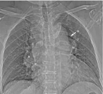
On physical examination, arterial blood pres-sure was 100 / 60 mmHg and radial pulse was regular with a rate of 100 / min. Cardiovascular sys-tem examination was normal except tachycardia. In teleradiography, a prominent pulmonary artery was detected (Figure 1). On transthoracic echocardiog-raphy, right and left ventricular size and functions were normal, but the main pulmonary artery and its branches were excessively dilated . Doppler echo-cardiography revealed mild tricuspid and pulmo-nary regurgitation. Pulmopulmo-nary artery systolic pres-sure meapres-sured through tricuspid regurgitant flow was 32 mmHg and diastolic and mean pressures of pulmonary artery were detected to be 14 and 18 mmHg respectively. In multislice CT angiography, main pulmonary artery was measured to be 55 mm at the level of the aorticopulmonary window. Left pulmonary artery was 32 mm and right pulmonary artery was 42 mm (Figure 2). The patient underwent endoscopic examination of the larynx and the left vocal cord was found to be fixed in the paramedian position.
Figure 1. Antero-posterior chest x-ray shows aneurysmal main pulmonary artery and its branches in central and pe-ripheral localizations
Figure 2. Axial contrast-enhanced CT image shows sac-cular aneurysm at the level of aorticopulmonary window
Surgery was not considered due to existence of Behçet ‘s disease, advanced age of the patient and extent of the aneurysm involving both pulmo-nary artery and its branches. Considering systemic symptoms and oral ulcers, the patient was thought to have an acute attack and medical therapy was started. Remission of fever, fatigue and oral ulcer was achieved with cyclophosphamide and cortico-steroid therapy. A mildly improvement in hoarseness was also observed. The patient was discharged with follow up recommendations. The patient maintained asymptomatic on medical follow up.
DISCUSSION
PAA is a quite rare clinical condition [6]. The most common type of vasculitis associated with pulmo-nary artery aneurysms are Behcet’s disease and Hughes-Stovin syndrome [7]. The patients with PAA are usually asymptomatic or may present with non-specific complaints such as exertional dyspnea, fe-ver, cough and hemoptysis. In some patients, com-pression of surrounding structures via the enlarged pulmonary artery or stretching of the pulmonary artery itself can cause symptoms. Hoarseness due to compression of the left recurrent laringeus nerve secondary to pathologies of the heart and intratho-racic great vessels is defined as Ortner’s syndrome. The most common cause of Ortner’s syndrome is left atrial enlargement due to mitral stenosis. Mitral valve prolapse, thoracic aortic aneurysm, aortic dis-section, cor pulmonale and pulmonary artery dilata-tion secondary to increased pulmonary artery pres-sure with any reason have also been described in etiology of Ortner’s syndrome [8,9].
Çelik A. et al. Ortner’s syndrome secondary to Behçet’s disease 71
J Clin Exp Invest www.jceionline.org Vol 6, No 1, March 2015
Several etiologies have been described in the pathogenesis of PAA, namely, pulmonary hyper-tension (PH), congenital heart disease, Behçet’ s disease, infections such as the formerly prevalent syphilis, arteriovenous fistulas, connective tissue diseases, atherosclerosis, and trauma [10, 11]. Our patient had none of these etiological factors except Behçet’ s disease. Therefore, the point that makes this paper worthy of reporting is development of Ort-ner’s syndrome due to pulmonary artery aneurysm probably resulting from Behçet’s disease. Absence of increased pulmonary arterial pressure leads to the diagnosis of pulmonary artery aneurysm in our patient and hoarseness was due to enlargement of pulmonary artery. Beyond these clinical char-acteristics, the patients with PAA are also at high risk for dissection or rupture which are mortal [12]. Therefore, corrective surgery is recommended, but the risks and long-term results of surgical treatment are not well defined. In our case, while the absence of pulmonary hypertension reduces the risk of an-eurysm rupture, the presence of Behçet’s disease increases the risk [13]. Remission of the systemic symptoms and improvement in hoarseness with medical therapy led us to avoid surgery in this case. The improvement in hoarseness was probably due to resolution of the edema of the pulmonary arterial wall.
Our report is one of the rare PAA cases second-ary to Behçet’s disease causing Ortner’s syndrome in the literature. Consequently, it should be taken into consideration that hoarseness in a patient with Behcet’s disease may develop due to a PAA and further evaluation should be performed. Additional-ly, although surgery seems to be the effective treat-ment modality of PAA, in Behçet’s disease steroids targeting the primary cause may provide clinical im-provements and eliminate the need for surgery.
REFERENCES
1. Türkcü FM, Yüksel H, Uçmak D, et al. Demographic char-acteristics and ocular involvement in Behçet patients in Southeast Anatolia. J Clin Exp Invest 2013;4:339-342. 2. Küçük A, Albayrak İ, Bağçacı S, Küçükşen S, Tunç R.
Upper extremity thrombosis in Behçet. Dicle Med J 2013;40:507-509.
3. Tüzün H, Hamuryudan V, Yıldırım S, et al. Surgical ther-apy of pulmonary arterial aneurysms in Behçet’s syn-drome. Ann Thorac Surg 1996;61:733-735.
4. Day JR, Walesby RK. A spontaneous ductal aneurysm presenting with left recurrent laryngeal nevre palsy. Ann Thorac Surg 2001;72:608-609.
5. Kokotsakis J, Misthos P, Athanassiou T, et al. Acute Ort-ner’s syndrome arising from ductus arteriosus aneu-rysm. Tex Heart Inst J 2008;35:216-217.
6. Shih HH, Kang PL, Lin CY, et al. Main pulmonary artery aneurysm. J Chin Med Assoc 2007;70:453-455. 7. Imazio M, Cecchi E, Giammaria M, et al. Main pulmonary
artery aneurysm: a case report and review of the litera-ture. Ital Heart J 2004;5:232-237.
8. Subramaniam V, Herle A, Mohammed N, et al. Ortner’s syndrome: case series and literature review. Braz J Oto-rhinolaryngol 2011;77:559-562.
9. Mulpuru SK, Vasavada BC, Punukollu GK, et al. Cardio-vocal syndrome: a systematic review. Heart Lung Circ 2008;17:1-4.
10. Sakuma M, Demachi J, Suzuki J, et al. Proximal pul-monary artery aneurysms in patients with pulpul-monary artery hypertension: Complicated cases. Intern Med 2007;46:1789-1793.
11. Bartter T, Irwin R, Nash G. Aneurysms of the pulmonary arteries. Chest 1988;94:1065-1075.
12. Smalcelj A, Brida V, Samarzija M, et al. Giant, dis-secting, high-pressure pulmonary artery aneurysm: case report of a 1-year natural course. Tex Heart Inst J 2005;32:589-594.
13. Graham JK, Shehata B. Sudden death due to dissecting pulmonary artery aneurysm: a case report and review of the literature. Am J Forensic Med Pathol 2007;28:342-344.