Mechanical Chest Compression Devices: Historical Evolution,
Classification and Current Practices, A Short Review
Mahmure Aygün1, Hacer Erten Yaman2, Aslı Genç1, Funda Karadağlı1, Nadiye Barış Eren1 1Department of Nursing, İstanbul Gelişim University College of Health Science, İstanbul, Turkey 2Department of Nursing, İstanbul Maltepe University College of Health Science, İstanbul, Turkey
Introduction
Standard cardiopulmonary resuscitation (S-CPR) refers to the entire body of techniques of external chest compression and se-curing positive pressure ventilation for the purpose of achieving adequate blood and oxygen flow into vital organs such as the he-art and brain following cardiac arrest (1). The current application of S-CPR is based on the technique of “external chest compression” that was defined by Kouwenhoven in 1960 and comprises the pha-ses of active compression and passive decompression. Despite the evolution of resuscitation medicine, the limited improvement in survival rates following cardiac arrest has led researchers to explore the possibility of different CPR techniques and also to develop de-vices that support ventilation and circulation (2-6). This manuscript was prepared to review the experimental and clinical studies con-ducted on the historical progress and effectiveness of mechanical chest compression devices (MCCD).
Limitations of S-CPR
The fundamental goal of effective CPR applications is to achieve return of spontaneous circulation (ROSC) and a good neurological outcome and the return of the patient to the patient’s previous
qu-ality of life and functional level of health. Guidelines emphasize the importance of effective chest compression for successful CPR. The effectiveness of chest compressions depends on a couple of para-meters (such as application of compressions to the right place, at an adequate depth and rate, on a regular and uninterrupted basis; let-ting the chest to fully recoil after each compression; avoiding over-ventilation; and maintaining a balance between compression and ventilation) (1, 5, 7, 8).
One of the basic problems related to S-CPR techniques is that even in the most effective chest compressions, a physiologically adequate amount of cardiac output may not be reached and be-cause the quality of compression may change over time, this may cause cerebral and coronary blood flow to reduce even further as a result of the interruptions (9-11). If the chest compressions could reach the needed depth, as it does in infants and children, a higher intrathoracic pressure and cardiac output would be possible (12, 13). Another problem is that the quality of CPR is limited to the degree of knowledge, experience, and endurance of the rescuer (9, 14-17). Transferring the patient into an ambulance, discontinuing CPR prior to defibrillation, the difficulty of effectively applying the technique in a moving ambulance, failure to maintain the relationship between compression and ventilation, and reduced elastic recoil of the chest Correspondence to: Mahmure Aygün e-mail: [email protected]
Received: 18.11.2015 Accepted: 02.03.2016
©Copyright 2016 by Emergency Physicians Association of Turkey - Available online at www.eajem.com DOI: 10.5152/eajem.2016.74936
Abstract
The standard treatment of cardiac arrest is cardiopulmonary resuscitation (CPR), performed with effective manual chest compressions. Although current CPR was developed 50 years ago, cardiac arrest still has a high mortality rate and manual chest compressions have some potential limitations. Because of these limitations, mechanical chest compression devices were developed to improve the efficiency of CPR. This CPR technology includes devices such as the mechanical piston load-distributing band, active compression–decompression CPR, simultaneous sterno-thoracic CPR, impedance threshold valve, phased thoracic-abdominal active compression–decompression CPR and active compression-decompression CPR with enhanced external counterpulsation, and the impedance threshold valve. The purpose of this manuscript was to draw attention to developments in this medical area and to examine studies on the effectiveness of these devices. (Eurasian J Emerg Med 2016; 15: 94-104)
wall in prolonged CPR applications could all have an adverse effect on the success of CPR (5, 7, 13-17).
When the importance of chest compression is considered and in the light of the issues experienced in currently employed techniques and the low survival rates following cardiac arrest, the necessity of further developing S-CPR techniques and increasing the effective-ness of chest compression becomes clear.
Mechanical Chest Compression Devices: Definition, History, and Classification
Definition
MCCDs are noninvasive circulation support devices that func-tion manually, pneumatically, or electrically and in accordance with CPR guidelines, provide uninterrupted and effective external chest compression to achieve an adequate blood flow to the heart and other vital organs during non-traumatic adult cardiac arrest. MCCDs can be used as an alternative to S-CPR in cases that may hinder effec-tive compressions such as prolonged CPR during the transport of the patients or in the shortage of personnel (2, 6, 10, 18, 19).
These devices are included in the guidelines of the American Heart Association (AHA) under the heading “circulatory support devi-ces” (6). They are described by the U.S. Food and Drug Administration (FDA) as “External cardiac compressors” (19). In the literature, the no-menclature varies, and “external cardiac compression devices,” “auto-matic chest-compression devices,” and “mechanical CPR devices” are some of the terms of reference (2, 10, 13, 18).
History
The advent of MCCDs is not new. These devices began to be developed in the beginning of the 1960s, when resuscitation medi-cine was merely in its infancy. Chronologically speaking, the “elect-ro-pneumatic machine” developed by Harkins and Bramson (20) in 1961; the “portable pneumatic pump” developed by Nachlas and Siedband (21) in 1962; the “Beck-Rand external cardiac compression machine” developed by Safar et al. (22) in 1963; the “cardiac massage machine” developed by Bailey and Browse in 1964 (23); and the “hos-pital mechanical pump” developed by Nachlas and Siedband (21) in 1965 were the first MCCDs to be invented. In later years, experiments were conducted with many manually operated devices such as the “cardio-massager,” “cardio-pulser,” pneumatical “iron heart,” and “Tra-venol LR50-90” (13, 24).
Many of the first developed and tested of these devices were very complex, too heavy, or ineffective for use in CPR; therefore, they were found to be nonfunctional and unacceptable for the clinical set-ting. On the other hand, since the 2000s, many devices have begun to be developed and have found a clinical area of use, and the lite-rature of the effects of these devices on CPR outcomes are steadily increasing.
Classification
These devices are different from one another in terms of their working principles, the energy they consume, and their electronic features. MCCDs used currently can be classified as follows:
1. Piston-driven CPR devices (PD-CPR),
2. Load-distributing band CPR devices (LDB-CPR).
In addition to these two fundamental groups, third-generation devices, which combine different working mechanisms and different
CPR techniques, have been used in recent years, aiming to increase the hemodynamic effects of S-CPR. These are as follows:
3. Active compression–decompression CPR devices (ACD-CPR), 4. Simultaneous sterno-thoracic cardiopulmonary resuscitation
devices (SST-CPR/S-CPR/X –CPR),
5. Inspiratory impedance threshold valve/devices and ResQCPR (ACD + ITD CPR),
6. Phased thoracic-abdominal active compression–decompressi-on CPR devices (PTACD-CPR), and
7. Active compression–decompression CPR with enhanced exter-nal counterpulsation and the inspiratory impedance threshold valve (AEI-CPR) (2, 6, 13, 25-29).
The devices in these groups and their working principles have been discussed below.
1. Piston-driven devices (PD-CPR)
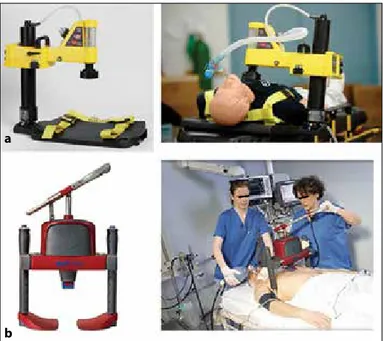
These are based on the “cardiac pump theory” and are first-ge-neration mechanical devices that use a piston to exert “single-point compression” on the sternum. One of the first examples in this group was the Pneumatically Run Thumper. A more developed model of this device is the Thumper Mechanical CPR Device Model 1007 and its updated model the Life-Stat. The Life-Stat consists of a backboard attached to a column and operates pneumatically with a piston. It has a ventilator that is meant to be used in conjunction with chest compression (Figure 1a) (Michigan Instruments, USA). Mechanical piston-driven devices that are operated manually work with a lever system and are marketed under brand names such as the “Animax Mono” (Figure 1b) (AAT Alber Antriebstechnik GmbH, Albstadt, Ger-many) and the “CPR RsQAssist,” which employs an audio-visual met-ronome (10, 13, 24, 26, 29).
2. Load-distributing band devices (LDB-CPR)
“Load-distributing band devices” are based on the “thoracic pump theory” and represent second-generation mechanical chest compression technology. These devices exert thoracic compression on the anterior-anterolateral thorax using a wide pneumatic band that wraps around the chest, inflating and deflating at cyclically. The basic equipment in these devices consists of a backboard, a chest compression band (load-distributing Life Band), and a power system. The first example of this type of device was the “Vest-CPR.” Currently, devices that work with this mechanism are marketed under commer-cial names such as the pneumatic Automated CPR Vest (Reax resus-citation device) and the pneumatic or electrical AutoPulse (Figure 2). Studies show that the chest compression achieved all around the chest with the AutoPulse creates higher coronary perfusion pressure than sternal pressure (13, 24, 25, 29-33).
3. Active compression–decompression CPR devices (ACD-CPR)
ACD-CPR devices are third-generation devices that work on the piston principle. These devices were developed based on a news article published in 1990 about a successful resuscitation at-tempt of a lay person performed with a toilet plunger to his father (34, 35). As is known, in S-CPR, the return of blood to the heart is dependent only on the passive recoil of the chest wall. The prin-ciple behind this technique may be summarized as the pumping of blood outside of the thorax through positive pressure in the active compression phase and then exerting an external negative
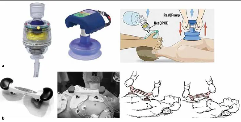
vacu-um, creating an intrathoracic negative pressure during the active decompression phase to increase the venous return of blood to the heart. Thus, in the next compression phase, an increase is achieved in cardiac output, coronary and cerebral blood flow, and in arteri-al blood pressure. The most widely known and commonly used of the ACD-CPR devices is the LUCAS (Lund University Cardiac Assist System). The original LUCAS 1 was a pneumatic device that was de-veloped in Sweden by Steen in 2002. The new model of the device, introduced in 2010 under its new name LUCAS 2 (LUCAS™ 2 Chest Compression System), runs on electricity and consists of a piston for compression, a silicone suction cup for decompression, a rechar-geable battery, a backboard, and connecting legs (Figure 3a). The device allows defibrillation without interrupting compression, and its X-ray translucent capability makes cardiac catheterization pos-sible. The ACDC Thumper is another pneumatic device. The manu-al devices that operate with the ACD-CPR technique are marketed under trade names such as CardioPump, ResQPump, and Ambu® Cardio Pump (Figure 3b) (6, 13, 24, 29, 36-40).
4. Simultaneous sterno-thoracic CPR devices (SST-CPR / X-CPR)
These devices were designed to benefit from both the cardiac pump and thoracic pump theories. These devices have two com-ponents: a piston (which depresses the sternum in the compressi-on phase) and a circumferential band (which ccompressi-onstricts the thorax simultaneously compressions).The “Life Belt” is a device that is opera-ted manually using the SST-CPR principle. Another such device is the pneumatic “Weil Mini Chest Compressor” (Figure 4) (Resuscitation International, USA) (13, 41-44).
5. Inspiratory Impedance Threshold Device (ITD) and ResQCPR (ACD+ITD CPR)
The inspiratory ITD is a pressure-sensitive one-way valve sys-tem that can be connected to a face mask or to any developed airways equipment such as endotracheal tubing. The valve closes in the decompression phase of CPR, temporarily blocking the more than necessary passage of passive air through the open airway into the patient’s lungs, thus decreasing intrathoracic pressure and cre-ating a small vacuum. This increases the flow of venous blood to the heart, and the increased venous return increases cardiac out-put in the next compression. ITD are marketed under the trademark “ResQPOD® ITD 16.” ITD can be used alone during S-CPR as well as it may be used in combination with manual ACD-CPR devices such as the CardioPump and the ResQPump. This system is known as ResQ-CPR. ResQCPR=ACD-CPR (ResQPUMP)+ITD (ResQPOD) (Figure 5a) (6, 27, 45-47).
Figure 2. Load-distributing band CPR devices
Auto Pulse (69)
Figure 3. a, b. Active compression–decompression CPR devices. (a)
Pneumatic/electrically driven ACD-CPR devices: LUCAS 2 (36). (b) Ma-nual driven ACD-CPR devices: ResQPump (18).
a
b
Figure 4. Simultaneous sterno-thoracic cardiopulmonary
resuscita-tion devices
Weil Mini Chest Compressor (http://www.resusintl.com/)
Figure 1. a, b. Piston-driven CPR devices (a) Pneumatic: Thumper
(Li-feStat) (https://www.michiganinstruments.com). (b) Manual: Animax Mono (http://www.aat-online.de/)
a
6. Phased thoracic-abdominal active compression– decompression CPR devices (PTACD-CPR)
Interposed abdominal compression CPR (IAC-CPR) activates the abdominal venous reservoir by increasing abdominal pressure; this CPR technique is based on forcing venous return, thereby increasing venous return to the heart. Abdominal compression is applied to the area midpoint between the xiphoid and umbilicus in the relaxa-tion phase of chest compression. Phased thoracic-abdominal active compression–decompression CPR constitutes the working principle behind ACD-CPR and IAC-CPR and is a new method that combines the two techniques. PTACD-CPR is applied by simultaneous chest compression (positive intrathoracic pressure) and active abdominal decompression and then following this phase, simultaneous active chest decompression (negative intrathoracic pressure) and abdomi-nal compression. The Lifestick™ was developed for use in this tech-nique; it is a manually controlled device. The device consists of a rigid central bar and two arms with adhesive pads that are connected to this rigid bar. The larger adhesive pad is placed over the abdomen and the smaller over the anterior chest wall. An implementer com-presses the two sides of the device just like a seesaw, applying pres-sure both on the chest and the abdomen alternately (Figure 5b) (11, 27, 48-50).
7. Active compression–decompression CPR with Enhanced External Counterpulsation and the Inspiratory Impedance Threshold Valve (AEI-CPR)
Enhanced external counterpulsation (EECP) is a circulatory sup-port system that achieves increased cardiac output using a method whereby cuffs applied to the lower extremities are inflated during diastole to increase coronary blood circulation and deflated at the early systole to reduce afterload and increase venous return. AEI-CPR is another experimental technique, which is a combination of active
compression–decompression CPR, EECP, and Inspiratory impedance threshold valve, aiming to improve CPR hemodynamics and increase survival rates. This technique, which is still in its theoretical and expe-rimental stage, is simply expressed as AEI-CPR=ACD-CPR+EECP+ITV (51, 52).
Studies Conducted on the Effectiveness of MCCDs
Theoretically, MCCDs appear to provide many practical advan-tages, such as the mechanical devices deliver compressions at the same frequency and depth which are recommended in the guide-lines, as opposed to the inter-rescuer variations and fatigue factors that affect the quality of chest compression; these devices allow the rescuers to perform other tasks (cannulation, airway, etc.) and defib-rillation without the need of interruption in CPR; and they provide consistent rate and depth of chest compressions during transport of the patient.
However, the main issue is to what degree these devices have an impact on survival in cardiac arrest, on hemodynamic parameters, and on the survival neurologically intact and whether they produ-ce a significant differenprodu-ce in in-hospital and out-of-hospital cardiac arrests (IHCA and OHCA, respectively) compared to S-CPR. Experi-mental studies conducted with some mechanical chest compression devices developed in recent years present strong evidence that the-se devices increathe-se the effectiveness and quality of CPR. Moreover, although they were first developed to achieve uninterrupted CPR, particularly in cases of OHCA, the studies on the use of these devi-ces in IHCAs help to expand their clinical usage area. This section will review some of the results of some MCCD-related experimental or clinical studies on in-hospital and out-of-hospital cases.
Experimental studies with LUCAS-CPR have shown that the device enables significantly higher cerebral blood circulation than S-CPR as well as higher rates of cardiac output, carotid artery blood
Figure 5. a, b. Other CPR devices. (a) Impedance threshold device (ITD) and ResQCPR: ResQPod+ResQPump (46). (b) Phased
thoracic-abdomi-nal compression–decompression CPR: Lifestick (48)
a
Title Study design Results Conclusion
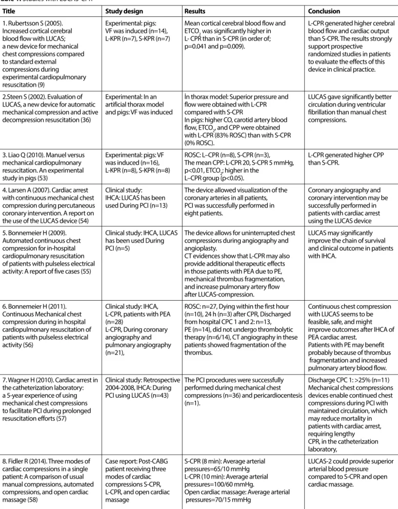
1. Rubertsson S (2005). Experimental: pigs: Mean cortical cerebral blood flow and L-CPR generated higher cerebral Increased cortical cerebral VF was induced (n=14), ETCO2 was significantly higher in blood flow and cardiac output blood flow with LUCAS; L-KPR (n=7), S-KPR (n=7) L- CPR than in S-CPR (in order of; than S-CPR. The results strongly
a new device for mechanical p=0.041 and p=0.009). support prospective
chest compressions compared randomized studies in patients
to standard external to evaluate the effects of this
compressions during device in clinical practice.
experimental cardiopulmonary resuscitation (9)
2.Steen S (2002). Evaluation of Experimental: In an İn thorax model: Superior pressure and LUCAS gave significantly better LUCAS, a new device for automatic artificial thorax model flow were obtained with L-CPR circulation during ventricular mechanical compression and active and pigs: VF was induced compared with S-CPR fibrillation than manual chest decompression resuscitation (36) In pigs: higher CO, carotid artery blood compressions.
flow, ETCO2, and CPP were obtained with L-CPR (83% ROSC) than with S-CPR (0% ROSC).
3. Liao Q (2010). Manuel versus Experimental: pigs: VF ROSC: L–CPR (n=8), S-CPR (n=3), L-CPR generated higher CPP mechanical cardiopulmonary was induced (n=16), The mean CPP: L-CPR 20, S-CPR 5 mmHg, than S-CPR.
resuscitation. An experimental L-KPR (n=8), S-KPR (n=8) p<0.01, ETCO2; higher in the
study in pigs (53) L–CPR group (p<0.05).
4. Larsen A (2007). Cardiac arrest Clinical study: The device allowed visualization of the Coronary angiography and with continuous mechanical chest IHCA: LUCAS has been coronary arteries in all patients, coronary intervention may be compression during percutaneous used During PCI (n=13) PCI was successfully performed in successfully performed in coronary intervention. A report on eight patients. patients with cardiac arrest
the use of the LUCAS device (54) using the LUCAS device
5. Bonnemeier H (2009). Clinical study: IHCA, LUCAS The device allows for uninterrupted chest LUCAS may significantly Automated continuous chest has been used During compressions during angiography and improve the chain of survival compression for in-hospital PCI (n=5) angioplasty. and clinical outcome in patients cardiopulmonary resuscitation CT evidences show that L-CPR may also with IHCA.
of patients with pulseless electrical provide additional therapeutic effects activity: A report of five cases (55) in those patients with PEA due to PE,
mechanical thrombus fragmentation, and increase pulmonary artery flow after LUCAS-compression.
6. Bonnemeier H (2011). Clinical study: IHCA, ROSC: n=27, Dying within the first hour Continuous chest compression Continuous Mechanical chest L-CPR, patients with PEA (n=10), 24 h (n=3) after CPR, Discharged with LUCAS seems to be compression during in hospital (n=28) from hospital CPC 1 and 2: n=13, feasible, safe, and might cardiopulmonary resuscitation of L-CPR, During coronary PE (n=14), did not undergo thrombolytic improve outcomes after IHCA of patients with pulseless electrical angiography and therapy (n=6/14), CT angiography in these PEA cardiac arrest.
activity (56) pulmonary angiography patients showed fragmentation of the Patients with PE may benefit
(n=21), thrombus. probably because of thrombus
fragmentation and increased
pulmonary artery blood flow. 7. Wagner H (2010). Cardiac arrest in Clinical study: Retrospective The PCI procedures were successfully Discharge CPC 1: >25% (n=11) the catheterization laboratory: 2004-2008, IHCA: During performed during mechanical chest Mechanical chest compressions a 5-year experience of using PCI using LUCAS (n=43) compressions (n=36) and pericardiocentesis devices enable continued chest
mechanical chest compressions (n=1). compressions during PCI with
to facilitate PCI during prolonged maintained circulation, which
resuscitation efforts (57) may reduce mortality in
patients with cardiac arrest, requiring lengthy
CPR, in the catheterization
laboratory,
8. Fidler R (2014). Three modes of Case report: Post-CABG S-CPR (8 min): Average arterial LUCAS-2 could provide superior cardiac compressions in a single patient receiving three pressures=65/10 mmHg arterial blood pressure patient: A comparison of usual modes of cardiac L-CPR (10 min): Average arterial compared to S-CPR and open manual compressions, automated compressions S-CPR, pressures=100/60 mmHg. cardiac massage.
compressions, and open cardiac L-CPR, and open cardiac Open cardiac massage: Average arterial
massage (58) massage pressures=70/15 mmHg
flow, end tidal CO2 (ETCO2), aortic and coronary perfusion pressure, and ROSC (9, 36, 53). The LUCAS device has been used in IHCA si-tuations, in cardiac catheterization laboratories, and intensive care units. Studies show that the LUCAS device is functional during per-cutaneous coronary interventions (54-58). The findings reported in the mentioned studies have been summarized in Table 1. The use and effectiveness of mechanical chest compression devices such as LUCAS in organ transplants from non-heart-beating donors, in situations where a decision to terminate life and execute an organ
transplant has been made, and where CPR is continued until the start of extracorporeal oxygenation (ECMO) are the subjects of on-going studies (59).
Details of LUCAS-CPR studies with nontraumatic adult OHCA patients are shown in Table 1. Axelsson et al. (60) and Smekal et al. (61) have reported no significant differences in their studies when compared with S-CPR, whereas the same team in another study (62) revealed that ETCO2 values, which are a prognostic value for cardiac output and survival, were significantly higher in the LUCAS group
Title Study design Results Conclusion Table 1. Studies with LUCAS-CPR (Continued)
9. Axelssona C (2006). OHCA, Non-randomized, No significant difference in ROSC No sufficient evidence to Clinical consequences of the Sweden, 2003-2005, (51% in both groups), support that mechanical CPR introduction of mechanical chest L-CPR (n=159), Survival to hospital admission: would improve outcomes. It is compression in the EMS system for S-CPR (n=169) L-CPR (38%) and S-CPR (37%), important to perform further treatment of out-of-hospital cardiac Hospital discharge: L-CPR 8% and S-CPR 10% randomized trials to investigate arrest- A pilot study (60) Discharge CPC 1: L-CPR 83%, S-CPR 76%. how to use mechanical chest
(device was used in only 105 cases (66%) compressions in accordance
with pre-hospital standards 10. Smekal D (2011). A pilot study of OHCA, prospective pilot ROSC with a palpable pulse: L-CPR 41%, Discharged: L-CPR 8%, mechanical chest compressions study, Sweden, 2005-2007 S-CPR 32 (p=0.30), S- CPR 10% (p=0.78). with the LUCAS device in L-CPR (n=75), S- CPR (n=73) ROSC with BP >80/50 mmHg > 5 min: In this pilot study, there was no cardiopulmonary resuscitation (61) L-CPR 31%, S-CPR 26%, p=0.59 difference in early survival
Hospitalized alive >4 h: L-CPR 24%, between L-CPR and S-CPR. S-CPR 21%, p=0.69
11. Axelssona C (2009). Mechanical OHCA, a prospective pilot ETCO2 was significantly higher in L- CPR L-CPR performed better than active compression–decompression study, Sweden, 2003-2005 than in S- CPR according to initial (p=0.01), S-CPR regarding cardiac output. cardiopulmonary resuscitation L-CPR (n=64), S- CPR (n=62) average (p=0.04), and minimum (p=0.01).
(ACD-CPR) versus manual CPR No differences in survival outcomes. according to pressure of end tidal
carbon dioxide (PETCO2) during CPR in out-of-hospital cardiacj arrest (62)
12. Rubertsson S (2014). Mechanical OHCA, randomized, ROSC:L-CPR 35.4%, S-CPR 34.6%, p: 0.68, There was no significant Chest Compressions and multicenter (Sweden, Four-hour survival: L-CPR 23.6%, difference between the two Simultaneous Defibrillation vs Netherlands, England), S-CPR 23.7%, p>0.99, groups. In clinical practice, Conventional Cardiopulmonary 2008–2012 Discharge with a CPC score of 1-2: mechanical CPR using the Resuscitation in Out-of-Hospital L-CPR (n=1300) L-CPR 8.3%, S-CPR 7.8%, p:0.61 presented algorithm did not Cardiac Arrest: The LINC S-CPR (n=1289) Surviving at 6 months with a CPC score result in improved effectiveness Randomized Trial (63) of 1-2: L-CPR 8.5%, S-CPR 8.1%, p:0.67 compared with manual CPR. 13. Perkins G (2015). Mechanical OHCA, randomized, ROSC (survived event): L-CPR 23%, There was no significant versus manual chest compression England, 2010-2013, S-CPR 23%, difference between two groups. for out-of-hospital cardiac arrest L-CPR (n=1652) 30 day survival: L-CPR 6%, S-CPR 7%, p: 0.86, This trial was unable to show (PARAMEDIC): a pragmatic cluster S-CPR (n=2819) Survival with CPC 1–2: L-CPR 5%, S-CPR 6%, any superiority of mechanical
randomized controlled trial (64) p: 0.72 CPR.
[device was used in only 985 cases (60%)]
14. Blomberg H (2011). Poor chest Experimental, evaluated the Adequate compressions: L-CPR 58%, The performance of trained compression quality with CPR performance of S-CPR 88%, ambulance crews (which uses mechanical compressions in ambulance crews The median compression depth: LUCAS) was found to be simulated cardiopulmonary (L-CPR and S-CPR) in a L-CPR 3.8 cm, S-CPR 4.7 cm remarkably poor.
resuscitation: A randomized, manikin setup Only 12 out of the 21 ambulance crews Poor chest compressions due to cross-over manikin study (65) (n=21) (57%) applied the mandatory stabilization failure in recognizing and
strap on the LUCAS device. correcting a malposition of the device reduced a potential benefit of mechanical chest
compressions.
VF: ventricular fibrillation; CO: cardiac output; CPP: coronary perfusion pressures; PCI: percutaneous coronary intervention; IHCA: in-hospital cardiac arrest; OHCA: out -hospital cardiac arrest; ROSC: return of spontaneous circulation; PE: pulmonary emboli; PEA: pulseless electrical activity; CPC: cerebral perfor-mance category; CABG: coronary artery bypass grafting; ETCO2: end-tidal CO2; L-CPR: LUCAS KPR; BP: blood pressure
than in the S-CPR group. In two large randomized studies (“LINC-LU-CAS in Cardiac Arrest” and “PARAMEDIC-The Prehospital Randomised Assessment of a Mechanical Compression Device In Cardiac Arrest”), no significant difference was found between LUCAS-CPR and S-CPR in terms of ROSC and survival with good neurological outcomes (63, 64). In another study in which the skills of healthcare personnel in using LUCAS were evaluated, it was determined that the rate and depth of compressions applied to a manikin using LUCAS were ina-dequate compared with S-CPR (65).
Table 2 displays the findings of studies conducted on the ef-fectiveness of AutoPulse in CPR (A-CPR). A study conducted with IHCA patients where AutoPulse was used reported that A-CPR pro-duced higher coronary perfusion pressure than S-CPR (66). In two non-randomized studies using AutoPulse in OHCA patients, it was shown that A-CPR produced better results than S-CPR (32, 67). In a multi-center randomized study, the results of the ASPIRE Trial (Assisted Prehospital International Resuscitation Research) indi-cated that survival to hospital discharge and good neurological outcomes were lower in A-CPR than in S-CPR (68). The results of two nonrandomized, small sample size studies support the effec-tiveness of A-CPR (69, 70). Another large, randomized study (30) reports that survival rates with ROSC and satisfactory neurological outcomes were better with A-CPR than with S-CPR. A review of the results of the “Circulation-Improving Resuscitation Care (CIRC) Trial,” another multicenter randomized study conducted with Au-toPulse, revealed that A-CPR is equal to S-CPR in terms of ROSC and survival rates (71).
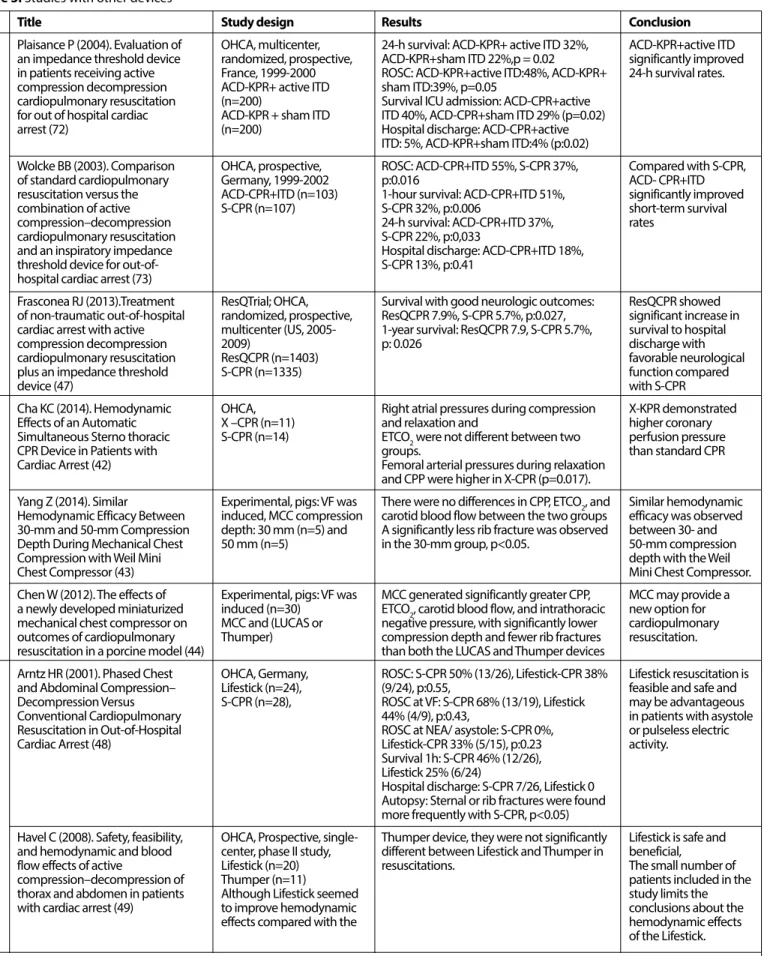
Table 3 presents the results of some studies that have reported on other MCCDs and techniques. Some trials on ResQCPR (ACD+ITD CPR) have reported short- and long-term survival rates to be higher than those for S-CPR (47, 72, 73). The results of trials with simulta-neous sterno-thoracic CPR devices (SST-CPR/X-CPR) reveal hope for the future of these devices. In a study that consisted of a small series of cases, X-CPR produced higher coronary perfusion pressure than S-CPR (42). In a study with the Lifestick, a phased thoracic-abdominal active compression–decompression CPR device, no difference was detected compared with S-CPR in terms of ROSC, but it was reported that this technique could be advantageous for patients with asystole or pulseless electrical activity (48). Another study conducted with a small sample (49) did not report any statistically significant difference between using the Lifestick and applying S-CPR.
In the literature, there are also simulation studies where CPR devices and techniques were compared. Zhang et al. (11) used a cir-culation computer model in an experimental study to compare five ITD-supported techniques (S-CPR, ACD‐CPR, IAC‐CPR, Lifestick‐CPR, and EECP‐CPR) in terms of their hemodynamic effects, and they found Lifestick-CPR to be the most effective. A similar simulation study of five CPR techniques [S-CPR, ACD-CPR, IAC-CPR, ACD-CPR+External counterpulsation (ECP), and S-CPR+ECP] had made a comparison and found that cardiac output, cerebral blood flow, coronary blo-od flow, and mean coronary perfusion pressure to be the lowest in S-CPR and highest in IAC-CPR, with ACD-CPR+ECP exhibiting values close to this (51).
MCCDs in the Guidelines
An evaluation was made of the recommendations for use and the levels of evidence cited in the AHA 2010 and 2015 guidelines based on large randomized trials. In the case of automatic ACD-CPR
devices such as LUCAS and LDB-CPR devices such as Autopulse, the guidelines state that the evidence to support or reject the routine use of these devices in the treatment of cardiac arrest is not sufficient and that manual chest compressions remain as the standard treat-ment of cardiac arrest; however, these devices may be a reasonable alternative for use by properly trained personnel (AHA 2015: Class IIb, LOE B-R). Furthermore, the guidelines state that the use of mechani-cal piston devices may be considered in specific settings where the delivery of high-quality manual compressions may be challenging or dangerous for the provider [e.g., limited rescuers available and pro-longed CPR during hypothermic cardiac arrest, in a moving ambulan-ce, in the angiography suite, and during preparation for extracorpo-real CPR (ECPR)], provided that rescuers strictly limit interruptions in CPR during deployment and removal of the devices (AHA 2015: Class IIb, LOE C-EO) (6, 74).
The AHA 2010 guideline states with regard to manual ACD-CPR devices that there is no adequate evidence to either recommend or reject the routine use of these devices and that the use of the devices may be considered in the event of properly trained personnel. The AHA 2015 guideline has not made any revision with regard to these devices, maintaining the same recommendations and evidence level specified in the 2010 guideline (Class IIb, LOE B) (6, 74).
With respect to the sole use of the ITD-CPR device, the AHA 2010 guideline’s recommendation and evidence level places the device in Class IIb, LOE B, whereas the AHA 2015 guideline has changed the re-commendation and evidence level, placing it in the category of “Not recommended for routine use S-CPR” (Class III: No benefit, LOE A) (6, 74).
There appears to be no evaluation in the AHA 2010 guideline for ITD+ACD-CPR (RESQCPR). In the 2015 guideline, however, it is stated that this combination is not recommended for routine use as an al-ternative for S-CPR but may be considered as an alal-ternative only in the presence of available equipment and trained personnel (Class IIb, LOE C-LD) (6, 74).
It can be seen that the AHA 2010 and 2015 guidelines do not include information and data on CPR devices and combinations such as the SST-CPR, PTACD-CPR, and AEI-CPR because these are still in the experimental stage and are not supported by adequate clinical research (6, 74).
Conclusion
In a general assessment, it may be stated that although large randomized trials have as yet highlighted the superiority of these de-vices over S-CPR in OHCA, they have at the same time not produced any evidence to prove their failure or harm.
The possibilities to be created by the harmonious coopera-tion of the disciplines of mathematics, biology, medicine, engi-neering, and the physical sciences in “the process of developing biomedical equipment technologies” and the role technology will play in constructing the future cannot be ignored. Therefore, an increase in the number of experimental and clinical research on CPR technologies and the evaluations & revisions performed ac-cording to the results of these studies will pave the way for the development of changes with respect to the application methods and areas of these devices. Ensuring that these devices become more functional, effective, and reliable will improve the effective-ness of CPR and may reduce the incidence of morbidity and mor-tality accompanying cardiac arrest.
Title Study design Results Conclusion
1. Timerman S (2004). Improved IHCA (at ICU), Brazil, Peak aortic pressure: A-CPR153, A-CPR demonstrated a clinically hemodynamic performance with a 2000-2001, A-CPR (n=8), S-CPR 115 mmHg, p<0.0001 significant improvement in novel chest compression device S-CPR (n=8) Peak right atrial pressure: A-CPR=129, hemodynamics compared to during treatment of in-hospital S-CPR=83 mmHg, p<0.0001 CPP: A-CPR=20 manual chest compressions.
cardiac arrest (66) mmHg, S-CPR=15 mmHg, p<0.015
2. Hock Ong ME (2006). Use of an OHCA, observational, ROSC: A-CPR 34.5%, S-CPR 20.2% A-CPR is better. AutoPulse was automated, load-distributing band A-CPR (n=284), (2003-2005) Survival to hospital admission: A-CPR 20.9%, improved survival to hospital chest compression device for S-CPR (n=499) (2001-2003) S-CPR 11.1% discharge when compared to out-of-hospital cardiac arrest device was used in only Survival to hospital discharge: S- CPR
resuscitation (32) 210 cases A-CPR 9.7%, S-CPR 2.9% No difference in CPC (p=0.360)
(device was used in only 210 cases)
3. Casner M (2005). The impact of OHCA, Retrospective, Arrival to an emergency department with A-CPR may improve ROSC and a new CPR device on rate of A-CPR (n=69), S-CPR (n=93) measurable spontaneous pulses: may particularly benefit patients spontaneous circulation in out-of A-CPR 39%, manual 29%, p: 0.003, with no shockable rhythms. hospital cardiac arrest (67) At shockable rhythms: A-CPR 44%,
manual 50%, p: 0.340,
At asystole: A-CPR 37%, manual 22%, p: 0.008,
At PEA: A-CPR 38%, manual 23%, p: 0.079).
4. Hallstrom A (2006). Manual chest OHCA, randomized, Survival to 4 h: A-CPR 29.5%, Use of an automated LDB-CPR compression vs. use of an multicenter (US, Canada) S-CPR 28.5%, p=0.74 device as implemented in this automated chest compression 2004-2005 Survival to hospital discharge: A-CPR 5.8%, study was associated with device during resuscitation A-CPR (n=554), S-CPR 9.9%, p=0.060 worse neurological outcomes following out-of-hospital cardiac S-CPR (n=517) CPC 1–2 at hospital discharge: A-CPR 3.1%, and a trend toward worse
arrest: A randomized trial S-CPR 7.5%, p=0.006 survival compared with manual
(ASPİRE) (68) CPR.
5. Krep H (2007). Out-of-hospital OHCA, prospective, ROSC: 54.3% (n=25/46), chest compression The AutoPulse is an effective cardiopulmonary resuscitation with Germany, 2004-2005 device (69) and safe mechanical CPR device the AutoPulseTM system: A-CPR (n=46), Admitted to ICU: 39.1% (n=18/46), useful in OHCA
A prospective observational study ACD-CPR (n=48) Discharged from ICU: 21.8% (n=10/46) with a new load-distributing band Discharged CPC 1=2; CPC 2=1; CPC 3=
7 patient (n=10)
ROSC ACD-CPR with use cardio pump: 52% (n=48)
6. Duchateau F-X (2010). Effect of OHCA, prospective, Median diastolic BP: A-CPR 23 mmHg, The use of the AuotoPulse is the AutoPulse™ automated band France (2008) S-CPR 17 mmHg, p<0.001 associated with increased chest compression device on A-CPR (n=29) (first S-CPR Median systolic BP: A-CPR 106 mmHg, diastolic BP compared to S-CPR. hemodynamics in out-of-hospital and then A-CPR same S-CPR 72 mmHg, p<0.02,
cardiac arrest resuscitation (70) groups) Mean BP: A-CPR 36 mmHg, S-CPR 29 mmHg, p<0.002,
ETCO2: did not increase with Autopulse (from 21 to 22 mmHg, p=0.80)
7. Jennings PA (2012). An automated OHCA, retrospective, Survival to hospital : A-CPR 26% (17/66), Further research is warranted, CPR device compared with standard Australia, 2006-2010 S-CPR 20% (43/220), p=0.23 which involves randomization chest compressions for A-CPR (n=66), Survived to hospital discharge: A-CPR 3% and larger number of cases to out-of-hospital resuscitation (25) S-CPR (n=220) (n=2/66), S-CPR 7% (15/220), p=0.38 investigate the potential
benefits of A-CPR, including survival to hospital discharge. 8. Hock Ong ME (2012). Improved OHCA, multicenter, ROSC: A-CPR 35.3% (n=195), The AutoPulse improved neurologically intact survival with randomized, Singapore S-CPR 22.4% (n=103) survival with intact neurological the use of an automated, load- S-CPR (n=459, 2004-2007) Survival to hospital discharge: A-CPR 3.3%, status on discharge in adults distributing band chest compression A-CPR (n=522, 2007-2009) S-KPR 1.3% with non-traumatic cardiac device for cardiac arrest presenting CPC 1 -2 at hospital discharge: A-CPR 81.3% arrest.
to the emergency department (30) (n=13/16), S-CPR 33.3% (n=2/6).
9. Wik L (2014). Manual vs. integrated OHCA, multicenter ROSC: A-CPR 28.6%, S-CPR 32.3%, CIRC Trial: Compared to high- automatic load-distributing band (US, Europe), randomized, no different quality A-CPR, S-CPR resulted in CPR with equal survival after out of (2009-2011), 24-h survival: A-CPR 21.8%, S-CPR 25%, statistically equivalent survival hospital cardiac arrest. The A-CPR (n=2099), no different survival to hospital discharge: to hospital discharge. randomized CIRC trial (71) S-CPR (n=2132) A-KPR 9.4%, S-KPR 11%, no different
CO: cardiac output; CPP: coronary perfusion pressure; IHCA: in-hospital cardiac arrest; OHCA: out -hospital Cardiac arrest; ROSC: return of spontaneous circulation; PEA: pulseless electrical activity; CPC: cerebral performance category; ETCO2: end-tidal CO2; A-KPR : AutoPulse CPR; ICU: intensive care unit; BP: blood pressure
Title Study design Results Conclusion
Plaisance P (2004). Evaluation of OHCA, multicenter, 24-h survival: ACD-KPR+ active ITD 32%, ACD-KPR+active ITD an impedance threshold device randomized, prospective, ACD-KPR+sham ITD 22%,p = 0.02 significantly improved in patients receiving active France, 1999-2000 ROSC: ACD-KPR+active ITD:48%, ACD-KPR+ 24-h survival rates. compression decompression ACD-KPR+ active ITD sham ITD:39%, p=0.05
cardiopulmonary resuscitation (n=200) Survival ICU admission: ACD-CPR+active for out of hospital cardiac ACD-KPR + sham ITD ITD 40%, ACD-CPR+sham ITD 29% (p=0.02)
arrest (72) (n=200) Hospital discharge: ACD-CPR+active
ITD: 5%, ACD-KPR+sham ITD:4% (p:0.02)
Wolcke BB (2003). Comparison OHCA, prospective, ROSC: ACD-CPR+ITD 55%, S-CPR 37%, Compared with S-CPR,
of standard cardiopulmonary Germany, 1999-2002 p:0.016 ACD- CPR+ITD
resuscitation versus the ACD-CPR+ITD (n=103) 1-hour survival: ACD-CPR+ITD 51%, significantly improved combination of active S-CPR (n=107) S-CPR 32%, p:0.006 short-term survival
compression–decompression 24-h survival: ACD-CPR+ITD 37%, rates
cardiopulmonary resuscitation S-CPR 22%, p:0,033
and an inspiratory impedance Hospital discharge: ACD-CPR+ITD 18%,
threshold device for out-of- S-CPR 13%, p:0.41
hospital cardiac arrest (73)
Frasconea RJ (2013).Treatment ResQTrial; OHCA, Survival with good neurologic outcomes: ResQCPR showed of non-traumatic out-of-hospital randomized, prospective, ResQCPR 7.9%, S-CPR 5.7%, p:0.027, significant increase in cardiac arrest with active multicenter (US, 2005- 1-year survival: ResQCPR 7.9, S-CPR 5.7%, survival to hospital
compression decompression 2009) p: 0.026 discharge with
cardiopulmonary resuscitation ResQCPR (n=1403) favorable neurological
plus an impedance threshold S-CPR (n=1335) function compared
device (47) with S-CPR
Cha KC (2014). Hemodynamic OHCA, Right atrial pressures during compression X-KPR demonstrated Effects of an Automatic X –CPR (n=11) and relaxation and higher coronary Simultaneous Sterno thoracic S-CPR (n=14) ETCO2 were not different between two perfusion pressure
CPR Device in Patients with groups. than standard CPR
Cardiac Arrest (42) Femoral arterial pressures during relaxation and CPP were higher in X-CPR (p=0.017).
Yang Z (2014). Similar Experimental, pigs: VF was There were no differences in CPP, ETCO2, and Similar hemodynamic Hemodynamic Efficacy Between induced, MCC compression carotid blood flow between the two groups efficacy was observed 30-mm and 50-mm Compression depth: 30 mm (n=5) and A significantly less rib fracture was observed between 30- and Depth During Mechanical Chest 50 mm (n=5) in the 30-mm group, p<0.05. 50-mm compression
Compression with Weil Mini depth with the Weil
Chest Compressor (43) Mini Chest Compressor.
Chen W (2012). The effects of Experimental, pigs: VF was MCC generated significantly greater CPP, MCC may provide a a newly developed miniaturized induced (n=30) ETCO2, carotid blood flow, and intrathoracic new option for mechanical chest compressor on MCC and (LUCAS or negative pressure, with significantly lower cardiopulmonary outcomes of cardiopulmonary Thumper) compression depth and fewer rib fractures resuscitation. resuscitation in a porcine model (44) than both the LUCAS and Thumper devices
Arntz HR (2001). Phased Chest OHCA, Germany, ROSC: S-CPR 50% (13/26), Lifestick-CPR 38% Lifestick resuscitation is and Abdominal Compression– Lifestick (n=24), (9/24), p:0.55, feasible and safe and Decompression Versus S-CPR (n=28), ROSC at VF: S-CPR 68% (13/19), Lifestick may be advantageous
Conventional Cardiopulmonary 44% (4/9), p:0.43, in patients with asystole
Resuscitation in Out-of-Hospital ROSC at NEA/ asystole: S-CPR 0%, or pulseless electric
Cardiac Arrest (48) Lifestick-CPR 33% (5/15), p:0.23 activity.
Survival 1h: S-CPR 46% (12/26), Lifestick 25% (6/24)
Hospital discharge: S-CPR 7/26, Lifestick 0 Autopsy: Sternal or rib fractures were found more frequently with S-CPR, p<0.05)
Havel C (2008). Safety, feasibility, OHCA, Prospective, single- Thumper device, they were not significantly Lifestick is safe and and hemodynamic and blood center, phase II study, different between Lifestick and Thumper in beneficial,
flow effects of active Lifestick (n=20) resuscitations. The small number of
compression–decompression of Thumper (n=11) patients included in the
thorax and abdomen in patients Although Lifestick seemed study limits the
with cardiac arrest (49) to improve hemodynamic conclusions about the
effects compared with the hemodynamic effects
of the Lifestick. Impedanc e thr eshold devic e (ITD+A CD -KPR) Simultaneous S ter no thor acic
Phased Chest and A
bdominal
Table 3. Studies wıth other devices
CO: cardiac output; CPP: coronary perfusion pressure; IHCA: in-hospital cardiac arrest; OHCA: out -hospital Cardiac arrest; ROSC: return of spontaneous circulation; PEA: pulseless electrical activity; CPC: cerebral performance category; ETCO2: end-tidal CO2; A-KPR : AutoPulse CPR; ICU: intensive care unit; BP: blood pressure
Peer-review: Externally peer-reviewed.
Conflict of Interest: No conflict of interest was declared by the authors. Financial Disclosure: The authors declared that this study has received no financial support.
References
1. Travers AH, Rea TD, Bobrow BJ, Edelson DP, Berg RA, Sayre MR, et al. 2010 American Heart Association Guidelines for Cardiopulmonary Resuscita-tion and Emergency Cardiovascular Care: Part 4: CPR Overview. Circulati-on 2010; 122: 676-84. [CrossRef]
2. Cooper JA, Cooper JD, Cooper JM. Contemporary reviews in cardiovas-cular medicine: cardiopulmonary resuscitation; history, current practice, and future direction. Circulation 2006; 114: 2839-49. [CrossRef] 3. Sasson C, Rogers MA, Dahl J, Kellermann AL. Predictors of survival from
out-of-hospital cardiac arrest: a systematic review and meta-analysis. Circ Cardiovasc Qual Outcomes 2010; 3: 63-81. [CrossRef]
4. Kozacı N, Ay MO, İçme F, Aktürk A, Satar S. Are we successful in cardiopul-monary resuscitation? Cukurova Med J 2013; 38: 601-9.
5. Kleinman ME, Brennan EE, Goldberger ZD, Swor RA, Terry M, Bobrow BJ, et al. 2015 American Heart Association Guidelines Update for Cardiopul-monary Resuscitation and Emergency Cardiovascular Care: Part 5: Adult Basic Life Support and Cardiopulmonary Resuscitation Quality. Circulati-on 2015; 132: 414-35. [CrossRef]
6. Cave DM, Gazmuri RJ, Otto CW, Nadkarni VM, Cheng A, Brooks SC, et al. 2010 American Heart Association Guidelines for Cardiopulmonary Re-suscitation and Emergency Cardiovascular Care: Part 7: CPR Techniques and Devices. Circulation 2010; 122: 720-8. [CrossRef]
7. Berg RA, Hemphill R, Abella BS, Aufderheide DC, Hazinski MF, Lerner B, et al. Part 5: adult basic life support: 2010 American Heart Association Guidelines for Cardiopulmonary Resuscitation and Emergency Cardio-vascular Care. Circulation 2010; 122(18 Suppl 3): S685-705. [CrossRef] 8. Nolan JP, Soar J, Zideman DA, Biarent D, Bossaert LL, Deakin C, et al.
Euro-pean Resuscitation Council Guidelines for Resuscitation 2010 Section 1. Executive summary. Resuscitation 2010; 81: 1219-76. [CrossRef] 9. Rubertsson S, Karlsten R. Increased cortical cerebral blood flow with
LUCAS; a new device for mechanical chest compressions compared to standard external compressions during experimental cardiopulmonary resuscitation. Resuscitation 2005; 65: 357-63. [CrossRef]
10. Keseg DP. The Merits of Mechanical CPR: Do mechanical devices impro-ve compression consistency and resuscitation outcomes. JEMS 2012; 37: 24-9.
11. Zhang Y, Wu X, Yuan H, Xu L, Karemaker J. Computer models in bedside physiology. Chapter 2: Optimal cardiopulmonary resuscitation as identi-fied by computer modeling. 2013: 12-32. Downloaded from UvA-DARE. Available at http://dare.uva.nl.
12. Topjian AA, Nadkarni AA, Berg VM. Cardiopulmonary resuscitation in children. Curr Opin Crit Care 2009; 15: 203-8. [CrossRef]
13. Gaxiola A, Varon J. Evolution and new perspective of chest compression mechanical devices. Am J Emerg Med 2008; 26: 923-31. [CrossRef] 14. Hightower D, Thomas SH, Stone CK, Dunn K, March JA. Decay in quality
of closed-chest compressions over time. Ann Emerg Med 1995; 26: 300-3. [CrossRef]
15. Ochoa FJ, Ramalle-Gomara E, Lisa V, Saralegui I. The effect of rescuer fa-tigue on the quality of chest compressions. Resuscitation 1998; 37: 149-52. [CrossRef]
16. Wik L, Kramer-Johansen J, Myklebust H, Sorebo H, Svensson L, Fellows B, et al. Quality of cardiopulmonary resuscitation during out-of-hospital cardiac arrest. JAMA 2005; 293: 299-304. [CrossRef]
17. Perkins GD, Boyle W, Bridgestock H, Davies S, Oliver Z, Bradburn S, et al. Quality of CPR during advanced resuscitation training. Resuscitation 2008; 77: 69-74. [CrossRef]
18. Wigginton JG, Miller AH, Benitez FL, Pepe PE. Mechanical devices for cardi-opulmonary resuscitation. Curr Opin Crit Care 2005; 11: 219-23. [CrossRef] 19. Classification of the External Cardiac Compressor (including CPR aid de-vices). FDA Executive Summary. Available at: http://www.fda.gov/down-loads/AdvisoryCommittees/CommitteesMeetingMaterials/Medical De-vices. September 11, 2013.
20. Harkins GA, Bramson ML. Mechanized external cardiac massage for car-diac arrest and for support of the failing heart. A preliminary communi-cation. J Surg Res 1961; 1: 197-200. [CrossRef]
21. Nachlas MM, Siedband MP. A simple portable pneumatic pump for exter-nal cardiac massage. Am J Cardiol 1962; 10: 107-9. [CrossRef]
22. Safar P, Harris LC. The Beck-Rand external cardiac compression machine. Anesthesiology 1963; 24: 586-8. [CrossRef]
23. Bailey, Browsw NL, Keating VJ. Automatic external cardıac massage: a portable pneumatic external cardıac compression machine. Brit Heart J 1964; 26: 481-9. [CrossRef]
24. Harrison-Poul R. A history of mechanical devices for providing external chest compressions. Resuscitation 2007; 73: 330-6. [CrossRef]
25. Jennings PA, Harris L, Bernard S, Bray J, Walker T, Spelman T, et al. An au-tomated CPR device compared with standard chest compressions for out-of-hospital resuscitation. BMC Emerg Med 2012; 12: 2-5. [CrossRef] 26. Xu L, Wu X, Zhang Y, Yuan H. The Optimization Study on Time Sequence
of Enhanced External Counter-Pulsation in AEI-CPR. Journal of Compu-ters 2009; 4: 1243-8. [CrossRef]
27. Lurie KG. Mechanical devices for cardiopulmonary resuscitation: an up-date. Emerg Med Clin N Am 2002; 20: 771-84. [CrossRef]
28. Perkins GD, Brace S, Gates S. Mechanical chest-compression devices: cur-rent and future roles. Curr Opin Crit Care 2010; 16: 203-10. [CrossRef] 29. Halperin H, Carver DJ. Mechanical CPR devices. SIGNA VITAE 2010; 5: 69-73. 30. Ong ME, Fook-Chong S, Annathurai A, Hu Ang S, Tiah L, Yong KL, et al.
Improved neurologically intact survival with the use of an automated, load-distributing band chest compression device for cardiac arrest pre-senting to the emergency department. Crit Care 2012; 16: 14-21. 31. Eroğlu SE. Türkiye’de satışa sunulan göğüs kompresyon cihazları:
Autopul-se. Available at: http://www.acilci.net/eroglu-turkiyede-satisa-sunulan-go-gus-kompresyon-cihazlari- Autopulse. Erişim tarihi: 10 Ağustos 2013. 32. Ong ME, Ornato JP, Edwards DP, Dhindsa HS, Best AM, Ines CS, et al. Use of an
automated, load-distributing band chest compression device for out-of-hos-pital cardiac arrest resuscitation. JAMA 2006; 295: 2629-37. [CrossRef] 33. Haydin S, Ündar A. Yaşam destek sistemlerinin Dünya’daki gelişmeleri ve
Türkiye’deki son durum. Anadolu Kardiyol Derg 2013; 13: 580-8. 34. Lurie KG, Lindo C, Chin J. CPR: the P stands for plumber’s helper. JAMA
1990; 264: 1661. [CrossRef]
35. Altman LK. Toilet Plunger Is the Model For Device to Restart Hearts. The New York Times. Published: June 3, 1992.
36. Steen S, Liao Q, Pierre L, Paskevicius A, Sjoberg T. Evaluation of LUCAS, a new device for automatic mechanical compression and active decomp-ression resuscitation. Resuscitation 2002; 55: 285-99. [CrossRef] 37. Eroğlu SE. Türkiye’de satışa sunulan göğüs kompresyon cihazları: LUCAS-2.
Available at: http://www.acilci.net/eroglu-turkiyede-satisa-sunulan-go-gus-kompresyon-cihazlari-LUCAS-2. Erişim tarihi: 16 Ağustos 2013. 38. Ju H. Technology Brief Update: Mechanical chest compression devices
for patients experiencing cardiac arrest. HealthPACT. Available at: htt-ps://www.health.qld.gov.au/healthpact/docs/updates/WP153_update. pdf. May 2013.
39. Smekal D. Safety with mechanical chest Compressions in CPR; clinical studies with the LUCAS™ device. Available at: http://www.diva-portal. org/smash/get/diva2:639365/FULLTEXT01.pdf. 2013
40. Cohen TJ, Goldner BG, Maccaro PC, Ardito AP, Trazzera S, Cohen MB, et al. A comparison of active compression-decompression cardiopulmonary resuscitation with standard cardiopulmonary resuscitation for cardi-ac arrests occurring in the hospital. N Engl J Med 1993; 329: 1918-21. [CrossRef]
41. Hwang SO, Lee KH, Lee JW, Lee SY, Yoo BS, Yoon J, et al. Simultaneous sterno-thoracic cardiopulmonary resuscitation improves short-term survival rate in canine cardiac arrests. Resuscitation 2002; 53: 209-16. [CrossRef]
42. Cha KC, Kim H, Lee DG, Cha YS, Kim OH, Kim TH, et al. Hemodynamic effe-cts of an automatic simultaneous sternothoracic CPR device in patients with cardiac arrest. Circulation 2014; 130: A108.
43. Yang Z, Gong P, Wu X, Qian J, Zhao S, Duggal N, et al. Similar hemodyna-mic efficacy between 30 mm and 50 mm compression depth during me-chanical chest compression with weil mini chest compressor. Circulation 2014; 130: A306.
44. Chen W, Yinlun W, Xiaobo W, Shijie S, Joe B. The effects of a newly developed miniaturized mechanical chest compressor on outcomes of cardiopulmonary resuscitation in a porcine model. Crit Care Med 2012; 40: 3007-12. [CrossRef] 45. Lurie KG, Voelckel WG, Zielinski T, Mc Knite S, Lindstrom P, Colleen P, et al.
Improving standard cardiopulmonary resuscitation with an inspiratory impedance threshold valve in a porcine model of cardiac arrest. Anesth Analg 2001; 93: 649-55. [CrossRef]
46. Thayne RC, Thomasa DC, Neville JD, Van Dellen A. Use of an impedance threshold device improves short-term outcomes following out-of-hospi-tal cardiac arrest. Resuscitation 2005; 67: 103-8. [CrossRef]
47. Frascone RJ, Wayneb MA, Sworc RA, Mahoneyd BD, Domeiere RM, Olin-ger ML, et al. Treatment of non-traumatic out-of-hospital cardiac arrest with active compression decompression cardiopulmonary resuscitation plus an impedance threshold device. Resuscitation 2013; 84: 1214-22. [CrossRef]
48. Arntz HR, Agrawal R, Richter H, Schmidt S, Rescheleit T, Menges M, et al. Phased chest and abdominal compression-decompression versus conventional cardiopulmonary resuscitation in out-of-hospital cardiac arrest. Circulation 2001; 104: 768-72. [CrossRef]
49. Havel C, Berzlanovich A, Sterz F, Domanovits H, Herkner H, Zeiner A, et al. Safety, feasibility, and hemodynamic and blood flow effects of active compression-decompression of thorax and abdomen in patients with cardiac arrest. Crit Care Med 2008; 36: 1832-37. [CrossRef]
50. Kern KB, Hilwig RW, Berg RA, Schock RB, Ewy GA. Optimizing ventilati-on in cventilati-onjunctiventilati-on with phased chest and abdominal compressiventilati-on–de- compression–de-compression (Lifestick™) resuscitation. Resuscitation 2002; 52: 91-100. [CrossRef]
51. Luo J, Zeng H. Modeling and Simulation of Hemodynamic effects du-ring different cardiopulmonary resuscitation (CPR) technologies. PeerJ PrePrints. Available at: http://dx.doi.org/10.7287/peerj.preprints.334v1. 31 Mar 2014.
52. Yannopoulos D, Halperin HR, Hopkins J. Lower extremity counterpulsation during the decompression phase of CPR improves hemodynamics and pro-vides continuous forward carotid blood flow. Circulation 2007; 116: II-485. 53. Liao Q, Sjöberg T, Paskevicius A, Wohlfart B, Steen S. Manual versus
me-chanical cardiopulmonary resuscitation. An experimental study in pigs. BMC Cardiovasc Disord 2010; 10: 2-8. [CrossRef]
54. Larsen AI, Hjornevik AS, Ellingsen CL, Dennis WT. Cardiac arrest with con-tinuous mechanical chest compression during percutaneous coronary intervention. A report on the use of the LUCAS device. Resuscitation 2007; 75: 454-9. [CrossRef]
55. Bonnemeier H, Olivecron G, Simonis G, Götberg M, Weitz G, Iblher P, et al. Automated continuous chest compression for in-hospital cardiopulmo-nary resuscitation of patients with pulseless electrical activity: A report of five cases. Int J Cardiol 2009; 136: 39-50. [CrossRef]
56. Bonnemeier H, Simonis G, Olivecron G. Continuous mechanical chest com-pression during in hospital cardiopulmonary resuscitation of patients with pulseless electrical activity. Resuscitation 2011; 82: 155-9. [CrossRef] 57. Wagner H, Terkelsen CJ, Friberg H, Harnek J, Kern K. Cardiac arrest in
the catheterisation laboratory: a 5-year experience of using mechanical chest compressions to facilitate PCI during prolonged resuscitation ef-forts. Resuscitation 2010; 81: 383-7. [CrossRef]
58. Fidler R, Hirsch J, Strechert M, Johnson M. Three modes of cardiac com-pressions in a single patient: A comparison of usual manual compressi-ons, automated compressicompressi-ons, and open cardiac massage. Resuscitation 2014; 85: 75-6. [CrossRef]
59. Rodríguez AM, Ferrara LP, Pascuala JMN, Alonsoa CB, Maldonadoa ME, Belmonte AA, et al. Kidney transplant function using organs from non-heart-beating donors maintained by mechanical chest compressi-ons. Resuscitation 2010; 81: 904-7. [CrossRef]
60. Axelsson C, Nestin J, Svensson L, Axelsson AB, Herlitz J. Clinical consequ-ences of the introduction of mechanical chest compression in the EMS system for treatment of out-of-hospital cardiac arrest- A pilot study. Re-suscitation 2006; 71: 47-55. [CrossRef]
61. Smekal D, Johansson J, Huzevka T, Rubertsson S. A. Pilot study of mecha-nical chest compressions with the LUCAS™ device in cardiopulmonary resuscitation. Resuscitation 2011; 82: 702-6. [CrossRef]
62. Axelsson C, Karlsson T, Axelsson AB, Herlitzb J. Mechanical active comp-ression decompcomp-ression cardiopulmonary resuscitation (ACD-CPR) versus manual CPR according to pressure of end tidal carbon dioxide (PETCO2) during CPR in out-of-hospital cardiac arrest (OHCA). Resuscitation 2009; 80: 1099-103. [CrossRef]
63. Rubertsson S, Lindgren E, Smekal D, Östlund O, Silfverstolpe J, Lichtveld RA, et al. Mechanical chest compressions and simultaneous defibrillation vs conventional cardiopulmonary resuscitation in out-of-hospital cardi-ac arrest: the LINC randomized trial. JAMA 2014; 311: 53-61. [CrossRef] 64. Perkins GD, Lall R, Quinn T, Deakin CD, Cooke MW, Horton J, et al.
Mecha-nical versus manual chest compression for out-of-hospital cardiac arrest (PARAMEDIC): a pragmatic, cluster randomized controlled trial. Lancet 2015; 385: 947-55. [CrossRef]
65. Blomberg H, Gedeborg R, Berglund L, Karlsten R, Johansson J. Poor chest compression quality with mechanical compressions in simulated cardi-opulmonary resuscitation: A randomized, cross-over manikin study. Re-suscitation 2011; 82: 1332-7. [CrossRef]
66. Timerman S, Cardoso LF, Ramires JA, Halperin H. Improved hemodynamic performance with a novel chest compression device during treatment of in-hospital cardiac arrest. Resuscitation 2004; 61: 273-80. [CrossRef] 67. Casner M, Andersen D, Isaacs SM. The impact of a new CPR device on
rate of spontaneous circulation in out-of-hospital cardiac arrest. Prehosp Emerg Care 2005; 9: 61-7. [CrossRef]
68. Hallstrom A, Rea TD, Sayre MR, Christenson J, Anton AR. Manual chest compression vs use of an automated chest compression device during resuscitation following out-of-hospital cardiac arrest: a randomized trial. JAMA 2006; 295: 2620-8. [CrossRef]
69. Krep H, Mamier M, Breil M, Heister U, Fischer M, Hoeft A, et al. Out-of-hos-pital cardiopulmonary resuscitation with the AutoPulseTM system: A prospective observational study with a new load-distributing band chest compression device. Resuscitation 2007; 73: 86-95. [CrossRef] 70. Duchateau FX, Gueye P, Curac S, Tubach F, Broche C, Plaisance P, et al.
Effect of the AutoPulse™ automated band chest compression device on hemodynamics in out-of-hospital cardiac arrest resuscitation. Intensive Care Med 2010; 36: 1256-60. [CrossRef]
71. Wik L, Olsen JA, Presse D, Sterz F, Lozano M, Brouwer MA, et al. Manual vs. integrated automatic load-distributing band CPR with equal survival after out of hospital cardiac arrest. The randomized CIRC trial. Resuscita-tion 2014; 85: 741-8. [CrossRef]
72. Plaisance P, Lurie KG, Vicaut E, Martin D, Gueugniaud PY, Petit JL, et al. Evaluation of an impedance threshold device in patients receiving acti-ve compression decompression cardiopulmonary resuscitation for out of hospital cardiac arrest. Resuscitation 2004; 61: 265. [CrossRef] 73. Wolcke BB, Mauer DK, Schoefmann MF, Teichmann H, Provo TA, Lindner
KH, et al. Comparison of standard cardiopulmonary resuscitation versus the combination of active compression-decompression cardiopulmo-nary resuscitation and an inspiratory impedance threshold device for out-of-hospital cardiac arrest. Circulation 2003; 108: 2201-5. [CrossRef] 74. Brooks SC, Anderson ML, Bruder E, Daya MR, Gaffney A, Otto CW, et al.
2015 American Heart Association Guidelines Update for Cardiopulmo-nary Resuscitation and Emergency Cardiovascular Care: Part 6: Alternati-ve Techniques and Ancillary Devices for Cardiopulmonary Resuscitation. Circulation 2015; 132: 436-43.[CrossRef]