Ankara Üniv Vet Fak Derg, 56, 149-151, 2009
Short Communication / Kısa Bilimsel Çalışma
Poliencephalomalacia in two donkeys (Equus asinus)
Çağdaş OTO1, Zafer ÖZYILDIZ2, Rıfkı HAZIROĞLU3, Şule Yurdagül ÖZSOY41Ankara University, Faculty of Veterinary Medicine, Department of Anatomy, Ankara; 2Kafkas University, Faculty of Veterinary
Medicine, Department of Pathology, Kars; 3Ankara University, Faculty of Veterinary Medicine, Department of Pathology, Ankara; 4Mustafa Kemal University, Faculty of Veterinary Medicine, Department of Pathology, Hatay, Turkey.
Summary: Polioencephalomalacia was described in two senile male donkeys. Necropsy revealed necrosis and cavitations of irregular shape and depth, extending from the cortex into the inner brain, on the left cerebral hemisphere in the first case and the right cerebral hemisphere in the second case. Microscopic examination demonstrated the grey matter of the brain to have disappeared in many areas, and cyctic structures to have formed in some areas. Some were oedematous and in these areas the presences of myelin-filled phagocytic gitter cells, astrocytes and also slight perivascular lymphocyte infiltration was determined.
Key words: Donkey, pathology, polioencephalomalacia
İki merkepte (Equus asinus) polioensefalomalasi
Özet: Yaşlı iki merkepte polioensefalomalasi olgusu tanımlandı. Nekropside; Birinci olguda sol serebral hemisfer, ikinci olguda sağ serebral hemisferde, korteksten iç kısımlara doğru uzanan, şekli ve derinliği düzenli bir yapı göstermeyen, nekroz ve kavitasyonlara rastlandı. Mikroskobik olarak serebral korteksin bir çok bölgesinde beyin gri maddesinin ortadan kalktığı, yer yer kistik oluşumların şekillendiği tespit edildi. Bazı alanlarda ödem ile birlikte myelin fagosite etmiş gitter hücreleri, astrositler ve hafif perivaskuler lenfosit infiltrasyonlarının bulunduğu belirlendi.
Anahtar sözcükler: Merkep, patoloji, polioensefalomalasi.
Polioencephalomalacia, which is also known as cerebrocortical necrosis, occurs primarily in cattle (1, 5), sheep (5), goats (9), horses (7) and less frequently in cats (10) and dogs, mings and pigs (6). However to the authors’ knowledge, no previous literature report exist on its occurence in the donkey.
Polioencephalomalacia lesions are generally of toxic and metabolic origin. These lesions are reported to may develop as a result of thiamine inadequacy or lead and sulphate intoxication, as well as due to cerebral infarction or anesthesia (6, 5, 3, 7).
Polioencephalomalacia, which among domestic mammals, affects primarily cattle and sheep, and rarely goats, horses, cats and dogs has been described in detail in two donkeys, in association with macroscopic and histopathological findings in the present study.
Two male donkeys, one of sixteen years of age and 74 kg body weight and the other of twelve years of age and 118 kg body weight, which were sedated with chloral hydrate so as to be fed to wild animals kept at the Atatürk Orman Çiftliği, were decapitated at the atlanto-occipital joint. The calvaria was lifted and the exposed brains were removed from the skull. Brains were fixed for 24 hours in 10% neutral buffered formalin solution. Tissue samples
taken from areas displaying macroscopic findings were first subjected to routine tissue examination and later embedded in paraffin blocks. Thin cross sections (5µ) were cut and stained with hematoxylin-eosin (HE) and toluidine blue.
The anamnesis taken excluded the presence of clinical findings suggestive of any disease, yet the animals were stated to be agitated and aggressive, and to display a tendency to bite animals nearby. The animals were indicated to not have received any medical treatment.
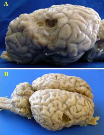
In the first case, upon lifting the calvaria, the brain was observed to be normal size. However the dura mater and arachnoidea were determined to be dissociated at the junction of the temporal and parietal lobes in the left cerebral hemisphere. In the same region, necrosis and cavitations, confined dorsally by the suprasylvian sulcus and ventrally by the lateral cerebral fissure, which displayed intense peripheral vascularization, were determined to be present (Figure 1-A). In the second case, it was noted that necrosis and cavitations existed in the temporal lobe of the right cerebral hemisphere, in the region situated between the diagonal sulcus and lateral cerebral fissure, which also includes the medial sylvian
Çağdaş Oto - Zafer Özyıldız - Rıfkı Hazıroğlu - Şule Yurdagül Özsoy 150
gyrus (Figure 1-B). Both of the brains’ transversal sections revealed lesions to be localized in an area lying between the substantia grisea neopalli, corpus medullare hemispherium and radiatio corporis callosi (Figure 2-A,B).
Figure 1. Necrosis and cavitations on the cerebral hemispheres, A-) Case 1-lateral view B-) Case 2-dorsolateral view
Şekil 1. Serebral hemisferlerdeki nekroz ve kavitasyonlar, A-) Olgu 1-lateral’den görünüm, B-) Olgu 2-dorsolateral’den görünüm
Figure 2. Caudal view of transversal sections with necrosis and cavitations, A-) Case 1-sol hemisphere, B-) Case 2-sağ hemisphere Şekil 2. Transversal kesitlerde nekroz ve kavitasyonların caudal’den görünümü, A-) Olgu 1-sol hemisfer, B-) Olgu 2-sağ hemisfer
In both cases, the meningeal blood vessels were determined to be congested and the dura mater and arachnoidea were ascertained to be dissociated. The grey matter of the brain had disappeared in many parts of the cerebral cortex and cystic structures were determined to have formed in certain areas. Such areas were determined to contain oedematous fluid, as well as newly formed blood vessels, myelin filled phagocytic gitter cells, astrocytes and slight perivascular lymphocyte infiltration (Figure 3-A,B).
Figure 3. Wide necrotic and malasic areas in grey matter, A-) HE X 40, B-) Toluidine blue X 40
Şekil 3. Substantia grisea’daki yaygın nekrotik ve malasik alanlar, A-) HE X 40, B-) Toluidine blue X 40
In horses, large malacic foci, extending from the globus pallidus and substantia nigra to the corpus mamillare, are reported to develop in the brain, particularly as a result of the long-term consumption of the yellow starthistle (Centaurea solstitialis) and the Russian knapweed (Centaurea repens) plants which remain green longer into the dry season, and thereby are readily consumed by animals (3). However the localization of the lesions determined in the cases examined did not comply with the localization of lesions resulting from intoxications with the indicated plants. In animals fed on the bracken fern (Pteris aquilinum) and horsetail (Equisetum arvense) plants, which are known to contain a high level of thiaminase, cerebrocortical
Ankara Üniv Vet Fak Derg, 56, 2009 151
necrosis and malacia develop as a result of thiamine inadequacy (4). Literature reports exist, which point out to the widespread presence of particularly the Russian knapweed and horsetail plants in the vicinity of Ankara (2). The localization of the lesions determined in both cases displays similarity with the localization of lesions resulting from bracken fern and horsetail intoxications. However, since blood and tissue thiamine levels were not able to be measured and the animals could not be proven to have eaten the indicated plants, a full evaluation was not able to be performed.
Severe cortical necrosis and malacia are reported to may develop during the first 24 hours of anaesthesia, particularly in horses (7). However due to the decapitation of the animals immediately after being sedated in the two cases examined, this option has been excluded.
Encephalomalacia is known to may also develop in case of cerebral ischemia. However, it is noted that such lesions may not always involve blood vessel lesions, and that in some cases, ischemic malacic lesions may also develop upon circulatory disorders (10, 3). Similarly, in the cases examined in the present study, lesions specific to blood vessels did not exist. The intense vascularization observed just below the meninges in the cerebral cortex was considered to may be a collateral blood vessel network possibly resulting from a cerebral infarction.
The present study constitutes the first case definition of polioencephalomalacia in the donkey in Turkey.
References
1. Berkin Ş, Haziroğlu R, Urman HK (1988). Bir buzağıda cerebrocortical necrosis. Ankara Üniv Vet Fak Derg, 35, 135-143.
2. Davis PH (1965). Flora of Turkey. Vol. 1. 33-50, Edinburgh at the University Press.
3. Haziroğlu R (2001). Sinir sistemi. In: Veteriner Patoloji Vol. 1, 282-286, Edited by. R. Haziroğlu, , Ü.H. Milli. Ankara: Tamer Matbaacılık.
4. Henderson JA, Evans EV, Mcintosh RA (1952). The antithiamine action of Equisetum. J Am Vet Med Assoc, 120, 375-378.
5. Jeffrey M, Duff JP, Higgins RJ, Simpson VR, Jackman R, Jones TO, Mechie SC, Livesey CT (1994). Polioencephalomalacia associated with the ingestion of ammonium sulphate by sheep and cattle. Vet Rec, 134, 343-348.
6. Markson LM, Giles N (1973). Cerebrocortical necrosis in a fallow deer (Dama dama). Vet Rec, 93, 243-246. 7. Mckay JS, Forest TW, Senior M, Kelly DF, Jones RS,
de Lahunta A, Summers BA (2002). Postanaesthetic cerebral necrosis in five horses. Vet. Rec, 150, 70-74. 8. Nakazato L, Lemos RAA, Correa FR (2000).
Polioencefalomalacia em bovinos nos estados de mato grosso do sul e sao paulo. Pesq Vet Bras, 20, 119-125. 9. Newsholme SJ, O'neill TP (1985). An outbreak of
cerebrocortical necrosis (polioencephalomalacia) in goats. J S Afr Vet Assoc, 56, 37-38.
10. Williams KJ, Summers BA, de Lahunta A. (1998). Cerebrospinal cuterebriasis in cats and its association with feline ischemic encephalopathy. Vet Pathol, 35, 330-343.
Geliş tarihi: 29.04.2008 / Kabul tarihi: 10.06.2008 Adress for correspondence :
Dr. Çağdaş Oto
Ankara University, Faculty of Veterinary Medicine, Department of Anatomy, 06110 - Ankara