Address for Correspondence: Dr. Mustafa Çalışkan, İstanbul Medeniyet Üniversitesi, Göztepe Eğitim ve Tıbbi Araştırma Merkezi, Kardiyoloji Kliniği, Doktor Erkin Cad. Kadıköy, İstanbul-Türkiye Phone: +90 216 280 33 33 Fax: +90 464 216 280 33 33
E-mail: [email protected], [email protected] Accepted Date: 08.11.2013 Available Online Date: 14.02.2014
©Copyright 2014 by Turkish Society of Cardiology - Available online at www.anakarder.com DOI:10.5152/akd.2014.4775
A
BSTRACTObjective: Idiopathic dilated cardiomyopathy (IDC) impairs and reduces coronary flow reserve (CFR). High level of red cell distribution width (RDW) is an independent risk factor for cardiovascular diseases. Therefore, in this observational case-control study we have aimed to deter-mine whether RDW level is associated with CFR impairment in patients with IDC.
Methods: We examined 36 patients with IDC and 35 healthy subjects formed as a control group. In addition to this, patients with IDC were divided into two subgroups according to their CFR levels [normal CFR group (CFR value ≥2) and lower CFR group (CFR value<2)]. Control and patients groups were compared using the student t-test for multiple comparisons. The subgroups were compared using the Mann-Whitney U test for continuous variables and chi-square for categorical variables. The Pearson’s and Spearman correlation analysis was used to test the possible associations between CFR and the study variables as appropriate. The receiver-operating characteristic (ROC) curve was determined to evaluate the predictive performance of RDW to detect low CFR.
Results: There were no significantly differences between the lower and higher CFR groups’ clinical data, baseline hemodynamic, medication and biochemical data except RDW and high-sensitivity C-reactive protein levels. We found that RDW level was a good predictor of low CFR at the receiver-operating characteristic curve. The area under the curve (AUC) was 73% (95% confidence interval between 0.56-0.90 is 95%, p: 0.018) After adjusting potential confounders include age, body-mass index, blood pressure, lipid and glucose, RDW independently associated with CFR level (Beta:-0.374; p=0.015) and hsCRP value (Beta:-0.520; p=0.001) were the independent predictors of lower CFR.
Conclusion: Results showed that there was an independent correlation between RDW level and CFR level in patients with IDC. (Anadolu Kardiyol Derg 2014; 14: 342-8)
Key words: dilated cardiomyophaty, coronary flow reserve, RDW, ROC analysis
Mehmet Özülkü, Mustafa Çalışkan, Hakan Güllü, Doğan Erdoğan, Zuhal Çalışkan, Haldun Müderrisoğlu
Department of Cardiology, Heart and Vascular Surgery, Başkent University Training and Research Center; Konya-Turkey
Interrelation of RDW and coronary flow reserve in patient with
idiopathic dilated cardiomyopathy
Introduction
The RDW is a marker of variation of the size of the circulating red blood cells. RDW is widely available, inexpensive and included in blood panel. Studies have been reported that the elevated RDW levels are associated with poor prognosis in the setting of stable angina (1, 2), acute myocardial infarction (3, 4), heart failure (5, 6), stroke (7), peripheral arterial disease (8), older age (9) and in the patient without coronary artery disease (10, 11). One study dem-onstrated that high RDW values are associated with an increased risk of all-cause mortality in the intensive care unit (12). RDW also predicts left ventricular ejection fraction, heart failure (HF) diag-nosis and readmission for HF among coronary artery disease (CAD) patients (13). Thus, RDW seems to be a strong and
indepen-dent predictor of mortality in a wide range of population of cardio-vascular patients across a spectrum of risk (14).
Higher levels of RDW may reflect an underlying inflammatory state, which is associated with adverse clinical outcomes and lead to impaired erythrocyte maturation. Several investigators have reported that oxidative stress is implicated in the pathogen-esis and progression of heart failure. Recent experimental and clinical studies have revealed that reactive oxygen species (ROS) are produced in the failing myocardium and that ROS induces the functional and structural damage of cardiomyocytes (15, 16).
CFR reflects coronary microvascular function. Despite the fact that coronary artery is normal, CFR is altered in patient with IDC due to coronary microcirculatory dysfunction. Reduced CFR has been reported to be a poor prognostic indicator in patients
with idiopathic left ventricle dysfunction (17). However, the mechanism of reduced CFR in IDC is not fully understood. Studies have shown that CFR measured by transthoracic sec-ond harmonic Doppler echocardiography (TTDE) has an excel-lent correlation with CFR measured by position emission tomog-raphy (18). Studies reported that there was an important and highly significant correlation between CFR and myocardial cap-illary density in patients with IDC.
In previous studies, although it was shown that high levels of RDW is the prognostic marker in heart failure cases, there was no data indicating the correlation between RDW and CFR in patients with IDC. For this reason, in the present study we hypothesized that subclinical inflammation, oxidative stress and increased RDW in IDC patients might affect coronary microcir-culation and impairs myocardial contractile function. Therefore, we aimed to investigate whether there is a relationship between coronary flow reserve and RDW levels in patient with IDC.
Methods
Study population
Thirty six patients with IDC and 35 healthy subjects were enrolled in this study. This an observational study was per-formed June 2008 and November 2013 at the cardiology and heart and vascular surgery department of Başkent University Training and Research Center in Konya, Turkey.
Study design
In an observational case-control study, patients with IDC were divided into two subgroups according to their CFR levels. To exclude the patients with IDC caused by ischemic heart disease we applied coronary angiography to all patients before the study. They have normal epicardial coronary arteries without any wall irregularities. Functional capacity of patients were class I-III according to New York Heart Association (NYHA) functional class. Left ventricular (LV) ejection fraction (LVEF) was less than 45% by echocardiography and left ventriculography. All patients were diagnosed as IDC at least before 6 months and treated with beta blocker, ACE inhibitor and diuretic agents. All patients were clinically stabilized under these treatments and had normal sinus rhythm at the time of study. Exclusion criteria were the following; any identifiable reason that might cause dilated cardiomyopathy, a valvular or congenital heart disease, cardiac rhythm other than sinus; chronic obstructive pulmonary diseases or cor pulmonale, chronic renal failure (serum creatinine >1.8 mg/dL), excessive alcohol consumption (>120 g/day) and smoking. Moreover, patients with any condition which can cause coronary microvas-cular dysfunction such as hemolytic, hepatic, inflammatory, auto-immune, coronary artery disease, diabetes mellitus, hyperten-sion, hyperlipidemia, smoking, having triglyceride levels greater than 4.56 mmol/L (400 mg/dL), BMI greater than 35 kg/m2, or left
ventricular mass index (LVMI) of 125 g/m2 or more for men and
110 g/m2 or more for women were excluded.
Study variables Biochemical analyses
Blood samples were drawn from antecubital vein at 08:00-10:00 AM after a fasting time period of 12 hours. An automatic blood counter was used. Blood samples were collected in dipo-tassium EDTA tubes. The measurements were performed within 30 minutes after sampling. Total cholesterol, high-density lipopro-tein (HDL), low-density lipoprolipopro-tein (LDL) and triglyceride were measured by enzymatic methods. Plasma levels of high sensitiv-ity C-reactive protein (hsCRP) were measured with a highly sen-sitive sandwich ELISA technique. Serum RDW levels determined using the (Cell-Dyne 3700, Abbott, Abbott Park, IL, USA).
Echocardiographic examination
Each subject was examined using an Acuson Sequoia C256 Echocardiography System equipped with 3V2c and 5V2c high-resolution transducers with second harmonic capability (Acuson, Mountain View, CA, USA). Each subject underwent 2-dimensional, M-mode, and subsequent standard and pulsed tissue Doppler echocardiographic examinations. The echocar-diographic images were recorded on VHS videotapes. Diastolic and systolic interventricular septal (IVS) thickness, posterior wall (PW) thickness, and left ventricular end-diastolic diameter (LVEDD), and left ventricular end-systolic diameter (LVESD) were measured on the parasternal long-axis views, and left ventricular mass was calculated according to the formula below (19): LVM=0.8x(1.04[(IVSd+PWd+LVEDD)3-(LVEDD)3])+0.6g. All
measurements were performed on M-mode images. The pulsed Doppler sample volume was positioned at the mitral leaflet tips. Early diastolic peak flow velocity (E), late diastolic peak flow velocity (A) and E/A ratio, and E wave deceleration time (DT) were measured from transmitral Doppler spectra. All diastolic parameters were measured in 3 consecutive cardiac cycles and averaged. The same-blinded investigator performed the cardiography, and two blinded cardiologists analyzed the echo-cardiogram recordings.
CFR measurement
Visualization of the distal left anterior descending (LAD) coro-nary artery was performed using a modified, foreshortened, two-chamber view obtained by sliding the transducer on the upper part and medially from an apical two-chamber view to reach the best alignment to the interventricular sulcus. Subsequently, coro-nary flow in the distal LAD was examined by color Doppler flow mapping over the epicardial part of the anterior wall, with the color Doppler velocity range of 8.9-24.0 cm/s. The color gain was adjusted to provide optimal images. The acoustic window was placed at approximately the midclavicular line, in the fourth and fifth intercostal spaces, with the subject in the left lateral decu-bitus position (20). The left ventricle was imaged on the long-axis cross-section, and the ultrasound beam was then inclined later-ally. Next, coronary blood flow in the LAD (middle to distal) was searched by color Doppler flow mapping. All subjects had
Doppler recordings of the LAD with a dipyridamole infusion at rate of 0.56 mg/kg over 4-6 min. Coronary diastolic peak velocities were measured at baseline and after dipyridamole infusion by averaging the highest 3 Doppler signals for each measurement. CFR was defined as the ratio of hyperemic to baseline diastolic peak velocities. CFR ≥2.0 was considered normal (20). To test the coefficient of repeatability of the CFR measurement, in 10 control subjects the measurement was repeated 2 days later. Intraobserver intraclass correlation coefficient for coronary flow measurement was 0.847, and for CFR value it was 0.903.
Our institutional Ethics Committee approved the study proto-col and each subject provided written informed consent.
Statistical analyses
All analyses were conducted using SPSS 9.0 (SPSS for Windows 9.0, Chicago, IL, USA). All group data are expressed as mean±standard deviation. The two groups were compared using the Student t-test for multiple comparisons. The sub-groups were compared using the Mann-Whitney U test for continuous variable and chi-square for categorical variables. The Pearson’s and Spearman correlation [fasting glucose, hsCRP, RDW, uric acid, blood urine nitrogen (BUN), creatinine and left ventricular EF] analysis was used to test the possible associations between CFR and the study variables as appropri-ate. Prediction of variables was obtained by stepwise, regres-sion including potential confounders (RDW, hsCRP, uric acid, BUN, creatinine and left ventricular EF). The receiver-operating characteristic (ROC) curve was determined to evaluate the pre-dictive performance of RDW to detect low CFR. The area under the ROC curve (AUC) and its standard error were calculated. A p value of <0.05 was considered significant.
Results
The general features and risk factors for coronary artery disease of the study population are demonstrated in Table 1. There was no significantly difference between the control and patient groups in regard to age, sex, BMI, heart rate, systolic blood pressure (BP), diastolic BP, lipid profiles (except total cho-lesterol level), hemoglobin and fasting blood glucose levels. However, high-sensitivity C-reactive protein levels were notably higher in the patients with IDC than those in the control group (3.52±2.17 vs. 1.94±1.37, p=0.006). Similarly, RDW ratio was statis-tically higher in patients with IDC than that in controls (16.14±1.83 vs. 15.11±0.96, p=0.030). Serum uric acid (5.98±2.26 vs. 4.21±1.12, p=0.001) and creatinine levels (1.02±0.29 vs. 0.87±0.11, p=0.017) were also significantly higher in patients with IDC than those in the control group.
As shown in Table 2, LVEDV and LVESV were notably higher in patients with IDC; Left ventricular EF was significantly lower in patients with IDC. LVMI was fairly higher in the IDC group compared to the controls. Echocardiographic diastolic function measurements (except mitral A-wave max), heart rate and blood
pressure values were comparable between the two groups. Heart rate and blood pressure responses to dipyridamole infu-sion were alike between two groups. Baseline DPFV (diastolic peak flow velocity) (27.2±7.1 vs. 22.2±2.8, p=0.001) was signifi-cantly higher and hyperemic DPFV (55.7±14.5 vs. 68.9±13.4, p=0.001) values were greatly lower in the IDC group compared to the controls. Therefore, statistically CFR was significantly lower in the IDC group (2.06±0.37 vs. 3.09±0.47, p=0.001).
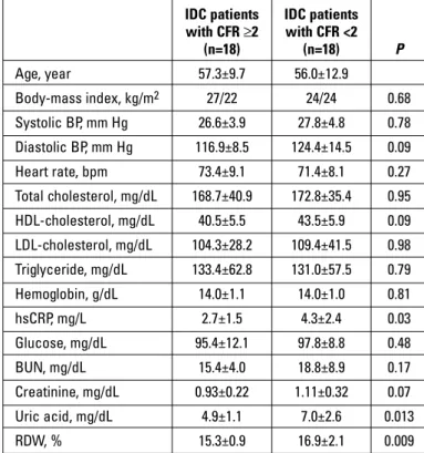
According to the median value of CFR (2.0) the patients were divided into two groups; the lower CFR group (<2.0) and the nor-mal CFR group (≥2.0). The demographic, biochemical and echo-cardiographic characteristics of two groups are summarized in Table 3. RDW levels were significantly higher in the lower CFR group than those in the normal CFR group (15.3±0.9 vs. 16.9±2.1, p=0.009). Left ventricular diastolic functions parameters were not significantly different in two groups. Baseline blood pres-sures were similar between the groups. Left ventricular EF was quite lower in the impaired CFR group than those in the normal CFR group (Table 4).
Subjects with Healthy
IDC controls (n=36) (n=35) P Clinical data Age, year 56.6±11.3 55.2±6.8 0.50 Male/female, % 20/16 18/17 0.43 Body-mass index, kg/m2 27.2±4.4 27.7±1.5 0.58 Baseline hemodynamics Systolic BP, mm Hg 120.6±12.3 122.0±12.7 0.64 Diastolic BP, mm Hg 77.4±7.1 75.6±6.0 0.25 Heart rate, beats/min 71.2±12.6 73.8±10.5 0.37 Hematological and biochemical data Total-cholesterol, mg/dL 170.7±37.8 190.3±27.4 0.02 HDL-cholesterol, mg/dL 42.0±6.7 42.8±9.6 0.65 LDL-cholesterol, mg/dL 106.9±35.0 115.5±19.5 0.21 Triglyceride, mg/dL 133.5±59.9 140.8±61.7 0.62 Glucose, mg/dL 96.7±10.4 93.5±4.9 0.24 hsCRP, mg/Lγ 3.52 (0.27-9.6) 1.94 (0.55-5.86) 0.006 Hemoglobin, g/dL 14.04±1.05 14.47±0.76 0.06 RDW, %γ 16.14 (13.5-22.6) 15.11 (13.5-17.0) 0.03 BUN, mg/dLγ 17.02 (10-36) 16.54 (9.0-24) 0.22 Creatinin, mg/dLγ 1.02 (0.6-2) 0.87 (0.67-1.10) 0.017 Uric acid, mg/dLγ 5.98 (2.6-12.5) 4.21 (2.5-7.9) 0.001 Statistics: The two groups were compared using the Student t-test or γMann-Whitney U test for multiple comparisons
BP - blood pressure; BUN - blood urea nitrogen; HDL - high-density lipoprotein; hsCRP - high-sensitivity C -reactive protein; IDC-idiopathic dilated cardiomyopathy; LDL - low-density lipoprotein; RDW - red cell distribution width; RDW - red cell distribution width Table 1. Demographic and biochemical characteristics, blood pressure values and medications in patients with IDC and control subjects
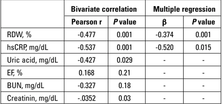
Hemoglobin, hematocrit, white blood cells were not different in the groups. However, patients with lower CFR had signifi-cantly higher RDW ratio as compared to patients with normal CFR, respectively. In addition, CFR is significantly and inversely correlated to RDW levels. We have no findings which show that patients with impaired CFR have different total cholesterol, LDL cholesterol and triglyceride values. hsCRP values were higher in the lower CFR group (2.7±1.5 vs. 4.3±2.4; p=0.03). CFR is signifi-cantly and inversely correlated to RDW level, serum uric acid, creatinine and hsCRP. Furthermore, in stepwise linear regression analysis, when CFR was taken as dependent, and RDW and other study variables including serum uric acid, BUN, creatinine and hsCRP, left ventricular EF as independent, we found that RDW value (Beta:-0.374, p=0.015) and hsCRP levels (Beta:-0.520, p=0.001) were significantly associated with CFR (Table 5). We also showed that RDW level was an accurate predictor of low CFR on the receiver operating characteristic (ROC) curve (Fig. 1). The area under the curve (AUC) was 73% (95% confidence inter-val 0.56-0.90), and RDW levels were significantly predictive of
low CFR (p=0.018). In addition, RDW significantly and positively correlated with serum uric acid (r=0.546, p=0.001) and BUN lev-els (r=0.471, p=0.005).
Discussion
In this study, we found that IDC patients have decreased CFR, increased RDW, uric acid and hsCRP compared to healthy controls. Previous studies revealed that coronary microvascular dysfunction, oxidative stress (as a surrogate marker of uric acid), subclinical inflammation, and increased RDW might take part in the development of myocardial contractile dysfunction (3, 21). In this respect, our results are consistent with the previous published data. Differing from the previous one, in this study we have searched for a possible role of increased RDW and serum uric acid levels, and whether subclinical inflammation might negatively contribute or not to impaired coronary microvascular function in patients with IDC. Hence, the patients with IDC were divided into two groups based on the median value of CFR, with the cut-off point two. The main findings of the study were that IDC patients with lower CFR had increased RDW, hsCRP and serum uric acid levels. Also it was evident that there was inde-pendent association between RDW levels and CFR in patients with IDC. To our knowledge this is the first study showing the
Subjects with Healthy
IDC controls (n=36) (n=35) P LVEDV, mL 199.6±64.5 87.1±8.4 0.001 LVESV, mL 129.0±51.9 35.6±6.7 0.001 EF, %γ 35.6 (23-44) 66.9 (63-70) 0.001 LVMI, g/m2 148.3±33.9 80.0±11.3 0.001
Mitral E-wave max, cm/s 70.3±26.3 77.5±12.6 0.14 Mitral A-wave max, cm/s 66.8±25.5 60.1±11.3 0.03
E/A ratio 1.48±1.44 1.31±0.21 0.64
Mitral E-wave 209.9±74.9 187.3±17.1 0.09 deceleration time, ms
Heart rate at rest, beats/min 70.8±12.6 73.5±12.3 0.23 Systolic BP at rest, mm Hg 120.6±12.3 122.0±12.6 0.64 Diastolic BP at rest, mm Hg 77.3±7.1 75.5±6.0 0.25 Heart rate after 94.4±11.4 98.3±12.9 0.18 dipyridamole, beats/min Systolic BP after 118.0±12.7 119.2±7.3 0.61 dipyridamole, mm Hg Diastolic BP after 75.5±6.4 76.1±5.5 0.69 dipyridamole, mm Hg Baseline DPFV, cm/s 27.2±7.1 22.2±2.8 0.001 Hyperemic DPFV, cm/s 55.7±14.5 68.9±13.4 0.001 CFR ratio 2.06±0.37 3.09±0.47 0.001
Statistics: The two groups were compared using the Student t-test or γ Mann-Whitney U test for multiple comparisons
BP - blood pressure; CFR - coronary flow reserve; DPFV - diastolic peak flow velocity; EF - ejection fraction; IDC-idiopathic dilated cardiomyopathy; LA - left atrium; LVEDV - left ventricular end-diastolic volume; LVESV - left ventricular end systolic volume; LVMI - left ventricular mass index
Table 2. Echocardiographic findings, and standard Doppler parameters of the left ventricle, coronary flow velocities and CFR values in patients with IDC and control subjects
IDC patients IDC patients
with CFR ≥2 with CFR <2 (n=18) (n=18) P Age, year 57.3±9.7 56.0±12.9 Body-mass index, kg/m2 27/22 24/24 0.68 Systolic BP, mm Hg 26.6±3.9 27.8±4.8 0.78 Diastolic BP, mm Hg 116.9±8.5 124.4±14.5 0.09 Heart rate, bpm 73.4±9.1 71.4±8.1 0.27 Total cholesterol, mg/dL 168.7±40.9 172.8±35.4 0.95 HDL-cholesterol, mg/dL 40.5±5.5 43.5±5.9 0.09 LDL-cholesterol, mg/dL 104.3±28.2 109.4±41.5 0.98 Triglyceride, mg/dL 133.4±62.8 131.0±57.5 0.79 Hemoglobin, g/dL 14.0±1.1 14.0±1.0 0.81 hsCRP, mg/L 2.7±1.5 4.3±2.4 0.03 Glucose, mg/dL 95.4±12.1 97.8±8.8 0.48 BUN, mg/dL 15.4±4.0 18.8±8.9 0.17 Creatinine, mg/dL 0.93±0.22 1.11±0.32 0.07 Uric acid, mg/dL 4.9±1.1 7.0±2.6 0.013 RDW, % 15.3±0.9 16.9±2.1 0.009
Statistics: Mann-Whitney U test was used for continuous variables and chi-square for categorical variables
bpm - beat per minute; BP - blood pressure; BUN - blood urea nitrogen; CFR - coronary flow reserve; HDL - high-density lipoprotein; hsCRP - high-sensitivity C-reactive protein; IDC-idiopathic dilated cardiomyopathy; LDL - low-density lipoprotein; RDW - red cell distribution width Table 3. Baseline clinical characteristics and laboratory analysis of study populations
presence of independent association between RDW and CFR in patients with IDC.
The red cell distribution width (RDW) is a marker of variation of the size of the circulating red blood cells. RDW is also defined as the standard deviation of erythrocyte size divided by the mean corpuscular volume (MCV) (22). MCV and RDW can help together to differentiate the several etiologies of anemia. RDW increases
in iron deficiency, B12 or folate deficiency, also in hemoglobin-opathies, hemolysis and after blood transfusions (15-17). Furthermore, a number of studies have been reported that the elevated RDW levels are associated with poor prognosis in the setting of stable angina (1, 2), acute myocardial infarction (3, 4), heart failure (5, 6), stroke (7), peripheral arterial disease (8), older age (9) and in the patients without CAD (10, 11). Chen at al. (11) concluded that high RDW values are associated with increased danger of all-cause mortality but not with the development of cardiovascular mortality in patients without CAD. On the other hand, Perlstein et al. (10) demonstrated that RDW strongly pre-dicted the all-cause and cardiovascular mortality in a large, com-munity based sample. Felker et al. (14) were the first to report the excess mortality and morbidity in patient with chronic heart fail-ure and elevated RDW. In another study, Allen et al. (23) also showed that RDW is significantly associated with adverse out-come in chronic HF and they suggested that the elevated RDW may indicate inflammatory stress and impaired iron mobilization. Correlation between RDW and cardiovascular diseases is as the following; in cardiovascular diseases inflammatory cytokines and neurohumoral mediators become activated. This causes suppressing the maturation of erythrocytes. Therefore, the increase in immature erythrocytes raises RDW levels (24).
In spite of angiographically normal coronary arteries, owing to coronary microvascular dysfunction CFR is impaired in patients with IDC (25). The association between hsCRP and coronary endothelial and/or microvascular dysfunction is evident. In our study, hsCRP levels were significantly higher in the patients with IDC than those in the control group. Also we found an important correlation between hsCRP and CFR in IDC patients. Chronic subclinical inflammatory state has been found to precede
IDC patients IDC patients
with CFR ≥2 with CFR <2 (n=18) (n=18) P LVEDV, mL 186.0±44.0 213.2±79.1 0.47 LVESV, mL 116.8±45.9 141.1±56.2 0.19 LA diameter, cm 3.32±0.30 3.33±0.28 0.76 EF, % 37.5±6.8 33.7±6.1 0.05 LVMI, g/m2 145.4±32.9 151.5±35.6 0.86 Mitral E max, cm/s 63.3±24.4 77.4±26.9 0.09 Mitral A max, cm/s 66.7±25.1 66.9±26.7 0.94 E/A ratio 1.23±1.31 1.76±1.56 0.16
Mitral E deceleration time, s 108.8±17.9 118.3±24.8 0.44 Baseline heart rate, bpm 73.2±6.9 72.1±11.2 0.47 Baseline systolic BP, mm Hg 148.9±8.4 150.1±7.3 0.24 Baseline diastolic BP, mm Hg 91.8±4.3 90.1±4.9 0.63 Peak heart rate, bpm 91.4±13.6 87.8±16.7 0.51 Peak systolic BP, mm Hg 116.1±11.3 119.7±13.9 0.50 Peak diastolic BP, mm Hg 74.6±5.0 76.5±7.5 0.35 Baseline DPFV, cm/s 25.8±4.9 28.7±8.7 0.38 Hyperemic DPFV, cm/s 60.3±14.1 51.1±13.7 0.06
CFR ratio 2.34±0.31 1.79±0.20 0.001
Statistics: Mann-Whitney U test was used for continuous variables and chi-square for categorical variables.
bpm - beat per minute; BP - blood pressure; CFR - coronary flow reserve; DPFV - diastolic peak flow velocity; EF - ejection fraction; IDC-idiopathic dilated
cardiomyopathy; LA - left atrium; LVEDV - left ventricular end-diastolic volume; LVESV - left ventricular end systolic volume; LVMI - left ventricular mass index
Table 4. Data from echocardiographic examinations of the study subject
Bivariate correlation Multiple regression Pearson r P value β P value
RDW, % -0.477 0.001 -0.374 0.001 hsCRP, mg/dL -0.537 0.001 -0.520 0.015 Uric acid, mg/dL -0.427 0.029 - -EF, % 0.168 0.21 - -BUN, mg/dL -0.327 0.18 - -Creatinin, mg/dL -.0352 0.03 -
-Statistics - The Pearson’s and Spearman correlation (hsCRP, uric acid, BUN, creatinine and left ventricular EF) and stepwise regression analysis was performed.
BUN - blood urea nitrogen; EF - ejection fraction; hsCRP - high-sensitivity C-reactive protein; RDW - red cell distribution width
Table 5. Correlation and regression analysis in patients with IDC
Figure 1. ROC curve analysis of RDW levels for low CFR
ROC Curve 1.0 0.8 0.6 0.4 0.2 0.0 1 - Specificity 0.0 0.2 0.4 0.6 0.8 1.0 Sensitivity CFR AUC: 0.731, P: 0.018
adverse cardiovascular outcome and hsCRP has been found to be a surrogate marker of inflammation (26). On the other hand, a strong positive correlation between RDW and markers of inflam-mation such as interleukin-6 and C-reactive protein (27). Also in our study, although it is poor we found a positive correlation between RDW and hsCRP in patients with IDC. Zalawadiya et al. (28) reported that in patients with HF taking statins RDW is lower. Anti-atherogenic property of statins has been shown via modu-lating inflammation and stabilizing atherosclerotic plaque. One could speculate that the inverse relation between the use of statin and RDW may be secondary to the anti-inflammatory prop-erty of statins, which further supports RDW as a marker of chronic inflammation in patients with IDC. Long-term treatment with beta blocker (carvedilol) can significantly increase coronary flow reserve and reduce the occurrence of stress-induced per-fusion defects, suggesting a favourable effect of the drug on coronary microvascular function in patients with IDC (29). The known effects of β-blocking agents in heart failure includes reversing LV remodelling, increasing myocardial oxygen to sup-ply ratio, additional mechanisms associated with vasodilatory and antioxidant properties. Furthermore, anti-inflammatory effects of beta blocker (carvedilol) and ACE inhibitor have been described. Though all the patients were treated with the same treatment it wasn’t completely explained why the level of some patients’ RDW is high and that of their CFR’s is low.
Studies suggest that oxidative stress might be involved in the pathogenetic processes in heart failure and myocardial oxida-tive stress and inflammation increase in hypertrophied or failing myocardium (30-32). Red blood cells (have a huge antioxidant capacity and serve as a primary oxidative sink, but they are prone to oxidative damage, which reduces cell survival and induces the release of immature erythrocytes into circulation (33). Altered CFR has been described in patients with dilated cardiomyopathy even when occlusive epicardial coronary arte-rial disease cannot be demonstrated angiographically (25). In addition, it has been shown that impaired CFR may lead to myo-cardial ischemia, progression of LV dysfunction, congestive heart failure (34) and increased mortality (17) in patients with dilated cardiomyopathy. The association of RDW with subclini-cal inflammation and oxidative stress markers (such as uric acid) suggests a close association with IDC patients. Since subclinical inflammation and oxidative stress markers that cause coronary microvascular dysfunction are effective in pathophysiological processes.
Study limitations
The main limitation of our study is the relatively small sample size.
Conclusion
The present study used second harmonic TTDE for CFR determination of LAD coronary artery to evaluate the possible
association between RDW and CFR in patients with IDC. The main findings of the study are that IDC patients with lower CFR have increased RDW and an independent association between RDW levels and CFR is evident in patients with IDC.
Conflict of interest: None declared. Peer-review: Partially peer-reviewed.
Authorship contributions: Concept - M.Ç.; Design - H.G., D.E.; Supervision - M.Ö., Z.Ç.; Resource - H.M.; Materials - M.Ö.; Data collection &/or processing - M.Ç., D.E.; Analysis &/or interpreta-tion - M.Ö.; Literature search - M.Ç.; Writing - M.Ç., M.Ö.; Critical review - D.E.
References
1. Tonelli M, Sacks F, Arnold M, Moye L, Davis B, Pfeffer M. Relation between red blood cell distribution width and cardiovascular event rate in people with coronary disease. Circulation 2008; 117: 163-8. [CrossRef]
2. Lappe JM, Horne BD, Shah SH. Red cell distribution width, C-reactive protein, the complete blood count, and mortality in patients with coronary disease and a normal comparison population. Clin Chim Acta 2011; 412: 2094-9. [CrossRef]
3. Uyarel H, Ergelen M, Çiçek G. Red cell distribution width as a novel prognostic marker in patients undergoing primary angioplasty for acute myocardial infarction. Coron Artery Dis 2011; 22: 138-44. [CrossRef]
4. Dabbah S, Hammerman H, Markiewicz W, Aronson D. Relation between red cell distribution width and clinical outcomes after acute myocardial infarction. Am J Cardiol 2010; 105: 312-7. [CrossRef]
5. Borné Y, Smith JG, Melander O, Hedblad B, Engström G. Red cell distribution width and risk for first hospitalization due to heart failure: a population-based cohort study. Eur J Heart 2011; 13: 1355-61. [CrossRef]
6. van Kimmenade RR, Mohammed AA, Uthamalingam S, van der Meer P, Felker GM, Januzzi JL Jr. Red blood cell distribution width and 1-year mortality in acute heart failure. Eur J Heart Fail 2010; 12: 129-36. [CrossRef]
7. Ani C, Ovbiagele B. Elevated red blood cell distribution width to predicts mortality in persons with known stroke. J Neurol Sci 2009; 277: 103-8. [CrossRef]
8. Ye Z, Smith C, Kullo IJ. Usefulness of red cell distribution width to predict mortality in patients with peripheral artery disease. Am J Cardiol 2011; 107: 1241-5. [CrossRef]
9. Patel KV, Semba RD, Ferruci L. Red cell distribution width and mortality in older adults: a meta-analysis. J Gerontol A Biol Sci Med Sci 2010; 65: 258-65. [CrossRef]
10. Perlstein TS, Weuve J, Pfeffer MA, Beckman JA. Red blood cell distribution width and mortality risk in a community-based prospective cohort. Arch Intern Med 2009; 169: 588-94. [CrossRef]
11. Chen PC, Sung FC, Chien KL, Hsu HC, Su TC, Lee YT. Red blood cell distribution width and risk of cardiovascular events and mortality in a community cohort Taiwan. Am J Epidemiol 2010; 171: 214-20. [CrossRef]
12. Bazick HS, Chang D, Mahadevappa K, Gibbons FK, Christopher KB. Red cell distribution width and all-cause mortality in critically ill patients. Crit Care Med 2011; 39: 1913-21. [CrossRef]
13. Horne BD, May HT, Kfoury AG, Renlund DG, Muhlestein JB, Lappé DL, et al. The Intermountain Risk Score (including the red cell distribution width) predicts heart failure and other morbidity endpoints. Eur J Heart Fail 2010; 12: 1203-13. [CrossRef]
14. Felker GM, Allen LA, Pocock SJ, Shaw LK, McMurray JJ, Pfeffer MA, et al. Red cell distribution width as a novel prognostic marker in heart failure: data from the CHARM Program and the Duke Databank. J Am Coll Cardiol 2007; 50: 40-7. [CrossRef]
15. Josephson RA, Silverman HS, Lakatta EG, Stern MD, Zweier JL. Study of the mechanisms of hydrogen peroxide and hydroxyl free radical-induced cellular injury and calcium overload in cardiac myocytes. J Biol Chem 1991; 266: 2354-61.
16. Wolfram R, Oguogho A, Palumbo B, Sinzinger H. Enhanced oxidative stres in coronary heart disease and chronic heart failure as indicated by an increased 8-epi-PGF(2alpha). Eur J Heart Fail 2005; 7: 167-72. [CrossRef]
17. Neglia D, Michelassi C, Trivieri MG, Sambuceti G, Giorgetti A, Pratali L, et al. Prognostic role of myocardial blood flow impairment in idiopathic left ventricular dysfunction. Circulation 2002; 105: 186-93. [CrossRef]
18. Saraste M, Koskenvuo J, Knutti J, Toikka J, Laine H, Niemi P, et al. Coronary flow reserve: Measurement with transthoracic Doppler echocardiography is reproducible and comparable with positron emission tomography. Clin Physiol 2001; 21: 114-22. [CrossRef]
19. Lang RM, Bierig M, Devereux RB, Flachskampf FA, Foster E, Pellikka PA, et al; American Society of Echocardiography’s Nomenclature and Standards Committee; Task Force on Chamber Quantification; American College of Cardiology Echocardiography Committee; American Heart Association; European Association of Echocardiography, European Society of Cardiology. Recommendations for chamber quantification. Eur J Echocardiogr 2006; 7: 79-108. [CrossRef]
20. Çalışkan M, Çiftçi O, Güllü H, Müderrisoğlu H. The effect of carvedilol therapy on coronary flow reserve in patients with idiopathic dilated cardiomyopathy. Turk Kardiyol Dern Ars 2008; 36: 247-52.
21. Özcan F, Turak O, Avcı S, Tok D, İşleyen A, Aras D, et al. Heart rate variability and red cell distribution width in patients with systolic left heart failure. Scand Cardiovasc J 2013; 47: 225-9. [CrossRef]
22. Bessman JD, Gilmer PR Jr, Gardner FH. Improved classification of anemias by MCV and RDW. Am J Clin Pathol 1983; 80: 322-6. 23. Allen LA, Felker GM, Mehra MR. Validation and potential
mechanisms of red cell distribution width as a prognostic marker in heart failure. J Card Fail 2010; 16: 230-8. [CrossRef]
24. Pierce CN, Larson DF. Inflammatory cytokine inhibition of erythropoiesis in patients implanted with a mechanical circulatory assist device. Perfusion 2005; 20: 83-90. [CrossRef]
25. Güllü H, Erdoğan D, Çalışkan M, Tok D, Kulaksızoğlu S, Yıldırır A, et al. Elevated serum uric acid levels impair coronary microvascular function in patients with idiopathic dilated cardiomyopathy. Eur J Heart Fail 2007; 9: 466-8. [CrossRef]
26. Ridker PM, Buring JE, Shih J, Matias M, Hennekens CH. Prospective study of C-reactive protein and the risk of future cardiovascular events among apparently healthy women. Circulation 1998; 98: 731-3.
[CrossRef]
27. Förhécz Z, Gombos T, Borgulya G, Pozsonyi Z, Prohászka Z, Jánoskuti L. Red cell distribution width in heart failure: prediction of clinical events and relationship with markers of ineffective erythropoiesis, inflammation, renal function, and nutritional state. Am Heart J 2009; 158: 659-66. [CrossRef]
28. Zalawadiya SK, Zmily H, Farah J, Daifallah S, Ali O, Ghali JK. Red cell distribution width and mortality in predominantly African-American population with decompensated heart failure. J Card Fail 2011; 17: 292-8. [CrossRef]
29. Neglia D, De Maria R, Masi S, Gallopin M, Pisani P, Pardini S, et al. Effects of long-term treatment with carvedilol on myocardial blood flow in idiopathic dilated cardiomyopathy. Heart 2007; 93: 808-13. [CrossRef]
30. Belch JJ, Bridges AB, Scott N, Chopra M. Oxygen free radicals and congestive heart failure. Br Heart J 1991; 65: 245-8. [CrossRef]
31. Mallat Z, Philip I, Lebret M, Chatel D, Maclouf J, Tedgui A. Elevated levels of 8-iso-prostaglandin F2 alpha in pericardial fluid of patients with heart failure: a potential role for in vivo oxidant stres in ventricular dilatation and progression to heart failure. Circulation 1998; 97: 1536-9. [CrossRef]
32. Inoue T, Sakai Y, Morooka S. Coronary flow reserve in patients with dilated cardiomyopathy. Am Heart J 1993; 125: 93-8. [CrossRef]
33. Kiefer CR, Snyder LM. Oxidation and erythrocyte senescence. Curr Opin Hematol 2000; 7: 113-6. [CrossRef]
34. van den Heuvel AF, van Veldhuisen DJ, van der Wall EE. Regional myocardial blood flow reserve impairment and metabolic changes suggesting myocardial ischemia in patients with idiopatic dilated cardiomyopathy. J Am Coll Cardiol 2000; 35: 19-28. [CrossRef]