A Rare Complication of Tonsillectomy: Subcutaneous
Emphysema
Ozan Erol, Erdinç Aydın
Department of Otorhinolaryngology, Başkent University School of Medicine Hospital, Ankara, Turkey Case Report
This study was presented at the 38th Turkish National Congress of Otorhinolaryngology Head and Neck Surgery, 26-30 October 2016, Antalya, Turkey.
Address for Correspondence: Ozan Erol
E-mail: [email protected] Received Date: 28.08.2016 Accepted Date: 06.11.2016
© Copyright 2016 by Official Journal of the Turkish Society of Otorhinolaryngology and Head and Neck Surgery Available online at www.turkarchotorhinolaryngol.org DOI: 10.5152/tao.2016.1888
172 Turkish Archives of Otorhinolaryngology
Türk Otorinolarengoloji Arşivi Turk Arch Otorhinolaryngol 2016; 54: 172-4
Abstract Tonsillectomy is one of the surgical procedures that are frequently performed by ear, nose, and throat sur-geons. The procedure is associated with many intra-operative and postintra-operative complications, and the nature of the operation site hampers surgical inter-ventions. Cervicofacial subcutaneous emphysema is characterized by the presence of air within the fascial planes of the head-neck region because of various re-asons. It may develop iatrogenically or
spontaneous-ly because of trauma. Herein, we report a 4-year-old male patient who presented to our clinic with comp-laints of frequent tonsillitis and snoring and who de-veloped subcutaneous emphysema involving only the maxillofacial region following tonsillectomy. In addi-tion, treatment strategies have been discussed, taking current literature into account.
Keywords: Tonsillectomy, complication, subcutaneo-us emphysema
Introduction
Subcutaneous emphysema is characterized by the presence of air between the fascial planes of the connective tissue. The etiology of subcutaneous em-physema involves traumatic and iatrogenic causes or spontaneous development. Head and neck sub-cutaneous emphysema is a life-threatening con-dition (1). When a large amount of air leaks into the fascial planes, in addition to the subcutaneous tissues, it may also spread to the retropharyngeal, mediastinal, pleural, and retroperitoneal spaces. Although it is known that many maxillofacial sur-gical procedures may be the cause of subcutaneous emphysema, they are very rare cases (1, 2). Aller-gic reactions, hematoma, angioedema, esophageal rupture, infection, and necrotizing fasciitis should be considered in the differential diagnosis. Mild cases can be followed up without treatment; how-ever, if anxiety, respiratory distress, severe pain, and infection are suspected, the patient should be hos-pitalized and followed up (3).
Case Report
Under general anesthesia, adenotonsillectomy was performed in a 4-year-old male patient who was brought to our polyclinic with complaints of
hav-ing tonsillitis more than five times per year, snor-ing, and apnea. After the patient was placed in a rose position and the Crowe–Davis mouth gag was inserted, adenoidectomy was first performed. Then, the upper poles of the left and right ton-sils were released using a sickle blade. The tonsillar capsule was inserted through the superior pharyn-geal constrictor muscle by making a blunt dissec-tion using a Kelly clamp. Tonsils were dissected from their lodges with cold steel elevators. After bleeding control was performed using bipolar cau-tery, a local anesthetic was injected into the bilat-eral anterior and posterior plicas.
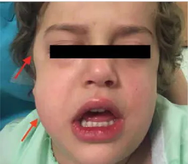
The patient had no complications and was tak-en to the day-care clinic. Ttak-en minutes after the operation, the swelling that started on the right side of the patient’s face after severe gagging and vomiting spread over the entire maxillofacial re-gion, including the eyelids (Figure 1). No swelling was observed in the neck region. The crepitation of emphysema was palpated. Except for fullness and swelling of the face, the patient had no other findings such as respiratory distress and cyanosis. There were no pathological findings on the anteri-or–posterior chest X-ray. In the cranial radiograph, air was observed under the skin on the right side of
the face (Figure 2). The blood gas findings were within normal limits. Except for postoperative routine treatment, there was no need for additional antibiotic treatment or hospitalization for follow-up. On follow-up, there was no regression in the swelling of his face, but he was discharged 4 hours after surgery because there were no oral intake problems and progress in his clinic. The findings completely regressed on the sixth day (Figure 3). Before the operation, a written informed consent was obtained from the relatives of the patient regarding the surgical procedure and the academic publications of clinical information, examina-tion, and visual material of the patient.
Discussion
Subcutaneous emphysema may rarely occur in the neck and face during the postoperative period. Emphysema may spontaneous-ly develop, but it may be usualspontaneous-ly observed after maxillofacial trauma, dental disease surgery, traumatic intubation, and ade-notonsillectomy, as in our case (4). A pneumomediastinum case that developed after tonsillectomy has been presented in a study conducted in our country (5).
Although the subcutaneous emphysema mechanism is not com-pletely understood, it is thought that descending and ascending mechanisms may play a role in its development. Descending mechanisms more frequently emerge (6). Emphysema may de-velop when positive pressure oxygen is supplied with a mask after the development of laryngospasm during tonsillectomy following extubation or because of subcutaneous air infiltration as a result of coughing and straining (1-9). The air may spread to the parapharyngeal region and neck through the fascial planes of the neck after entering the superior constrictor muscle fibers via the soft tissue spaces in the tonsillar region and gradual-ly becomes evident under the skin. Emphysema may spread to the supraclavicular region, mandible, cheek, under the eye, and temporal region (1-9). Occasionally, if mucosal rupture occurs in the epiglottis petiolus region or pyriform sinuses with the tip of laryngoscope, emphysema may occur through the same mecha-nism (7). The softness and crepitation of emphysema are palpat-ed under the skin. Because the parapharyngeal and retropharyn-geal regions are connected, air can migrate to the mediastinum. In these patients, pneumomediastinum and pneumothorax may develop, and problems such as respiratory distress and low satu-ration may occur (1, 3, 5-9).
In the ascending mechanism, after anesthesia or extubation, a rupture occurs in the alveoli with an increase in the intra-alveo-lar pressure and a corresponding increase in the intrapulmonary pressure. The air passes to the mediastinum through the perivas-cular interstitial spaces. The air in the pneumomediastinum can compress the heart, causing venous congestion, and the cardiac output may decrease. As a result, circulatory arrest may devel-op (1, 3, 5-9). If the parietal pleura, which is thin, is ruptured, pneumothorax may occur. In addition, the intense subcutane-ous emphysema in the neck may cause a collapse in tracheal rings, particularly in children, by compressing the trachea (7-9). Patients may have dyspnea, dysphagia and back pain, cyanosis,
Figure 1. Swelling that postoperatively occurred on the right side of the patient’s face (arrows)
Figure 3. View of the patient on the postoperative sixth day
Figure 2. Posteroanterior cranial radiograph showing the air under the skin on the right side of the face (arrow)
and Hamman’s sign (taking synchronized crepitation during the systole) (3, 5-9). Infection and abscess may develop in the cervi-cal and mediastinal tissues with the infection of oropharyngeal secretions (7). Because this complication, which postoperatively occurred in our patient, had a limited spread, it did not cause any findings other than swelling of the face. Thus, we believe that the descending mechanism played an important role because there were no serious findings, as observed in the ascending mecha-nism. Moreover, this finding, also observed in the postoperative period, supports the descending mechanism that first begins in the face region and then creates swelling around the eye and neck region. The development of emphysema on the right side of the face can be explained by the fact that the surgeon per-forming the operation had difficulty in entering the right plane because of being left-handed; thus, the traumatic process in the right location was higher. Conversely, we believe that this appli-cation will not cause emphysema because the local anesthetic injection at the end of the operation was submucosally adminis-tered only in the plicas and not in the tonsillar region.
Subcutaneous emphysema is a case that usually recovers sponta-neously within a few days without the need for treatment. How-ever, in rare cases, the disease may require treatment, which is proportional to its severity. Mild cases are followed up without treatment, but if anxiety, respiratory distress, severe pain, and infection are suspected, the patient should be hospitalized and followed up with oral intake restrictions (1-9). If the condition of the patient is stable, as in our case, monitoring is sufficient. In the case of a progressive condition, if there is a mucosal dam-age in the pharynx or tonsillar region, it should be treated. A cold compress should be applied to the region of swelling. The patient in whom respiratory distress develops should be admin-istered 100% oxygen. Patients should be admitted to a hospital, and broad spectrum antibiotics and drugs to prevent cough and vomit should be initiated (1-9). It should also be remembered that tracheotomy may be necessary because emphysema may be progressive in the neck and retropharyngeal area and may fur-ther result in airway obstruction (8, 9). The presence of a stable postoperative general condition and normal chest X-ray and blood gas findings did not suggest hospitalization or additional treatment options for our patient.
Conclusion
We believe that the high-pressure noninvasive ventilation per-formed in our patient resulted in subcutaneous emphysema after pharyngeal wall damage that occurred because of dissection or
intubation during the surgery. Otorhinolaryngologists should note that subcutaneous emphysema may rarely develop after adenotonsillectomy and that the treatment strategy should be planned by taking necessary precautions according to the gener-al condition of the patient.
Informed Consent: : Written informed consent was obtained from pa-tients’ parents who participated in this study.
Peer-review: Externally peer-reviewed.
Author contributions: Concept - E.A.; Design - O.E., E.A.; Supervi-sion - E.A.; Resource - O.E.; Materials - O.E.; Data Collection and/ or Processing - O.E.; Analysis and/or Interpretation - E.A.; Literature Search - O.E.; Writing - O.E.; Critical Reviews - E.A.
Conflict of Interest: No conflict of interest was declared by the authors. Financial Disclosure: The authors declared that this study has received no financial support.
References
1. Bizaki A, Kääriäinen J, Harju T, Rautiainen M. Facial subcutane-ous emphysema after tonsillectomy. Head Face Med 2014; 10: 11.
[CrossRef]
2. Villagra Siles EJ, Rodríguez Perales MA, García Mendoza A, Za-iden Torrez A. Cervical emphysema after tonsillectomy. A case report. Acta Otorhinolaryngol Esp 2006; 57: 251-2. [CrossRef]
3. Kim JP, Park JJ, Kang HS, Song MS. Subcutaneous emphysema and pneumomediastinum after tonsillectomy. Am J Otolaryngol 2010; 31: 212-5. [CrossRef]
4. Torgay A, Aydin E, Cilasun U, Durmaz L, Arslan G. Subcuta-neous emphysema after dental treatment: a case report. Paediatr Anaesth 2006; 16: 314-7. [CrossRef]
5. Düzgün N, Esme H, Övet E, Çalık M, Kurtipek E. Tonsillekto-minin nadir bir komplikasyonu: Pnömomediastinum. Respir Case Rep 2014; 3: 163-5.
6. Miman MC, Ozturan O, Durmus M, Kalcioglu MT, Gedik E. Cer-vical subcutaneous emphysema: an unusual complication of adeno-tonsillectomy. Paediatr Anaesth 2001; 11: 491-3. [CrossRef]
7. Gillot C, Tombu S, Crestani V, Huvelle P, Moreau P. Subcutaneo-us emphysema and mediastinitis: unSubcutaneo-usual complications of tonsil-lectomy. B-ENT 2005; 1: 197-200.
8. Yammine NV, Alherabi A, Gerin-Lajoie J. Post-tonsillectomy su-bcutaneous emphysema and pneumomediastinum. J Otolaryngol 2004; 33: 403-4. [CrossRef]
9. Tran DD, Littlefield PD. Late presentation of subcutaneous emp-hysema and pneumomediastinum following elective tonsillectomy. Am J Otolaryngol 2015; 36: 299-302.[CrossRef]
Turk Arch Otorhinolaryngol 2016; 54: 172-4 Erol and Aydın. A Rare Complication of Tonsillectomy