Anadolu University Journal of Science and Technology B – Theoretical Sciences 2017 - Volume: 5 Number: 1
Page: 56 - 69
DOI: 10.20290/aubtdb.289631
Received: 15 June 2016 Revised: 12 October 2016 Accepted: 14 October 2016
INVESTIGATION OF ELECTRONIC AND MOLECULAR PROPERTIES OF 5-(3-
METHYL-3-PHENYLCYCLOBUTYL)-N-PHENYL-3,6-DIHYDRO-2H-1,3,4-THIADIAZIN-2-IMINE BY EXPERIMENTAL AND THEORETICAL METHODS
Buse FERAH 1,*, İbrahim YILMAZ 2, Muharrem DİNÇER 1, Alaaddin ÇUKUROVALI 3
1 Department of Physics, Faculty of Arts and Sciences, Ondokuz Mayıs University, 55139- Kurupelit, Samsun, Turkey 2 Department of Chemistry, Faculty of Science, University of Karamanoglu Mehmet Bey, 70200 Karaman, Turkey,
3 Department of Chemistry, Faculty of Sciences, Fırat University, 23200 Elazığ, Turkey.
ABSTRACT
In this work, 5-(3-methyl-3-phenylcyclobutyl)-N-phenyl-3,6-dihydro-2H-1,3,4-thiadiazin-2-imine (C20H21N3S), MPDT, was prepared and characterized by X – ray single crystal diffraction and IR and 13C-NMR spectroscopic techniques. The molecular geometry, vibrational frequencies, gauge including atomic orbital (GIAO) 13C chemical shift values of MPDT in the ground state have been calculated by using the Hartree-Fock (HF) and Density Functional Theory (B3LYP) with 6-31G(d) basis set. The scaled B3LYP/6-31G(d) results shows the best agreement with the experimental values over the other method. B3LYP is applied to explore the Mulliken atomic charges of the title molecule. In addition to frontier molecular orbital (FMO) and molecular electrostatic potential (MEP) of the title molecule have been calculated by using the HF and B3LYP with 6-31G(d) basis set.
Keywords: Cyclobutane, X-ray structure determination, IR and (13C) NMR spectroscopy, Hartree-Fock (HF) Density Functional Theory (DFT).
5-(3-METHYL-3-PHENYLCYCLOBUTYL)-N-PHENYL-3,6-DİHYDRO-2H-1,3,4-THİADİAZİN-2-İMİNENİN DENEYSEL VE TEORİK METOTLARLA MOLEKÜLER VE
ELEKTRONİK ÖZELLİKLERİNİN İNCELENMESİ ÖZET
Bu çalışmada, 5-(3-methyl-3-phenylcyclobutyl)-N-phenyl-3,6-dihydro-2H-1,3,4-thiadiazin-2-imine (C20H21N3S), MPDT, IR ve 13C-NMR spektroskopik teknikleriyle ve X-ışını tek kristal kırınımı ile karakterize edilip hazırlandı. Temel halde molekülün, moleküler geometri, titreşim frekansları, GIAO 13C kimyasal değişim değerleri 6-31G(d) baz seti ile HF ve B3LYP metotları kullanılarak hesaplandı. B3LYP/6-31G(d) de elde edilen sonuçları diğer metoda kıyasla deneysel veriler ile daha uyumlu sonuçlar gösterdi. Bu sebeple molekülün Mulliken yük dağılımları B3LYP ile hesaplandı. Ek olarak molekülün moleküler orbital ve moleküler elektrostatik potensiyeli de HF/6-31G(d) ve B3LYP/6-31G(d) kullanılarak hesaplandı.
Anahtar Kelimeler: Siklobütan, X-ışını ile yapı belirleme, IR ve (13C) NMR spektroskopisi, Hartree-Fock (HF), Yoğunluk Fonksiyoneli Teorisi (DFT)
1. INTRODUCTION
5-(3-methyl-3-phenylcyclobutyl)-N-phenyl-3,6-dihydro-2H-1,3,4-thiadiazin-2-imine is a novel compound firstly prepared in our laboratories by us so there is no information about it in the literature. In order to support the experimental data, various computational techniques have been developed all over the world. Nowadays structural and electronic properties of atoms, molecules and solids can be calculated with great accuracy with the Density Functional Theory. The electronic structure and spectroscopic assignments of a molecule with given molecular geometry, have been calculated with following methods. DFT provides a variety of methods such as LSDA, BPV86, and B3LYP. B3LYP stands for Becke 3-Parameter (Exchange), Lee, Yang and Parr (Correlation; density functional theory) [1, 2].
In this paper, MPDT was described and characterized by 13C-NMR, IR and single-crystal X-ray diffraction methods as well as theoretical studies in the ground state which have been calculated using the HF and B3LYP with 6-31G(d) basis set.
2. MATERIALS AND METHODS 2.1. Synthesis
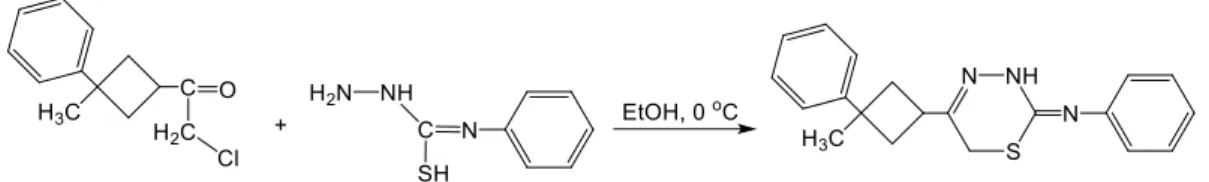
To a stirred solution of 4-phenyl thiosemicarbazide (1.6723 g, 10 mmoL) in 20 mL absolute ethanol, a solution of 2-chloro-1-(3-methyl-3-phenylcyclobutyl)ethanone (2.2271 g, 10 mmoL) in 10 mL absolute ethanol was added at 0 ºC and stirred for additional fifteen minutes (TLC). Light yellow precipitate separated by suction, washed with copious water and crystallized from acetone. Yield: 95%, melting point: 438 K. (see Figure 1).
Figure 1. Synthetic pathway for the synthesis of the target compound 2.2. General Remarks
All chemicals were of reagent grade and used as in commercially available forms without further purification. Melting point was determined by Gallenkamp melting point apparatus. The IR spectra of
the compound were recorded in the range of 4000–400 cm-1 using a Mattson 1000 FT-IR spectrometer
with KBr pellets. The 13C-NMR spectra were recorded on a Varian-Mercury-Plus 400 MHz
spectrometer using TMS as internal standard and CDCl3 (chloroform) as solvent.
2.3. Crystallography
All diffraction measurements were performed at room temperature (293 K) using graphite monochromatic MoKα radiation (k = 0.71073 Å) radiation and STOE IPDS (II) diffractometer. A suitable sample with the size of 0.62 × 0.42 × 0.13 mm was selected for the crystallographic study. Reflection data was recorded in the rotation mode using the ω scan technique by using X-AREA software [3]. Intensity parameters were collected in the θ range 2.7˚ ≤ θ ≤ 27.1˚. The structure was solved by direct methods using SHELXS-97 [4] implemented in the WinGX [5] program suite. The refinement was carried out by full-matrix least-squares method on the positional and anisotropic temperature parameters of the non-hydrogen atoms, or equivalently corresponding to 222 crystallographic parameters, using SHELXL-97 [6]. Computing data collection: X-AREA, cell refinement: X-AREA, computing data reduction: X-RED. The general-purpose crystallographic tool PLATON [7] was used for the structure analysis and presentation of the results. The structure was refined to Rint = 0.085 with 1689 observed reflections using I >2σ (I) threshold. The
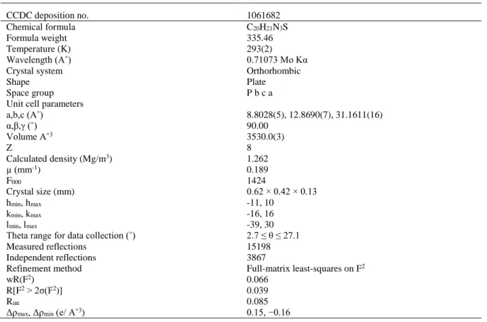
molecular graphic were displayed using ORTEP-3 for Windows [8]. Details of the data collection conditions and the parameters of the refinement process are given in Table 1.
Table 1. Crystal data and structure refinement parameters for the title compound
CCDC deposition no. 1061682
Chemical formula C20H21N3S
Formula weight 335.46
Temperature (K) 293(2)
Wavelength (A˚) 0.71073 Mo Kα
Crystal system Orthorhombic
Shape Plate
Space group Unit cell parameters a,b,c (A˚) α,β,γ (˚) Volume A˚3 Z Calculated density (Mg/m3) µ (mm-1) F000 Crystal size (mm) hmin, hmax kmin, kmax lmin, lmax
Theta range for data collection (˚) Measured reflections Independent reflections Refinement method wR(F2) R[F2 > 2σ(F2)] Rint
Δρmax, Δρmin (e/ A˚3)
P b c a 8.8028(5), 12.8690(7), 31.1611(16) 90.00 3530.0(3) 8 1.262 0.189 1424 0.62 × 0.42 × 0.13 -11, 10 -16, 16 -39, 30 2.7 ≤ θ ≤ 27.1 15198 3867 Full-matrix least-squares on F2 0.066 0.039 0.085 0.15, −0.16
2.4. Quantum Chemical Calculations
All the calculations were performed without specifying any symmetry for the title molecule by using Gauss View molecular visualization program [8] and Gaussian 03 program package [9]. For modeling, the initial guess of the compound was first obtained from the X-ray coordinates and the structure was optimized by Hartree–Fock (HF) and Density Functional Theory (DFT)/B3LYP methods [10, 1] with
the 6-31G(d) basis set. 13C-NMR chemical shift are calculated within GIAO approach [11, 12] applying
HF and B3LYP method with 6-31G(d) basis set. In addition to frontier molecular orbitals and molecular electrostatic potential map analysis were investigated by theoretical calculations
3. RESULTS AND DISCUSSION 3.1. Geometrical Structure
An ORTEP view of the title compound and the B3LYP optimized structure of the title compound are shown in Figure 2, where the compound crystallizes in the orthorhombic space group Pbca with eight molecules in the unit cell (Z = 8). The unit cell dimensions are a = 8.8028(5) Å, b = 12.8690(7) Å, c = 31.1611(16) Å and V = 3530.0(3) Å3. The title molecule is composed of a cyclobutane ring and two
benzene rings.
Figure 2. (a) A view of the title compound showing the atom-numbering scheme. Displacement ellipsoids are drawn at the 30% probability level and H atoms have been omitted for clarity. (b) The theoretical geometric structure of the title compound (B3LYP/6-31 G(d) level) The crystal structure could be described as being built from essentially planar fragments, the aromatic ring A (C1—C6), N1 atom is linking B (C7—S1), the cyclobutane plane C (C10—C13) and the other aromatic ring D (C15—C20). The dihedral angles between A, B, C and D are 50.16(0.08)˚ (A/B), 48.88(0.08)˚ (B/C), 87.82(0.08)˚ (C/D).
It is well known that 3-substituted cyclobutane carboxylic acid derivatives exhibit anti-inflammatory and antidepressant activities [13] as well as liquid crystal properties [14]. The popular anticancer drug, carboplatin, also contains a cyclobutane ring [15]. Cyclobutane ring has adopted a puckered conformation and although the value for the puckering of the cyclobutane ring found in the literature is 19.8(3)˚ [16], there is a negligible puckering in the cyclobutane ring (torsion angles; C12—C11—C10— C13 and C10—C13—C12—C11; 15.07(15)˚ and 14.90(16)˚, respectively). When the bond lengths and angles of the cyclobutane ring in the title molecule are compared with the previously reported cyclobutane derivatives [17-20], it is seen that there are no significant differences.
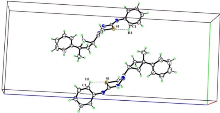
Perspective view of the crystal packing in the unit cell is shown in Figure 3. In the crystal packing, there is a C—H•••S hydrogen bonding, and the details of which are given in Table 2. The crystal structure of MPDT shows an intramolecular bond-interaction. In this hydrogen bonding, atom S1 acts as a donor to related C1 at (2-x,-y,-z).
Figure 3. Part of the crystal structure of the title compound, showing the C1–H1···S1 interactions Table 2. Hydrogen bond geometries in crystal structure (Å, ˚)
D—H···A D—H H···A D···A D—H···A
C1–H1···S1 0.93 2.60 3.045 (2) 110
Symmetry codes: 2-x,-y,-z. 3.2. Optimized Structures
The optimized parameters (bond lengths, bond angles and torsion angles) of the title compound have been obtained using HF and B3LYP methods with the 6-31G(d) basis set. Some selected geometrical parameters which have been experimentally gathered and theoretically calculated are listed in Table 3. From Table 3, it can be seen that there are some deviations in the computed geometrical parameters from those data, and these differences are probably due to the intramolecular interaction.
Table 3. Some selected experimental and optimized geometrical parameters of the title compound
Geometric parameters Experimental (X-ray)
Calculated [6-31G(d)] HF B3LYP
Bond lengths (A˚)
C1˗C6 1.386(3) 1.3897 1.4062 C6˗N1 S1˗C7 N2˗N3 C8˗C10 C10˗C11 C11˗C12 C12˗C14 C15˗C16 R2 1.408(3) 1.759(2) 1.396(2) 1.489(3) 1.534(3) 1.543(3) 1.530(3) 1.379(3) 1.4078 1.7757 1.3854 1.5007 1.5396 1.5592 1.5307 1.3972 0.9953 1.4006 1.7933 1.3859 1.5002 1.5469 1.5708 1.5332 1.4066 0.9889 Bond angles (˚) C2—C3—C4 119.5(2) 119.3454 119.3759 C5—C6—N1 N1—C7—S1 C7—N2—N3 N2—N3—C8 C8—C10—C13 C10—C11—C12 C14—C12—C15 R2 117.96(18) 126.66(17) 125.02(18) 116.56(18) 116.50(17) 90.13(16) 113.63(19) 119.3491 125.8496 122.8985 117.4256 116.9861 89.7584 113.4319 0.9908 117.949 127.0803 126.0701 117.2056 117.2379 90.072 113.677 0.9989 Torsion angles (˚) C1—C2—C3—C4 C5—C6—N1—C7 N1—C6—C5—C4 C10—C11—C12—C14 R2 0.8(4) 143.0(2) 176.7(2) 95.96(19) 0.3333 108.6945 177.63 95.5586 0.9499 0.7463 133.419 178.62 96.0979 0.9954
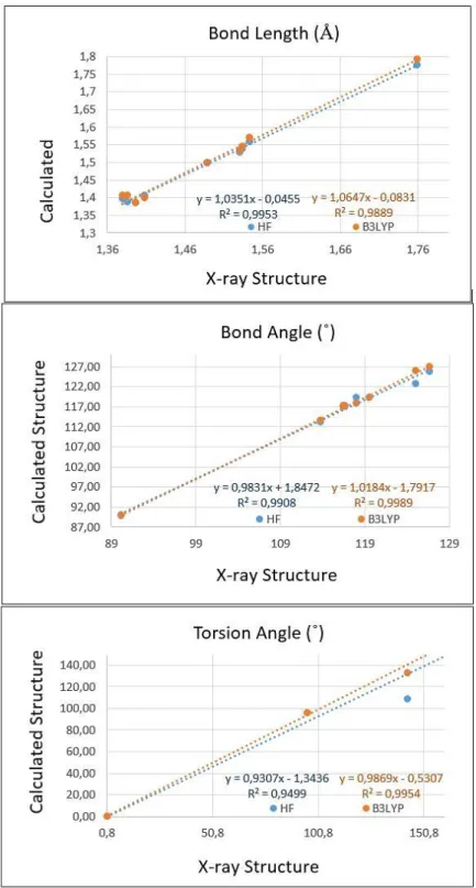
We have displayed the correlation graph in Figure 4 based on the correlation calculations. The correlation values (R2) have been found as 0.9953 and 0.9889 for bond lengths, 0.9908 and 0.9989 for
bond angles, 0.9499 and 0.9954 for torsion angles at HF/6-31G(d) and B3LYP/6-31G(d) levels, respectively. According to correlations values, the B3LYP/6-31G(d) method gave accurate results for the bond angles, when torsion angles compared with the HF/6-31G(d) method.
Figure 4. Correlation graphics between the experimental and theoretical geometric parameters of the title compound
A global comparison was performed by superimposing the molecular skeletons obtained from the X-ray diffraction and the theoretical calculations atom by atom (Figure 5), obtaining RMSE values of 0.232 and 0.107 Å for HF/6-31G(d) and B3LYP/6-31G(d), respectively. According to these results, the smallest RMSE value is acquired for B3LYP/6-31G(d) and the geometry obtained from this method coincides better with the crystalline structure than HF/6-31G(d) method. For that reason, we used the geometry from B3LYP/6-31G(d) method, to calculate molecular electrostatic potential (MEP) and frontier molecular orbitals (FMOs).
Figure 5. Atom-by atom superimposition of the calculated structures calculated (green) (a = HF/6-31 G(d), b = B3LYP/6-31 G(d)) on the X-ray structure (red) of the title compound
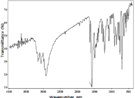
3.3. IR Spectroscopy
The experimental IR spectra of the title compound are shown in Figure 6. The vibrational bands have been assigned using GaussView molecular visualization program and calculated bands with both HF and B3LYP approach along with selected experimental counterparts which are indicated in Table 4.
Table 4. Comparisons of the observed and calculated vibrational spectra of the title compound Assignment Experimental FT-IR (cm-1) Calculated [6-31 G(d)] HF B3LYP νsN–H 3259 3584 3477 νasC–Haromatic 3067 3026 2868 νasC–H2cyclobutane 2961 3036 3015 νC–Hcyclobutane 2855 2861 2834 cyclobutane 1036 946 918 R2 0.9835 0.9784
Vibrational modes ν, stretching; s, symmetric; as, asymmetric; ,ring breathing.
Aromatic C—H vibrations
The C—H asymmetric stretching vibrations are observed at 3105 cm-1 in FT-IR spectrum [21]. In the
title compound, the C—H asymmetric stretching vibration is observed at 3067 cm-1, which has been
calculated at 3026 cm-1 for HF level, 2868 cm-1 for B3LYP level. There are no peaks observed in FT-IR
spectrum for C—H symmetric stretching vibrations.
N—H vibrations
The N—H aromatic stretching vibrations were occurred at 3300-3500 cm-1 [22]. In previous study, N—
H stretching vibration was founded at 3330-3481 cm-1 [23]. In here, the N—H stretching vibration is
observed at 3259 cm-1 as a very strong band in FT-IR spectrum and its corresponding calculated
frequencies are 3584 and 3477 cm-1 using HF/6-31G(d) and B3LYP/6-31G(d), respectively. Cyclobutane vibrations
In the title molecule, there are many cyclobutane ring vibrations. The asymmetric stretching of C—H2
was observed at 2939 and 2855 cm-1 [24]. The symmetric and asymmetric stretching are observed at
2855 and 2961 cm-1 in FT-IR spectrum. These modes have been calculated at 2861, 2834 cm-1 with
HF/6-31G(d) and 3036, 3015 cm-1 with B3LYP/6-31G(d), respectively.
The correlation values (R2) have been found as 0.9835 and 0.9784 for HF/6-31G(d) and
B3LYP/6-31G(d), respectively. According to the correlations values, the B3LYP/6-31G(d) method gave accurate results for the vibration modes compared with the HF/6-31G(d) method.
3.4. NMR Spectra
The isotropic chemical shifts are frequently used as an aid in identification of organic compounds and accurate predictions of molecular geometries that are essential for reliable studies of magnetic properties [25]. The experimental 13C-NMR spectra of the title compound recorded using TMS as an internal
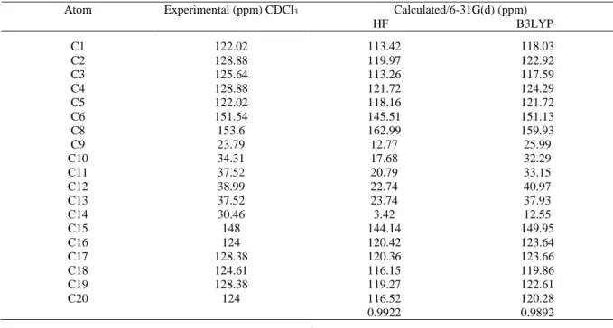
standard and chloroform (CDCl3) as solvent are shown in Figure 7. The theoretical 13C chemical shifts
are calculated for the optimized geometry obtained from HF/6-31G(d) and B3LYP/6-31G(d) by using Gaussian program with the standard GIAO approach.
Figure 7. Experimental 13C chemical shift spectra of the title compound
In the experimental 13C-NMR spectrum of MPDT, the signals at 153.6 and 23.79 ppm are assigned to
the atoms C8 and C9, respectively. In the cyclobutane ring the 13C-NMR signals of C12 and C13 carbons
are at 44.21 ppm [26]. In the present work, the experimental results are attained at 37.52 ppm. The other calculated chemical shifts values can be seen in Table 5.
Table 5. The experimental and calculated 13C isotropic chemical shifts (with respect to TMS, all values
in ppm) for title compound
Atom Experimental (ppm) CDCl3 Calculated/6-31G(d) (ppm) HF B3LYP C1 122.02 113.42 118.03 C2 128.88 119.97 122.92 C3 C4 C5 C6 C8 C9 C10 C11 C12 C13 C14 C15 C16 C17 C18 C19 C20 125.64 128.88 122.02 151.54 153.6 23.79 34.31 37.52 38.99 37.52 30.46 148 124 128.38 124.61 128.38 124 113.26 121.72 118.16 145.51 162.99 12.77 17.68 20.79 22.74 23.74 3.42 144.14 120.42 120.36 116.15 119.27 116.52 0.9922 117.59 124.29 121.72 151.13 159.93 25.99 32.29 33.15 40.97 37.93 12.55 149.95 123.64 123.66 119.86 122.61 120.28 0.9892
The large difference between the experimental and calculated values of the cyclobutane ring may be due to the fact that theoretical calculations of the title molecule have been done in gaseous phase.
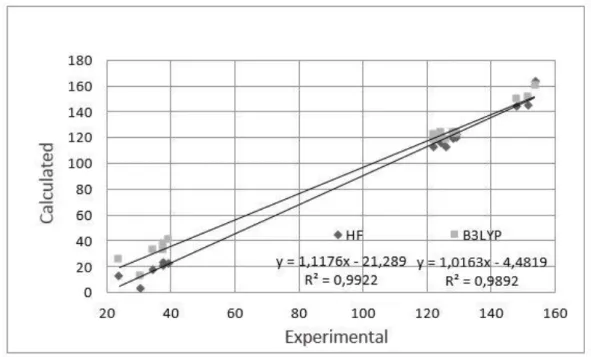
In order to compare the chemical shifts, correlation graph between the observed and calculated 13
C-NMR chemical shifts is shown in Figure 8. Therefore, the experimentally obtained values are in good agreement with the theoretically computed values by HF/6-31G(d) method when compared to B3LYP/6-31(G) method.
Figure 8. Correlation graphics between the experimental and theoretical NMR chemical shift values of the title compound
Quantum-Chemical Studies
3.5. Molecular Eectrostatic Potential (MEP)
The molecular electrostatic potential (MEP) is related to the electronic density and is a very useful descriptor in understanding sites of electrophilic attack and nucleophilic reactions as well as hydrogen bonding interactions [27, 28]. It is very useful to study the relationship between molecular structures with its physiochemical property.
Different values of electrostatic potential at the surface are represented by different colors. The color scheme for the MEP surface is red-electron rich, partially negative charge (electrophilic reactive centre); blue-electron deficient, partially positive charge; light blue-slightly electron deficient region (nucleophilic reactive centre); yellow-slightly electron rich region; green-neutral, respectively [29]. In the present study, MEP maps of MPDT has been found by HF/6-31G(d) and B3LYP/6-31G(d) methods and shown in Figure 9. The color code of these maps is in the range between -4.834 a.u. to 4.834 a.u. for HF/6-31G(d) and -4.043 a.u. to 4.043 a.u. for B3LYP/6-31G(d).
Figure 9. Molecular electrostatic potential map (MEP) (in a.u.) calculated at B3LYP/6-31 G(d) level Areas of low potential (red) are characterized by an abundance of electrons. Areas of high potential (blue) are characterized by a relative absence of electrons. The region around the nitrogen atom (N1) linked with carbon (C7) through double bond represents the most negative potential region in MPDT. Nitrogen has a higher electronegativity value which would consequently have a higher electron density around them. Thus the spherical region that corresponds to nitrogen atom would have a red portion on it. The MPDT of the compound clearly indicates the electron rich centers of nitrogen atom.
3.6. Frontier Molecular Orbital Analysis (FMO)
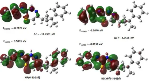
The most important frontier molecular orbital (FMO) such as highest occupied molecular orbital (HOMO) and lowest unoccupied molecular orbital (LUMO) plays a crucial part in the chemical stability of the molecule [30]. The HOMO represents the ability to donate an electron and LUMO represents the ability of accept an electron [31]. The energy gap between HOMO and LUMO also determines the chemical reactivity, optical polarizability and chemical hardness-softness of a molecule [30]. A molecule with a small frontier orbital gap is more polarizable and is generally associated with a high chemical reactivity, low kinetic stability and is also termed as soft molecule [32]. By examining the frontier orbitals of a molecule the optical properties and the steps to react with other molecules can be determined. The calculations indicate that the title compound has 89 occupied molecular orbitals. The plots of highest occupied molecular orbitals (HOMOs) and lowest unoccupied molecular orbitals (LUMOs) are shown in Figure 10.
Figure 10. Molecular orbital surfaces and energy levels given for the HOMO and LUMO of the title compound
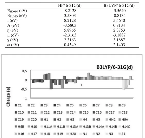
By using HOMO and LUMO energy values for a molecule, the global chemical reactivity descriptors of molecules such as hardness (ƞ), chemical potential (µ), softness (S), electronegativity (χ) and electrophilicity index (ω) have been defined [33, 34]. On the basis of EHOMO and ELUMO, these are
calculated using the below equations. Using Koopman’s theorem [35] for closed-shell molecules, The hardness of the molecule is ƞ = (I – A) / 2,
The chemical potential of the molecule is µ = - (I + A) / 2, The electronegativity of the molecule is χ = (I + A) / 2, The electrophilicity index of the molecule is ω = µ2 / 2ƞ.
Where A is the ionization potential and I is the electron affinity of the molecule. I and A can be expressed through HOMO and LUMO orbital energies as I = - EHOMO and A = - ELUMO. The calculated values of
the hardness, chemical potential, electronegativity and electrophilicity index of our molecule in gas phase are given in Table 6.
Table 6. The calculated values of the title molecule using both HF/ 6-31G(d) and B3LYP/ 6-31G(d) levels HF/ 6-31G(d) B3LYP/ 6-31G(d) EHOMO (eV) -8.2128 -5.5640 ELUMO (eV) I (eV) A (eV) 3.5803 8.2128 -3.5803 -0.8134 5.5640 0.8134 ƞ (eV) 5.8965 2.3753 µ (eV) -2.3163 -3.1887 χ (eV) 2.3163 3.1887 ω (eV) 0.4549 2.1403
Figure 11. The charge distribution calculated by the Mulliken method for the title molecule 4. CONCLUSIONS
Crystal structure of MPDT was determined and its geometry was optimized by HF and B3LYP method with 6-31G(d) basis set. Theoretical IR and NMR spectroscopies for optimized molecular structure were calculated. Compared to the theoretical and experimental results, compatible results have been observed. Investigations by B3LYP/6-31G(d) level for both the bond angles and torsion angles are performed
better. Mulliken atomic charge values are also calculated using same method with basis set. Besides of these properties of the molecule, we have calculated MEP, HOMO-LUMO, hardness, chemical potential, electronegativity and electrophilicity index of the molecule with HF and B3LYP.
ACKNOWLEDGMENTS
We wish to thank Prof. Dr. Orhan Büyükgüngör for his help with the data collection and acknowledge the Faculty of Arts and Sciences, Ondokuz Mayıs University, Turkey, for the use of the STOE IPDS II diffractometer.
SUPPLEMENTARY DATA
CCDC-1061682 contains the supplementary crystallographic data for the compound reported in this article. These data can be obtained free of charge at www.ccdc.cam.ac.uk/conts/retrieving.html [or from the Cambridge Crystallographic Data Centre (CCDC), 12 Union Road, Cambridge CB2 1EZ, UK; fax: +44(0)1223-336033; e-mail: [email protected]].
REFERENCES
[1] Becke AD. Density functional thermochemistry. III. The role of exact exchange. J Chem Phys 1993; 98: 5648.
[2] Lee C, Yang W, Parr RG. Development of the Colle-Salvetti correlation-energy formula into a functional of the electron density. Phys Rev 1988; B37: 785-789.
[3] Stoe & Cie. X-AREA Version 1.18 and X-RED32 Version 1.04 2002; Darmstadt, Germany. [4] Sheldrick GM. SHELXS-97; Program for the Solution of Crystal Structures, University of Gottingen, 1997.
[5] Farrugia LJ. WinGX suite for smallmolecule single-crystal crystallography. J Appl Crystallogr 1999; 32: 837–838.
[6] Sheldrick GM. SHELXL-97; Program for Crystal Structures Refinement, University of Gottingen, 1997.
[7] Spek AL. Structure validation in chemical crystallography. Acta Crystallogr 2009; D65: 148–155. [8] Dennington II R, Keith T, Millam J, Gauss View, Version 4.1.2, Semichem Inc, Shawnee Mission, KS, 2007.
[9] Frisch MJ, Trucks GW, Schlegel HB, Scuseria GE, Robb MA, Cheeseman JR, Montgomery Jr JA, Vreven T, Kudin K.N, Burant J.C, Millam J.M, Iyengar SS, Tomasi J, Barone V, Mennucci B, Cossi M, Scalmani G, Rega N, Petersson GA, Nakatsuji H, Hada M, Ehara M, Toyota K, Fukuda R, Hasegawa J, Ishida M, Nakajima T, Honda Y, Kitao O, Nakai H, Klene M, Li X, Knox JE, Hratchian HP, Cross JB, Bakken V, Adamo C, Jaramillo J, Gomperts R, Stratmann RE, Yazyev O, Austin AJ, Cammi R, Pomelli C, Ochterski JW, Ayala PY, Morokuma K, Voth GA, P Salvador, Dannenberg JJ, Zakrzewski VG, Dapprich S, Daniels AD, Strain MC, Farkas O, Malick DK, Rabuck AD, Raghavachari K, Foresman JB, Ortiz JV, Cui Q, Baboul AG, Clifford S, Cioslowski J, Stefanov BB, Liu G, Liashenko A, Piskorz P, Komaromi I, Martin RL, Fox DJ, Keith T, Al-Laham MA, Peng CY, Nanayakkara A, Challacombe
M, Gill PMW, Johnson B, Chen W, Wong MW, Gonzalez C, Pople JA, Gaussian 03, Revision E.01, Gaussian, Inc: Wallingford CT, 2004.
[10] Lee C, Yang W, Parr RG. Development of the Colle-Salvetti correlation-energy formula into a functional of the electron density. Phys Rev 1988; B37: 785.
[11] Ditchfield R. Molecular Orbital Theory of Magnetic Shielding and Magnetic Susceptibility. J Chem Phys 1972; 56: 5688.
[12] Wolinski K, Hinton JF, Pulay P. Efficient implementation of the gauge-independent atomic orbital method for NMR chemical shift calculations. J Am Chem Soc 1990; 112: 8251-8260.
[13] Dehmlow EV, Schmidt S. Synthesis of Stereoisomeric 3-Substituted cyclobutanecarboxylic acid-derivatives. Liebigs Ann Chem 1990; 5: 411–414.
[14] Coghi L, Lanfredi AMM, Tiripicchio A. Crystal and molecular structure of thiosemicarbazide hydrochloride. J Chem Soc Perkin Trans 1976; 2: 1808-1810.
[15] Wheate NJ, Walker S, Craig GE and Oun R. The status of platinum anticancer drugs in the clinic and in clinical trials. Dalton Transactions 2010; 39: 8113-8127.
[16] Dincer M, Ozdemir N, Cukurovali A, Yilmaz I, Buyukgungor O. "1-(3-Mesityl-3- methylcyclobutyl)-2-(pyrrolidin-1-yl) ethan-1-one. Acta Cryst 2004; E60: o1523-o1524.
[17] Sen F, Dincer M, Cukurovali A, Yilmaz I. (Z)-1-(3-Mesityl-3-methylcyclobu-tyl)-2-(morpholin-4-yl) ethanone oxime. Acta Cryst 2011; E67: o958-o959.
[18] Sen F, Dincer M, Cukurovali A, Yilmaz I. 1,1'-Bis(3-methyl-3-phenylcyclobutyl)-2,2'-(azanedi-yl)diethanol. Acta Cryst 2012; E68: o1052.
[19] Dincer M, Ozdemir N, Yılmaz I, Cukurovali A, Buyukgungor O. 1-Methyl-1-phenyl-3-[1- hydroxyimino-2-(succinimido)ethyl]cyclobutane. Acta Cryst 2012; C60: o674-o676.
[20] Ozdemir N, Dincer M, Yilmaz I, Cukurovali A. 1-Methyl-1-phenyl-3-(phthalimidoacetyl) cyclobutane. Acta Cryst 2004; E60: o14-o16.
[21] Sen F, Dincer M, Cukurovali A, Yilmaz I. N-[4-(3-methyl-3-mesityl-cyclobutyl)-thiazol-2-yl]-succinamic acid: X-ray structure, spectroscopic characterization and quantum chemical computational studies. J Mol Struc 2013; 1046: 1–8.
![Table 4. Comparisons of the observed and calculated vibrational spectra of the title compound Assignment Experimental FT-IR (cm -1 ) Calculated [6-31 G(d)] HF B3LYP ν s N–H 3259 3584 3477 ν as C–H aromatic 3067 3026 2868](https://thumb-eu.123doks.com/thumbv2/9libnet/4585055.84469/8.892.146.743.177.304/comparisons-observed-calculated-vibrational-compound-assignment-experimental-calculated.webp)