The frequency of type 2 second-degree and third-degree
atrioventricular block induced by blunt chest trauma
in the emergency department: A multicenter study
Banu Şahin Yıldız, M.D.,1 Mehmet Ali Astarcıoğlu, M.D.,2 Nazire Başkurt Aladağ, M.D.,1Ahmet Çağrı Aykan, M.D.,3 Hakan Hasdemir, M.D.,4 Alparslan Şahin, M.D.,5 Mustafa Yıldız, M.D.3 1Department of Internal Medicine, Dr. Lütfi Kırdar Kartal Training and Research Hospital, İstanbul
2Department of Cardiology, Dumlupınar University Evliya Çelebi Training and Research Hospital, Kütahya 3Department of Cardiology, Kartal Koşuyolu Yüksek İhtisas Training and Research Hospital, İstanbul
4Department of Cardiology, Dr. Siyami Ersek Cardiovascular and Thoracic Surgery Training and Research Hospital, İstanbul 5Department of Cardiology, Dr. Sadi Konuk Training and Research Hospital, İstanbul
ABSTRACT
BACKGROUND: Conduction disturbances including type 2 second-degree atrioventricular block (Mobitz II) and third-degree atrioventricular block following blunt chest trauma are probably rare. Moreover, the pathophysiological mechanisms responsible for this rare dysrhythmia following trauma are not well understood yet. In this study, it was aimed to identify the frequency of this dysrhythmia associated with trauma.
METHODS: Two hundred and fifty-three consecutive Mobitz II block and third-degree atrioventricular block patients admitted to the Emergency Department of Internal Medicine between January 2012 and March 2013 were evaluated. Only four patients with Mobitz II block and third-degree atrioventricular block associated with trauma were enrolled into the present study. The level of atrioventricular block was defined according to electrocardiographic characteristics.
RESULTS: Only four (mean age: 40.2±19.7 years, two male) of 253 patients were associated with trauma. All patients had normal coronary arteries in coronary angiography or multislice computed tomography. Permanent pacemaker was performed in two patients with third-degree atrioventricular block. None of the patients had coronary artery disease or hypertension.
CONCLUSION: Rare clinical cases in the literature confirm that blunt chest trauma can cause conduction defects, which are usually transient. However, patients with blunt chest trauma must need an electrocardiographic evaluation for atrioventricular block upon admission and in the follow-up period.
Key words: Blunt chest trauma; electrocardiography; third-degree atrioventricular block; Type 2 second-degree atrioventricular block (Mobitz II).
ity including the clavicles, ribs, esophagus, lungs, and heart.
[1] It may cause cardiac lesions in coronary arteries, valves
and pericardium. Depending on the extent of trauma, such injuries can cause varying amounts of mechanical or electrical dysfunction including high degree atrioventricular block and ventricular fibrillation.[2] There are still limited data
regard-ing the incidence, prevalence and clinical characteristics of important dysrhythmia including type 2 second-degree atrio-ventricular block (Mobitz II) and third-degree atrioatrio-ventricular block following trauma.[2-4] Moreover, the pathophysiological
mechanisms responsible for this rare dysrhythmia following trauma are not well understood yet.
The aim of this present study was to identify the frequency of the Mobitz II block and third-degree atrioventricular block
ORIGINAL ARTICLE
Ulus Travma Acil Cerrahi Derg, May 2015, Vol. 21, No. 3 193
Address for correspondence: Banu Şahin Yıldız, M.D. Dr. Lütfi Kırdar Kartal Eğitim ve Araştırma Hastanesi, Kardiyoloji Anabilim Dalı, İstanbul, Turkey
Tel: +90 274 - 230 66 60 E-mail: [email protected] Qucik Response Code Ulus Travma Acil Cerrahi Derg
2015;21(3):193-196 doi: 10.5505/tjtes.2015.04763 Copyright 2015
TJTES
INTRODUCTION
Blunt chest trauma is a significant cause of morbidity and mortality in life. It can affect chest wall and thoracic
cav-Şahin Yıldız et al. Type 2 second-degree and third-degree atrioventricular block
associated with trauma in the Emergency Department of In-ternal Medicine. The dysrhythmia was also assessed for the necessity of permanent pacemaker.
MATERIALS AND METHODS
Two hundred and fifty-three consecutive Mobitz II block and third-degree atrioventricular block patients admitted to the Emergency Department of Internal Medicine between Janu-ary 2012 and March 2013 were evaluated. Only four patients with Mobitz II block (two patients) and third-degree atrio-ventricular block (two patients) were associated with trauma, and those four patients were enrolled into the present study. The study was approved by the local committee. All subjects gave their consent for inclusion into the study. The investiga-tion conformed to the principles outlined in the Declarainvestiga-tion of Helsinki. Patients with electrolyte abnormalities, digitalis toxicity, vasovagal syncope, myocardial infarction history, and patients using drugs, which might affect atrioventricular node, were excluded from the study.
The level of atrioventricular block was defined according to the following electrocardiographic characteristics: Type 2 second-degree atrioventricular block (Mobitz II): Mobitz II heart block is characterized on a surface electrocardiogra-phy by intermittently non-conducted P waves not preceded by PR prolongation and not followed by PR shortening. The block may progress rapidly to third-degree atrioventricular block.
Third-degree atrioventricular block or complete heart block: This is a medical condition in which the impulse generated in the sinoatrial node in the atrium does not propagate to the ventricles. The PR interval will be variable as the hallmark of complete heart block is no apparent relationship between P waves and QRS complexes.
RESULTS
Only four patients (mean age: 40.2±19.7 years, two male) with Mobitz II block (two female patients) (Fig. 1) and third-degree atrioventricular block (two male patients) were asso-ciated with trauma. All patients had normal coronary arteries in coronary angiography or multislice computed tomography. Permanent pacemaker was performed in two patients with
Ulus Travma Acil Cerrahi Derg, May 2015, Vol. 21, No. 3 194
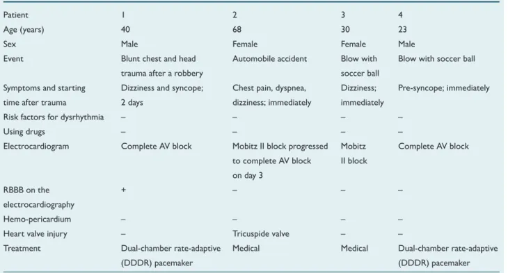
Table 1. Baseline clinical and electrocardiographic characteristics and treatment of patients with atrioventricular block
Patient 1 2 3 4
Age (years) 40 68 30 23
Sex Male Female Female Male
Event Blunt chest and head Automobile accident Blow with Blow with soccer ball
trauma after a robbery soccer ball
Symptoms and starting Dizziness and syncope; Chest pain, dyspnea, Dizziness; Pre-syncope; immediately
time after trauma 2 days dizziness; immediately immediately
Risk factors for dysrhythmia – – – –
Using drugs – – – –
Electrocardiogram Complete AV block Mobitz II block progressed Mobitz Complete AV block
to complete AV block II block on day 3
RBBB on the + – – –
electrocardiography
Hemo-pericardium – – – –
Heart valve injury – Tricuspide valve – –
Treatment Dual-chamber rate-adaptive Medical Medical Dual-chamber rate-adaptive
(DDDR) pacemaker (DDDR) pacemaker
AV: Atrioventricular; RBBB: Right bundle branch block.
Figure 1. The patient with Mobitz II block after blowing the soccer ball.
Şahin Yıldız et al. Type 2 second-degree and third-degree atrioventricular block
third-degree atrioventricular block. None of the patients had history of any disease like coronary artery disease and hyper-tension. Baseline clinical and electrocardiographic character-istics and treatment of patients with atrioventricular block are shown in Table 1.
DISCUSSION
In this study, it was found that only four of 253 consecu-tive Mobitz II block and third-degree atrioventricular block patients, admitted to the Emergency Department of Internal Medicine, were associated with blunt chest trauma. The oc-currence of myocardial contusion after blunt chest trauma varies between 8 and 76% according to the clinical criteria used for diagnosis. Traffic accidents are the most common cause, although it has also been described in sport accidents, kicks from animals, falls and cardiac resuscitation maneuvers.
[1] These injuries may result from lacerations to valve
rup-ture, myocardial rupture with tamponade, acute myocardial infarction following coronary arteries thrombosis and/or dis-section, arrhythmia like ventricular fibrillation and myocardial contusion which may result in dysrhythmias and blocks gen-erally bifascicular or right bundle branch; however, constitut-ing complete AV block is a rare complication.[2-7] Although the
occurrence of complete AV block is common in traumatic experimental models, it is an exceptional observation in clini-cal practice. Although the exact cause of these disorders is unknown, various mechanisms have been proposed, such as the appearance focal hemorrhages in the region of the atrio-ventricular node, localized hypoxia specific conduction tis-sue, increased vagal tone or release of depressants of the conduction system. Pathologic changes associated with blunt trauma induced by conduction disturbances are variable.[8,9]
Generally, no pathologic changes were found, despite find-ings of repolarization changes, conduction defects and dys-rhythmias. Conduction disturbances might be associated with inflammation. Several inflammatory mediators such as inter-leukin-6, tumor necrosis factor, and prostanoids are released following blunt chest trauma.[10-12] Moreover, scar tissue
for-mation during the healing process may cause late complica-tions like atrioventricular block in myocardial blunt trauma.[13]
Case 1 had normal sinus rhythm in electrocardiography before the event and demonstrated symptomatic complete atrioven-tricular block with right bundle branch morphology after blunt chest trauma. His echocardiography was normal in terms of trauma. Atrioventricular block may be associated with fibrosis affecting the conduction system as part of the healing process after myocardial injury. While complete atrioventricular block reoccurred after four months and became permanent, the dual-chamber rate-adaptive (DDDR) -mode pacemaker was implanted and the patient had an uneventful recovery. In Case 2, electrocardiography was consistent with Mobitz II block on admission, which subsequently progressed to complete atrio-ventricular block on day 3. Furthermore, severe tricuspid re-gurgitation with chordae tendineae rupture was seen in the
transthoracic echocardiography. During follow-up with close hemodynamic monitoring, her symptoms disappeared and se-quential echocardiographic evaluations revealed a regression in the severity of tricuspid regurgitation. Finally, she was dis-charged with medical therapy about one week later. During her follow-ups, electrocardiography revealed normal sinus rhythm. In Case 3 (a female footballer), admission electrocardiography was consistent with Mobitz II block alternating sinus rhythm after receiving a blow with the soccer ball. Holter rhythm monitoring, echocardiography and electrophysiological study revealed in normal range. After 12 months of follow-up, as she was athletic, she had no specific cardiovascular symptoms. In addition, she had normal electrocardiography. Case 4 was also admitted with complete atrioventricular block after re-ceiving a blow with the soccer ball. His echocardiography was normal in terms of trauma. While complete atrioventricular block reoccurred after two weeks and became permanent, the DDDR mode pacemaker was implanted and the patient had an uneventful recovery. Baseline clinical and electrocar-diographic characteristics and treatment of the patients with atrioventricular block were shown in Table 1.
Rare clinical cases in the literature confirm that blunt chest trauma can cause conduction defects and that these defects are usually transient, as they were in our study. However, patients with blunt chest trauma must need an electrocardio-graphic evaluation for atrioventricular block upon admission and in the follow-up period.
Conflict of interest: None declared.
REFERENCES
1. Calhoon JH, Grover FL, Trinkle JK. Chest trauma. Approach and management. Clin Chest Med 1992;13:55-67.
2. Brennan JA, Field JM, Liedtke AJ. Reversible heart block following non-penetrating chest trauma. J Trauma 1979;19:784-8. CrossRef
3. Hasdemir H, Arslan Y, Alper A, Osmonov D, Güvenç TS, Poyraz E, et al. Severe tricuspid regurgitation and atrioventicular block caused by blunt thoracic trauma in an elderly woman. J Emerg Med 2012;43:445-7. CrossRef 4. Aykan AC, Oguz AE, Yildiz M, Özkan M. Complete atrioventricular
block associated with non-penetrating cardiac trauma in a 40-year-old man. J Emerg Med 2013;44:41-3. CrossRef
5. Kumagai H, Hamanaka Y, Hirai S, Mitsui N, Kobayashi T. Mitral valve plasty for mitral regurgitation after blunt chest trauma. Ann Thorac Car-diovasc Surg 2001;7:175-9.
6. Sakka SG, Hüttemann E, Reinhart K. Left ventricular aneurysm after myocardial contusion caused by blunt chest trauma. [Article in German] Anasthesiol Intensivmed Notfallmed Schmerzther 2000;35:412-6. [Ab-stract] CrossRef
7. Ildstad ST, Tollerud DJ, Weiss RG, Cox JA, Martin LW. Cardiac con-tusion in pediatric patients with blunt thoracic trauma. J Pediatr Surg 1990;25:287-9. CrossRef
8. Carr KW, Johnson AD, Gregoratos G. Transient bifascicular block fol-lowing blunt chest trauma. West J Med 1982;137:245-9.
9. Cobanoglu U. Göğüs travmalı 70 olguda kardiyolojik değişikliklerin ince-lenmesi. Toraks Dergisi 2007;8:59-68.
10. Knöferl MW, Liener UC, Seitz DH, Perl M, Brückner UB, Kinzl L, et al. Cardiopulmonary, histological, and inflammatory alterations after lung contusion in a novel mouse model of blunt chest trauma. Shock 2003;19:519-25. CrossRef
11. Perl M, Kieninger M, Huber-Lang MS, Gross HJ, Bachem MG, Brau-müller S, et al. Divergent effects of activated neutrophils on inflamma-tion, Kupffer cell/splenocyte activainflamma-tion, and lung injury following blunt
chest trauma. Shock 2012;37:210-8. CrossRef
12. Majetschak M, Obertacke U, Schade FU, Bardenheuer M, Voggenreiter G, Bloemeke B, et al. Tumor necrosis factor gene polymorphisms, leu-kocyte function, and sepsis susceptibility in blunt trauma patients. Clin Diagn Lab Immunol 2002;9:1205-11. CrossRef
13. Liedtke AJ, DeMuth WE Jr. Nonpenetrating cardiac injuries: a collective review. Am Heart J 1973;86:687-97. CrossRef
Ulus Travma Acil Cerrahi Derg, May 2015, Vol. 21, No. 3 196
Şahin Yıldız et al. Type 2 second-degree and third-degree atrioventricular block
OLGU SUNUMU
Acil serviste künt göğüs travmasına bağlı tip 2 ikinci derece ve üçüncü derece
atriyoventriküler blok sıklığı: Çok merkezli çalışma
Dr. Banu Şahin Yıldız,1 Dr. Mehmet Ali Astarcıoğlu,2 Dr. Nazire Başkurt Aladağ,1
Dr. Ahmet Çağrı Aykan,3 Dr. Hakan Hasdemir,4 Dr. Alparslan Şahin,5 Dr. Mustafa Yıldız3
1Dr. Lütfi Kırdar Kartal Eğitim ve Araştırma Hastanesi, Kardiyoloji Kliniği, İstanbul;
2Dumlupınar Üniversitesi Evliya Çelebi Eğitim ve Araştırma Hastanesi, Kardiyoloji Anabilim Dalı, Kütahya; 3Kartal Koşuyolu Yüksek İhtisas Eğitim ve Araştırma Hastanesi, Kardiyoloji Kliniği, İstanbul;
4Dr. Siyami Ersek Kardiyovasküler ve Torasik Cerrahi Eğitim ve Araştırma Hastanesi, Kardiyoloji Kliniği, İstanbul; 5Dr. Sadi Konuk Eğitim ve Araştırma Hastanesi, Kardiyoloji Kliniği, İstanbul
AMAÇ: Künt göğüs travmasını takiben tip 2 ikinci derece atriyoventriküler blok (Mobitz II) ve üçüncü derece atriyoventriküler blok iletim bozukluk-ları oldukça nadirdir. Dahası, travmayı takiben oluşan bu disritmilerden sorumlu patofizyolojik mekanizmalar henüz tam olarak anlaşılamamıştır. Biz travma ile ilişkili bu disritmilerin sıklığını tespit etmeyi amaçladık.
GEREÇ VE YÖNTEM: Acil servise başvuran ardışık Mobitz II ve üçüncü derece atriyoventriküler bloklu 253 hasta, Ocak 2012 ile Mart 2013 tarihleri arasında değerlendirildi. Mobitz II ve üçüncü derece atriyoventriküler blok ve travma ile ilişkili sadece dört hasta mevcut çalışmaya alındı . Atriyovent-riküler blok düzeyi elektrokardiyografik özelliklere göre tanımlandı.
BULGULAR: 253 hastanın sadece dördü (ortalama yaş: 40.2±19.7 yıl, iki erkek) travma ile ilişkili bulundu. Tüm hastaların koroner anjiyografi veya çok kesitli bilgisayarlı tomografi ile koroner arterleri normal saptandı. Kalıcı kalp pili üçüncü derece atriyoventriküler bloğu olan iki hastaya takıldı. Hastaların hiçbirinde koroner arter hastalığı ve hipertansiyon yoktu.
TARTIŞMA: Literatürde künt göğüs travmasına bağlı iletim kusurları nadir görülen klinik olgulardır ve genellikle geçici olmaktadır. Bu nedenle künt göğüs travması olan hastaların başvuru sırasında ve takiplerde atriyoventriküler blok açısından elektrokardiyografik değerlendirilmesi gerekir.
Anahtar sözcükler: Elektrokardiyografi; künt göğüs travması; üçüncü derece atriyoventriküler blok; tip 2 ikinci derece atriyoventriküler blok (Mobitz II).
Ulus Travma Acil Cerrahi Derg 2015;21(3):193-196 doi: 10.5505/tjtes.2015.04763