ABSTRACT
Objectives: This study aims to evaluate the degree of conversion (DC) and microlekage scores of three different composite resins polymerized with a LED curing device in standard and extra-power mode. Material and Methods: One bulk-fill (Tetric EvoCeram Bulk-Fill – TECBF) and two conventional composite materials (Clearfil Majesty Posterior – CMP and Tetric EvoCeram – TEC) were evaluated. A total of 30 specimens were prepared for six groups (N = 5). These groups were polymerized with a LED curing device as follows: TECBF-6: 3200mW/cm2 for six seconds, TECBF-20: 1000mW/cm2 for 20 seconds, CMP-6: 3200mW/cm2 for six seconds, CMP-20: 1000mW/cm2 for 20 seconds, TEC-6: 3200mW/cm2 for six seconds, TEC - 20: 1000 mW/cm2 for 20 seconds. After 24 hours of water storage, DC was measured by Raman spectroscopy. Microleakage scores of the six groups were bonded to various adhesive systems (Clearfil SE Bond or Adhese Bond Universal) were also evaluated at Class II box cavities (N = 10). Results: While the highest DC was found at the top (TECBF-20= 79.92% and TECBF-6= 79.02%) and bottom surfaces (TECBF-20 = 68.94% and TECBF-6= 71.04%) for TECBF groups, TEC groups (TEC-20top = 59.06%, TEC-6top=49.66%, TEC-20bottom = 43.72% and TEC-6bottom= 40.68%) showed the lowest DC for the both surfaces (p < 0.05). Polymerization of materials in standard or extra-power mode was similar to DC (p > 0.05). Microleakage scores were found to be similar (p > 0.05). Conclusion: According to the results of the study, different power densities of LED curing light did not affect the DC of composite resins and microleakage values of restorations at small Class II cavities.
RESUMO
Objetivo: este estudo tem como objetivo avaliar os graus de conversão (DC) e microinfiltração de três diferentes resinas compostas polimerizáveis com um dispositivo de luz por LED nos modos padrão e no modo de alta-potência. Material e métodos: uma resina bulk-fill (Tetric EvoCeram Bulk-Fill - TECBF) e dois materiais resinosos convencionais (Clearfil Majesty Posterior - CMP e Tetric EvoCeram - TEC) foram avaliados. Um total de 30 espécimes foram preparados e divididos em seis grupos (N = 5). Esses grupos foram polimerizados com um dispositivo de luz de LED da seguinte forma: TECBF-6: 3200mW / cm2 por seis segundos, TECBF-20: 1000mW / cm2 por 20 segundos, CMP-6: 3200mW / cm2 por seis segundos, CMP-20: 1000mW / cm2 por 20 segundos, TEC-6: 3200mW / cm2 por seis segundos, TEC - 20: 1000 mW / cm2 por 20 segundos. Após 24 horas de armazenamento em água, a DC foi medida por espectroscopia Raman. Os escores de microinfiltração dos seis grupos dos vários sistemas adesivos (Clearfil SE Bond ou Adhese Bond Universal) também foram avaliados nas cavidades Classe II (N = 10). Resultados: embora a maior CD tenha sido encontrada nas partes superior (TECBF-20 = 79,92% e TECBF-6 = 79,02%) e inferiores (TECBF-20 = 68,94% e TECBF-6 = 71,04%) para grupos TECBF, os grupos TEC (TEC-20top = 59,06%, TEC-6top = 49,66%, TEC-20bottom = 43,72% e TEC-6bottom = 40,68%) apresentaram as menores CD para as ambas as partes (p < 0,05). A polimerização de materiais no modo padrão ou alta-potência foi semelhante à DC (p > 0,05). Os escores de microinfiltração foram semelhantes (p > 0,05). Conclusão: de acordo com os resultados do estudo, diferentes densidades de potência da luz de LED não afetaram as CD das resinas compostas e os valores de microinfiltração das restaurações em pequenas cavidades da Classe II.
O R I G I N A L A R T I C L E
Evaluation of microleakage and the degree of conversion of three
composite resins polymerized at different power densities
Avaliação da microinfiltração e do grau de conversão de três resinas compostas polimerizadas em diferentes densidades de potência
Kadir DEMIR1, Yusuf BAYRAKTAR2
1 - Lokman Hekim University Faculty of Dentistry - Department of Restorative Dentistry - Ankara - Turkey. 2 - Kırıkkale University Faculty of Dentistry - Department of Restorative Dentistry - Kırıkkale - Turkey.
Doi: 10.14295/bds.2020.v23i2.1948
KEYWORDS
Bulk-fill composite; degree of conversion; LED curing device; microleakage; Raman spectroscopy.
PALAVRAS-CHAVE
Resina de inserção em bloco; Grau de conversão; Dispositivo de polimerização por LED; Microinfiltração; Espectroscopia Raman.
INTRODUCTION
C
omposite resin materials have been usedmore often today as a restorative material, and the DC of these materials of these soft monomers to rigid polymers appears to be an important situation for clinical success [1]. DC is an important determinant of the physical, chemical and mechanical properties [2]. Adequate DC is also clinically significant for all physical properties such as hardness, elastic modulus, strength, dimensional stability, solubility, water sorption and color stability [3,4]. However, it should also be noted that there are other factors affecting the success of the restoration. DC is influenced by some factors such as filler amount, resin type, resin shade, spectrum and power of the polymerization light. [5]. Composite resin materials should be biocompatible, having good mechanical characteristics and low polymerization shrinkage. DC is an equally important consideration for any restorative material and filling technique [6]. While conventional composite resins reach a DC of approximately 50%-75% [7,8], this rate is reported to range from 50%-79% for bulk-fill composites [9, 10]. It has been reported in the literature that the DC should be at least 55% for clinical success [10,11]. Fourier Transform Infrared (FTIR) spectroscopy [12] and Raman spectroscopy [13] commonly uses spectroscopic and accurate methods to evaluate DC. In Raman spectroscopy, since the samples are not damaged, it allows multiple measurements on the same sample. In addition, samples are easier to prepare for Raman spectroscopy [14].
Marginal integrity/adaptation, polymerization shrinkage and therefore
microleakage are important factors affecting the clinical success of composite restorations. DC and shrinkage stresses are directly dependent on each other. As a consequence, the reduction in DC would result in lower shrinkage and lower shrinkage stress. The volumetric shrinkage of currently used composites is between 2% and 6% [15]. The polymerization shrinkage stresses can be reduced by slowly polymerizing the composite
resin material without affecting the final DC. A slower growing polymer network can delay the formation of shrinkage, resulting in reduced shrinkage forces via molecular rearrangements [16]. This leads to an increased time for the polymerization procedure. Whereas the rapid polymerization of composite resins leads to high polymerization shrinkage and offers considerable time-savings. It also leads to cuspal deflection, enamel cracks, gap formation, microleakage, secondary caries and post-operative sensitivity [17,18].
Short-time polymerization of composite resins with high-intensity LED curing devices saves considerable time and this provides an advantage both for the patient and the clinician. However, it is of interest to see how the DC of the material and the microleakage scores of the restoration will be affected after rapid polymerization.
The aim of this study was to evaluate the degree of conversion of three different composite resins polymerized with a LED light device in the standard (1000 mW/cm2)
and extra-power (3200mW/cm2) modes.
Microleakage scores of composites were also investigated in Class II box cavities after the standard and rapid polymerization procedures. The tested null hypotheses were: 1. There is no difference between microleakage scores of aproximal box cavities restored with composite resin, which are polymerized at different power densities. 2. There is no difference between the DC of composite materials that are polymerized at different power densities.
MATERIAL AND METHODS
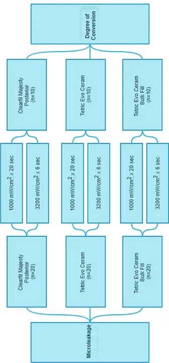
Degree of conversion (DC)One bulk-fill (Tetric EvoCeram Bulk-Fill - TECBF) and two conventional (Clearfil Majesty Posterior - CMP and Tetric EvoCeram - TEC) composite resin materials were evaluated in a total of six groups. For each material ten cylindrical specimens were prepared with a diameter of 8 mm and an adjustable depth using a teflon mold. The depth of the specimens was 4 mm for bulk-fill composites and 2 mm for conventional composites. The composite resin
materials were inserted into single increments for all groups. Polyester strip bands were used before polymerizing to eliminate the oxygen inhibition layer and to obtain flat surfaces. Two different polymerization modes for each material were assigned two different subgroups (N = 5). A third-generation LED device (Valo Cordless, Ultradent, USA) was used for sample polymerization. Specimens were polymerized
as follow: TECBF-6: 3200mW/cm2 for six
seconds, TECBF-20: 1000mW/cm2 for 20
seconds, CMP-6: 3200mW/cm2 for six seconds,
CMP-20: 1000mW/cm2 for 20 seconds,
TEC-6: 3200mW/cm2 for six seconds and TEC-20:
1000mW/cm2 for 20 seconds. After that, the
polymerized samples were stored in distilled water at a dark ambit for 24 hours before testing. For each sample two Raman spectra were obtained, one for the top surface and the other for the bottom.
Raman spectra of samples were obtained by using a computer-controlled laser Raman spectroscopy (Renishaw InVia, Renishaw plc, UK) integrated with an optical microscope with x100 magnification (NA 0.9; N Plan EPI objective; Leica, Germany). A 785 nm diode laser was used for Raman scattering effect. The exposure time was 40 seconds for each scan, and the power of the diode-laser was 8mW. Raman spectra were obtained from both polymerized (N = 5) and non-polymerized (N = 5) forms of the materials for calculating the DC of the polymerized specimens. DC was calculated by comparing the changes of polymerized and non-polymerized specimens
in the 1,640 cm-1 band, which is the C = C
reference band that represents the C = C stretching mode. For Tetric EvoCeram, Tetric EvoCeram Bulk-Fill and Clearfil Majesty Posterior the aromatic C=C band at 1,610 cm-1 was used as a reference [19]. To calculate the DC, the following equation was used: DC = 1 - Rpolymerized/Runpolymerized x 100, where R =
(C=C band area)/(reference band area) [20].
Microleakage and marginal adaptation
Thirty caries-free human third molars extracted for clinical reasons were selected for the study. The teeth were obtained from the oral surgery department after obtaining the
patients’ consent and approval of the ethics committee (Decision No: 12/10). Initially, the samples were checked with x40 magnification (OPMI Pico, Carl Zeiss Meditec, Germany) with an operating microscope to see if any developmental defects or cracks were present in the crowns of the teeth. Possible debris and residues on the teeth were removed with a scaler. The teeth were disinfected by immersion in 0.1% thymol for seven days and were kept in distilled water.
Teeth were randomly divided into six groups, and only two Class II box cavities were prepared with mesial and distal surfaces using an aerator under water cooling and diamond fissure burs (Dia-burs SF-13, Mani Co., Japan). Each cavity had a cervical limit of 1 mm above the cementoenamel junction, gingival seat thickness of 2 mm and buccolingual width of 4 mm. Marginal edges were not beveled. (Figure 1).
Figure 1 - Cavity parameters
All cavities were prepared by one operator and the dimensions were checked using a digital caliper (Mitutoyo, Kawasaki, Japan) and periodontal probe during and after the cavity preparation. After each of the four preparations, the bur was changed with a new one. Incorrect samples were not included in the study.
For the CMP-20 group restorations, the adhesive material (Clearfill SE Bond, Kuraray, Japan) was applied to the cavities using a disposable brush according to the manufacturer’s instructions and polymerized for 10 seconds with the LED curing (Valo Cordless, Ultradent, USA) device at 1000
mW/cm2 power. After that, metal matrix
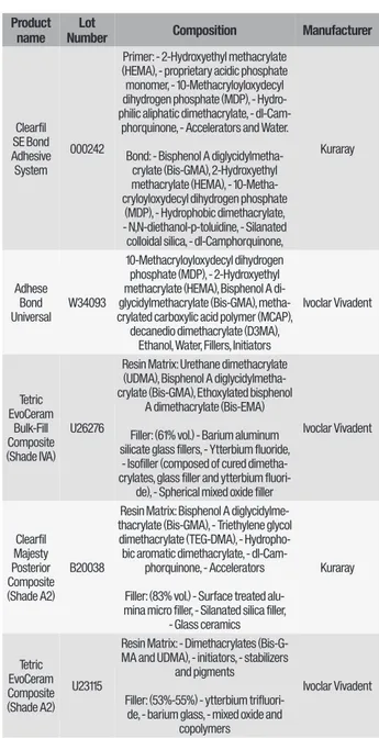
Kerr-Table I - Materials used in the study Product
name NumberLot Composition Manufacturer
Clearfil SE Bond Adhesive System
000242
Primer: - 2-Hydroxyethyl methacrylate (HEMA), - proprietary acidic phosphate monomer, - 10-Methacryloyloxydecyl dihydrogen phosphate (MDP), - Hydro-philic aliphatic dimethacrylate, -
dl-Cam-phorquinone, - Accelerators and Water. Bond: - Bisphenol A
diglycidylmetha-crylate (Bis-GMA), 2-Hydroxyethyl methacrylate (HEMA), - 10-Metha-cryloyloxydecyl dihydrogen phosphate
(MDP), - Hydrophobic dimethacrylate, - N,N-diethanol-p-toluidine, - Silanated colloidal silica, - dl-Camphorquinone,
Kuraray Adhese Bond Universal W34093 10-Methacryloyloxydecyl dihydrogen phosphate (MDP), - 2-Hydroxyethyl methacrylate (HEMA), Bisphenol A di-glycidylmethacrylate (Bis-GMA), metha-crylated carboxylic acid polymer (MCAP),
decanedio dimethacrylate (D3MA), Ethanol, Water, Fillers, Initiators
Ivoclar Vivadent Tetric EvoCeram Bulk-Fill Composite (Shade IVA) U26276
Resin Matrix: Urethane dimethacrylate (UDMA), Bisphenol A diglycidylmetha-crylate (Bis-GMA), Ethoxylated bisphenol
A dimethacrylate (Bis-EMA) Filler: (61% vol.) - Barium aluminum silicate glass fillers, - Ytterbium fluoride, - Isofiller (composed of cured dimetha-crylates, glass filler and ytterbium
fluori-de), - Spherical mixed oxide filler
Ivoclar Vivadent Clearfil Majesty Posterior Composite (Shade A2) B20038
Resin Matrix: Bisphenol A diglycidylme-thacrylate (Bis-GMA), - Triethylene glycol dimethacrylate (TEG-DMA), -
Hydropho-bic aromatic dimethacrylate, - dl-Cam-phorquinone, - Accelerators Filler: (83% vol.) - Surface treated alu-mina micro filler, - Silanated silica filler,
- Glass ceramics Kuraray Tetric EvoCeram Composite (Shade A2) U23115
Resin Matrix: - Dimethacrylates (Bis-G-MA and UD(Bis-G-MA), - initiators, - stabilizers
and pigments Filler: (53%-55%) - ytterbium
trifluori-de, - barium glass, - mixed oxide and copolymers
Ivoclar Vivadent filled 2 mm horizontal incremental technique
with a posterior nano-hybrid composite resin (Clearfil Majesty Posterior, Kuraray, Japan) and each increment polymerized with the LED device at 1000mW/cm2 for 20 seconds. For the
CMP-6 group, each process was carried out in the same manner, while the adhesive system was polymerized for three seconds at 3200
mW/cm2 and each layer was polymerized for
six seconds at 3200 mW/cm2.
For the TECBF-20 group, the adhesive material (Adhese Bond Universal, Ivoclar Vivadent, Liechtenstein) was applied to the cavities using a disposable brush according to the manufacturer’s instructions and polymerized for ten seconds with the LED curing (Valo Cordless, Ultradent, USA) device
at 1000 mW/cm2 power. After that, metal
matrix bands (Adapt SuperCap Matrix no 2182, Kerr-Hawe, Switzerland) were placed. Cavities were filled with a 4 mm horizontal bulk technique with a bulk-fill nanohybrid composite resin (Tetric EvoCeram Bulk-Fill, Ivoclar Vivadent, Liechtenstein) and each increment polymerized with the LED device at 1000mW/cm2 for 20 seconds. For the TECBF-6
group, each process was carried out in the same manner, while the adhesive system was polymerized for three seconds at 3200 mW/
cm2 and each layer was polymerized for six
seconds at 3200 mW/cm2.
For the TEC-20 group, the adhesive material (Adhese Bond Universal, Ivoclar Vivadent, Liechtenstein) was applied to the cavities using a disposable brush according to the manufacturer’s instructions and polymerized for ten seconds with the LED curing (Valo Cordless, Ultradent, USA) device
at 1000 mW/cm2 power. After that, metal
matrix bands (Adapt SuperCap Matrix no 2182, Kerr-Hawe, Switzerland) were placed. Cavities were filled with a 2 mm horizontal incremental technique with a nanohybrid composite resin (Tetric EvoCeram, Ivoclar Vivadent, Liechtenstein) and each increment polymerized with the LED device at 1000mW/ cm2 for 2 seconds. For the TEC-6 group, each
process was carried out in the same manner, while the adhesive system was polymerized
for three seconds at 3200 mW/cm2 and each
mW/cm2.
Finishing and polishing procedures to all groups were performed using an aluminum oxide finishing and polishing kit (Super Snap Rainbow Technique Kit, Shofu, Japan). Each group consisted of ten restorations (n = 10). The materials used in the study are listed in Table I.
After the restoration process, the teeth were stored in distilled water for 24 hours. All the teeth were then thermocycled 10,000 times in 5-55 °C water bathes (Thermal Cycler, Esetron Smart RoboTechnologies, Turkey) with a dwell time of 15 seconds for each bath. After the thermocycle process, marginal edges of the restorations were evaluated under a stereomicroscope (S100 OPMI pico, Carl Zeiss Meditec, Germany) with x40 magnification. After that, one specimen from each group was selected randomly and impressions of the specimens were taken using a polyvinyl siloxane material (S4 Suhy, Bisico, Germany) and epoxy resin replicas were obtained. Replicas were evaluated under a Scanning Electron Microscope (JSM-5600, Jeol Ltd., Japan).
Apical foramen of the teeth were plugged with sticky wax and each tooth was covered with two coats of nail varnish as well as 1 mm from the restoration margins. Coated teeth were immersed in 1% methylene blue solution for 24 hours and then separated longitudinally in a mesiodistal direction though the middle of the restorations using a low-speed cutting device (Microcut 126, Metkon, Turkey). Longitudinally separated samples were examined using a stereomicroscope (S100 OPMI pico, Carl Zeiss Meditec, Germany) with x40 magnification and the occlusal/gingival microleakage scores of each restoration were evaluated by two calibrated investigators who blinded to groups. The microleakage evaluation criteria are shown in Table II and the detailed steps of the study design are explained in Figure 2. Data were analyzed using the SPSS 22.0 program with Kruskal-Wallis and Man-Whitney U tests.
Table II - Microleakage evaluation criteria Microleakage
scores Occlusal microleakage description Gingival microleakage description
0 No dye penetration No dye penetration
1 Dye penetration for half extension Dye penetration for half extension of the cervical wall 2 Dye penetration more than half Dye penetration for more than half of all of the cervical wall 3 Dye penetration for the pulpal wall Dye penetration into the pulp
RESULTS
Degree of Conversion
Bulk-fill groups had significantly higher DC than other groups (p < 0.05). The mean level of polymerization degree ranged from highest to lowest TECBF-6, TECBF-20, CMP-20, CMP-6, TEC-CMP-20, TEC-6 respectively.
The same composites showed similar DC, as a result of rapid and standard polymerization (p > 0.05). Top surfaces of the composite specimens showed statistically higher DC values than bottom surfaces (p < 0.05), except the CMP-20 Group. DC results are shown in Figure 3.
DISCUSSION
This study was performed to investigate the DC values and microleakage scores in Class 2 cavities of three composite resin material after rapid and standard polymerization procedures. The microleakage scores of all groups were found to be similar according to the findings of this study and the first null hypothesis were accepted.
The presence of marginal gap can be considered as the first sign of failure of the restoration [21]. While some studies evaluate the gap width, presence or absence of a gap is more important [22]. Evaluating the adaptation of the restoration margins with SEM has been accepted as a reliable method [23]. For this purpose, epoxy resin replica of a randomly selected specimen from each group was examined under SEM. Additionally, all specimens were examined under a stereomicroscope. If a visual discrepancy was detected after these examinations, it could be interpreted that restorations would fail in the early period, but it was observed that all
Figure 3 - Bar graphs of “Top Surface DC”, “Bottom Surface
DC” and “Mean DC” of the composite materials.
Marginal adaptation and microleakage scores
No marginal maladaptation was observed in any of the samples examined under the stereomicroscope. In addition, no marginal maladaptation was observed in the examination of epoxy resin replicas under SEM. Gingival microleakage scores were found to be significantly higher than occlusal microleakage scores in TECBF-6 and TEC-6 (p < 0.05). Microleakage values at the occlusal regions of the groups were similar, and no significant differences were found between the groups (p > 0.05). For gingival microleakage scores, polymerized with extra-power mode groups showed higher microleakage values and no statistically significant information was found (p > 0.05). The distribution of the microleakage scores of the occlusal and gingival edges are shown in Figure 4.
Figure 4 - Microleakage scores of the occlusal and gingival
specimens showed good marginal adaptation. Gamarra et al. reported that marginal adaptation of the restorations is quite good when cavity margins are limited to enamel [24] and Campos et al. reported that dentin margins were more incompatible than enamel margins [25]. In this study, cavity margins were limited to enamel and the results were consistent with the studies above.
In dental composite resins, as in all polymeric materials, polymerization shrinkage that occurs during the conversion of monomers to polymers is one of the most important clinical problems encountered today [26]. As a result of polymerization shrinkage, clinical problems such as postoperative sensitivity, margin coloration, secondary caries, fractures in restorative material, loss of restoration, pulp inflammation and pulp necrosis occur due to microleakage at the tooth-restoration margin. Considering all these reasons, it can be said that microleakage is one of the most important criteria for evaluating the clinical success of restorations [27]. Polymerization shrinkage stress depends on many factors, such as the size of the cavity, the form of the cavity, the type of the substrate, the localization of the cavity margins, the restorative material used, and the placement and the polymerization technique [28]. In large restorations, it is important to reduce microleakage at the tooth/restoration interface [29]. For this reason, some studies have been conducted such as the development of adhesive systems, different light application protocols or the use of different layering methods in order to prevent microleakage [26].
According to the findings of this study, microleakage could not be prevented completely in any group. Although rapid polymerized groups showed slightly more microleakage, this was not significant. Methylene blue solution was used in many studies related to microleakage in terms of its low cost and easy preparation [30]. Methylene blue is smaller in size than bacteria. Therefore, it can show microleakage even in areas that are too small for bacteria to penetrate [31].
Placing composite resin material into the cavity in layers of 2 mm thickness allows better light transmission and therefore better polymerization of the composite resin. However, by using the layering technique, advantages such as reduction of configuration factor and cuspal deflection, and consequently reduction of bonding failures, can be achieved [28] In bulk-fill composite resins, some changes have been made to the filler content of the composite in order to increase the polymerization depth. Compared to conventional composite resins, bulk-fill composite resins have larger fillers (> 20 μm) and less filler-matrix contact. In this case, the light is less scattered and penetrates deeper [32]. In Tetric EvoCeram Bulk-Fill a new initiator named Ivocerin also allows polymerization of higher increments [33]. According to the literature, polymerization shrinkage of bulk-fill composites was regulated with modifications in an organic matrix [34,35]. As a result of this study, similar microleakage scores can be explained by similar polymerization shrinkage characteristics.
Kader et al. restored Class II cavities in human molar teeth by incremental (Filtek Z250) and bulk (Filtek bulk-fill) techniques and evaluated the microleakage. According to the data obtained, they showed that there is no statistically significant difference in microleakage scores, irrespective of the insertion technique [36]. According to the findings of this study polymerization of dental composite materials with standard or extra-power mode and the placement of the composite materials with incremental or bulk technique did not significantly affect the microleakage scores between the groups. Cavity size and composite volume are important parameters in terms of microleakage [37,38]. In this study, approximal box-only cavities that are relatively small and limited to enamel were prepared. These small cavities were filled with a small amount of composite therefore less stress may have occurred due to polymerization shrinkage at the composite/ tooth interface. This contributes to the reduction of microleakage values in rapidly
polymerized groups. Although the size of the cavity and the resulting shrinkage stress are the subjects of another study, relatively small and enamel-limited box-only restorations can be polymerized by rapid polymerization methods according to findings of this study. Bayraktar et al. reported that rapid polymerization procedures can be used at small Class V cavities with selective enamel etching and sixth-generation adhesive systems [39].
The polymerization procedure of dental composites can be divided into pre-gel and post-pre-gel phases. During the pre-pre-gel phase, the internal stresses formed in the composite material during polymerization may dissipate and the tension is reduced. After the pre-gel phase, the material becomes more rigid and continuing the polymerization procedure results in tension at the tooth/ restoration interface [40]. Therefore, it has been suggested that the polymerization should start with increasing low energy to extend the pre-gel phase [41,42]. However, Asmussen et al. revealed that polymerization shrinkage depends not only on the total amount of energy but also the combination of energy density and exposure time that affects the shrinkage pattern [43]. In the literature, different results were reported about the relationship between power density and polymerization shrinkage [43-47]. Further studies are needed to demonstrate the flow properties of the composite material during rapid or slow polymerization.
According to the findings of this study, the DC of composite materials were found to be similar between the groups that were polymerized at different power densities. The second null hypothesis was accepted. DC is an important factor for composite resin materials and refers to the conversion of carbon-carbon double bonds to monomeric carbonic to carbonic simple polymer [48]. DC can be analyzed through mechanical and spectroscopic methods. Spectroscopic methods provide direct measurements of DC values with more reliable results [49]. While higher DC leads to a better clinical performance of composite resin materials, a lower DC could cause unreacted monomers that lead to
degradation and that may affect the longevity of the composite restoration [50].
Light transmission depends on the opacity of the material. In general, bulk-fill composites exhibit higher translucency than conventional composites [51]. In addition, the ‘Ivocerin’ based initiator system has an absorption spectrum very close to CQ. It has been reported that photopolymerized activation is higher than CQ due to the absorption of visible radiation [32]. According to the results of this study, the bulk-fill composite resin with an ‘Ivocerin’ based photoinitiator system was found to have a higher DC than either of the conventional composites. The different polymerization modes used did not have any significant effect on the DC of the composites. These results are consistent with many studies in the literature [49,52,53].
According to the study, Clearfil Majesty Posterior showed a higher degree of conversion than Tetric EvoCeram. The differences in the organic matrix structure of the two composite resins have an effect on their degree of conversion. While the Tetric EvoCeram composite did not contain TEGDMA, the presence of this monomer in the Clearfil Majesty Posterior may have caused higher DC for the CMP group [54, 55]. TEG-DMA increases DC and crosslinking when mixed with Bis-GMA [55]. This finding can also be explained by the high organic matrix rate of Tetric EvoCeram (without TEGDMA monomer) as volume against Clearfil Majesty Posterior. [54,56,57].
Although there is no clear information on the clinically required DC value, it has been reported that this rate should not be below 55% [11]. Both the top and bottom surfaces of TEC-6 Group specimens showed less DC values than 55%. While the TEC-20 Group showed fewer DC at the bottom sides, the other groups showed higher DC levels.
In this study, DC and microleakage scores of the composites were evaluated but the interaction between the polymerization shrinkage, DC and microleakage scores could not be compared. Additionally, oral conditions could not be simulated properly. These factors
are the limitations of the study. The interaction between DC, polymerization shrinkage and microleakage is a complex relationship, and further studies are needed.
CONCLUSION
Polymerizing composite resins with high-power density LED device in a short time did not affect DC. Small composite restorations with low C-factor may be polymerized in a shorter time with high-power LED devices. Further studies are needed to evaluate the relationship between DC, polymerization shrinkage and microleakage values of composite resins.
REFERENCES
1. Moore BK, Platt JA, Borges G, Chu TM, Katsilieri I. Depth of cure of dental resin composites: ISO 4049 depth and microhardness of types of materials and shades. Oper Dent. 2008 Jul-Aug;33(4):408-12. Doi:10.2341/07-104 2. Bouschlicher MR, Rueggeberg FA, Wilson BM. Correlation of bottom-to-top
surface microhardness and conversion ratios for a variety of resin composite compositions. Oper Dent. 2004 Nov-Dec;29(6):698-704.
3. Leprince JG, Palin WM, Hadis MA, Devaux J, Leloup G. Progress in dimethacrylate-based dental composite technology and curing efficiency. Dent Mater. 2013 Feb;29(2):139-56. Doi:10.1016/j.dental.2012.11.005 4. Krifka S, Seidenader C, Hiller KA, Schmalz G, Schweikl H. Oxidative stress and
cytotoxicity generated by dental composites in human pulp cells. Clin Oral Investig. 2012 Feb;16(1):215-24. Doi:10.1007/s00784-010-0508-5 5. Campodonico CE, Tantbirojn D, Olin PS, Versluis A. Cuspal deflection and
depth of cure in resin-based composite restorations filled by using bulk, incremental and transtooth-illumination techniques. J Am Dent Assoc. 2011 Oct;142(10):1176-82. Doi:10.14219/jada.archive.2011.0087
6. Jang JH, Park SH, Hwang IN. Polymerization shrinkage and depth of cure of bulk-fill resin composites and highly filled flowable resin. Oper Dent. 2015 Mar-Apr;40(2):172-80. Doi:10.2341/13-307-L
7. Vasudeva G. Monomer systems for dental composites and their future: a review. J Calif Dent Assoc. 2009 Jun;37(6):389-98.
8. Tarle Z, Knezevic A, Demoli N, Meniga A, Sutaloa J, Unterbrink G, et al. Comparison of composite curing parameters: effects of light source and curing mode on conversion, temperature rise and polymerization shrinkage. Oper Dent. 2006 Mar-Apr;31(2):219-26. Doi:10.2341/05-15
9. Czasch P, Ilie N. In vitro comparison of mechanical properties and degree of cure of a self-adhesive and four novel flowable composites. J Adhes Dent. 2013 Jun;15(3):229-36. Doi:10.3290/j.jad.a29530
10. Alshali RZ, Silikas N, Satterthwaite JD. Degree of conversion of bulk-fill compared to conventional resin-composites at two time intervals. Dent Mater. 2013 Sep;29(9):e213-7. Doi:10.1016/j.dental.2013.05.011
11. Ferracane JL, Mitchem JC, Condon JR, Todd R. Wear and marginal breakdown of composites with various degrees of cure. J Dent Res. 1997 Aug;76(8):1508-16. Doi:10.1177/00220345970760081401
12. Yokesh CA, Hemalatha P, Muthalagu M, Justin MR. Comparative Evaluation of the depth of cure and degree of conversion of two bulk fill flowable composites. J Clin Diagn Res. 2017 Aug;11(8):ZC86-ZC9. Doi:10.7860/ JCDR/2017/28004.10444
13. Pianelli C, Devaux J, Bebelman S, Leloup G. The micro-Raman spectroscopy, a useful tool to determine the degree of conversion of light-activated composite resins. J Biomed Mater Res. 1999 48(5):675-81. Doi:10.1002/(sici)1097-4636(1999)48:5<675::aid-jbm11>3.0.co;2-p
14. Yu P, Yap A, Wang XY. Degree of conversion and polymerization shrinkage of bulk-fill resin-based composites. Oper Dent. 2017 Jan/Feb;42(1):82-9. Doi:10.2341/16-027-L
15. Braga RR, Ballester RY, Ferracane JL. Factors involved in the development of polymerization shrinkage stress in resin-composites: a systematic review. Dent Mater. 2005 Oct;21(10):962-70. Doi:10.1016/j.dental.2005.04.018 16. Kaisarly D, Gezawi ME. Polymerization shrinkage assessment of dental
resin composites: a literature review. Odontology. 2016 Sep;104(3):257-70. Doi:10.1007/s10266-016-0264-3
17. Elsharkasi MM, Platt JA, Cook NB, Yassen GH, Matis BA. Cuspal deflection in premolar teeth restored with bulk-fill resin-based composite materials. Oper Dent. 2018 Jan/Feb;43(1):E1-E9. Doi:10.2341/16-072-L
18. Ferracane JL, Hilton TJ. Polymerization stress–is it clinically meaningful? Dental Materials. 2016 32(1):1-10.
19. Par M, Gamulin O, Marovic D, Klaric E, Tarle Z. Raman spectroscopic assessment of degree of conversion of bulk-fill resin composites--changes at 24 hours post cure. Oper Dent. 2015 May-Jun;40(3):E92-101. Doi:10.2341/14-091-L
20. Shin WS, Li XF, Schwartz B, Wunder SL, Baran GR. Determination of the degree of cure of dental resins using Raman and FT-Raman spectroscopy. Dent Mater. 1993 Sep;9(5):317-24. Doi:10.1016/0109-5641(93)90050-z
21. Hilton TJ. Can modern restorative procedures and materials reliably seal cavities? In vitro investigations. Part 2. Am J Dent. 2002 Aug;15(4):279-89. 22. Alonso RCB, Correr GM, Cunha LG, Borges AFS, Puppin-Rontani RM, Sinhoreti
MAC. Dye staining gap test: an alternative method for assessing marginal gap formation in composite restorations. Acta Odontol Scand. 2006 Jun;64(3):141-5. Doi:10.1080/00016350500474565
23. Manhart J, Chen HY, Mehl A, Weber K, Hickel R. Marginal quality and microleakage of adhesive class V restorations. J Dent. 2001 Feb;29(2):123-30. Doi:10.1016/s0300-5712(00)00066-x
24. Gamarra VSS, Borges GA, Junior LHB, Spohr AM. Marginal adaptation and microleakage of a bulk-fill composite resin photopolymerized with different techniques. Odontology. 2018 Jan;106(1):56-63. Doi:10.1007/s10266-017-0294-5
25. Campos EA, Ardu S, Lefever D, Jasse FF, Bortolotto T, Krejci I. Marginal adaptation of class II cavities restored with bulk-fill composites. J Dent. 2014 May;42(5):575-81. Doi:10.1016/j.jdent.2014.02.007
26. Benetti AR, Havndrup-Pedersen C, Honore D, Pedersen MK, Pallesen U. Bulk-fill resin composites: polymerization contraction, depth of cure, and gap formation. Oper Dent. 2015 Mar-Apr;40(2):190-200. Doi:10.2341/13-324-L 27. Rocca GT, Gregor L, Sandoval MJ, Krejci I, Dietschi D. In vitro evaluation of marginal and internal adaptation after occlusal stressing of indirect class II composite restorations with different resinous bases and interface treatments. “Post-fatigue adaptation of indirect composite restorations”. Clin Oral Investig. 2012 Oct;16(5):1385-93. Doi:10.1007/s00784-011-0632-x 28. Han SH, Sadr A, Tagami J, Park SH. Internal adaptation of resin composites at
two configurations: Influence of polymerization shrinkage and stress. Dent Mater. 2016 Sep;32(9):1085-94. Doi:10.1016/j.dental.2016.06.005
29. Eick JD, Gwinnett AJ, Pashley DH, Robinson SJ. Current concepts on adhesion to dentin. Crit Rev Oral Biol Med. 1997;8(3):306-35. Doi:10.1177/104544119700 80030501
30. Agrawal VS, Parekh VV, Shah NC. Comparative evaluation of microleakage of silorane-based composite and nanohybrid composite with or without polyethylene fiber inserts in class II restorations: an in vitro study. Oper Dent. 2012 Sep-Oct;37(5):E1-7. Doi:10.2341/11-353-L
Dr. Yusuf Bayraktar (Corresponding address)
Kırıkkale University Faculty of Dentistry, Department of Restorative Dentistry, Kırıkkale, Turkey
E-mail: [email protected]
Date submitted: 2019 Nov 22 Accept submission: 2020 Jan 21
31. de Almeida JB, Platt JA, Oshida Y, Moore BK, Cochran MA, Eckert GJ. Three different methods to evaluate microleakage of packable composites in Class II restorations. Oper Dent. 2003 Jul-Aug;28(4):453-60.
32. Bucuta S, Ilie N. Light transmittance and micro-mechanical properties of bulk fill vs. conventional resin based composites. Clin Oral Investig. 2014 Nov;18(8):1991-2000. Doi:10.1007/s00784-013-1177-y
33. Almeida Junior L, Lula ECO, Penha KJS, Correia VS, Magalhaes FAC, Lima DM, et al. Polymerization shrinkage of bulk fill composites and its correlation with bond strength. Braz Dent J. 2018 May-Jun;29(3):261-7. Doi:10.1590/0103-6440201801838
34. Al Sunbul H, Silikas N, Watts DC. Polymerization shrinkage kinetics and shrinkage-stress in dental resin-composites. Dent Mater. 2016 Aug;32(8):998-1006. Doi:10.1016/j.dental.2016.05.006
35. Ilie N, Bucuta S, Draenert M. Bulk-fill resin-based composites: an in vitro assessment of their mechanical performance. Oper Dent. 2013 Nov-Dec;38(6):618-25. Doi:10.2341/12-395-L
36. Kader M, Altheeb A, Al-Asmry A, Luqman M. Microleakage evaluation of class II composite restoration with incremental and bulk fill technique. J Dent Res Rev. 2015 Oct;2(4):153-5. Doi:10.4103/2348-2915.176678
37. Braga RR, Boaro LC, Kuroe T, Azevedo CL, Singer JM. Influence of cavity dimensions and their derivatives (volume and ‘C’ factor) on shrinkage stress development and microleakage of composite restorations. Dent Mater. 2006 Sep;22(9):818-23. Doi:10.1016/j.dental.2005.11.010
38. Costa Pfeifer CS, Braga RR, Cardoso PE. Influence of cavity dimensions, insertion technique and adhesive system on microleakage of Class V restorations. J Am Dent Assoc. 2006 Feb;137(2):197-202. Doi:10.14219/jada. archive.2006.0145
39. Bayraktar Y, Hatırlı H. Rapid polymerization and etching procedure effect on microleakage of Class V restorations. Clin Dent Res. 2019 Aug;43(2):72-8. 40. El-Korashy DI. Post-gel shrinkage strain and degree of conversion of preheated
resin composite cured using different regimens. Oper Dent. 2010 Mar-Apr;35(2):172-79. Doi:10.2341/09-072-L
41. Rueggeberg F. Contemporary issues in photocuring. Compend Contin Educ Dent Suppl. 1999 (25):S4-15; quiz S73.
42. Chan DC, Browning WD, Frazier KB, Brackett MG. Clinical evaluation of the soft-start (pulse-delay) polymerization technique in Class I and II composite restorations. Oper Dent. 2008 May-Jun;33(3):265-71. Doi:10.2341/07-120 43. Asmussen E, Peutzfeldt A. Polymerization contraction of resin composite vs.
energy and power density of light-cure. Eur J Oral Sci. 2005 Oct;113(5):417-21. Doi:10.1111/j.1600-0722.2005.00239.x
44. Davidson-Kaban SS, Davidson CL, Feilzer AJ, de Gee AJ, Erdilek N. The effect of curing light variations on bulk curing and wall-to-wall quality of two types and various shades of resin composites. Dent Mater. 1997 Nov;13(6):344-52. Doi:10.1016/s0109-5641(97)80105-4
45. Neves AD, Discacciati JA, Orefice RL, Yoshida MI. Influence of the power density on the kinetics of photopolymerization and properties of dental composites. J Biomed Mater Res B Appl Biomater. 2005 Feb 15;72(2):393-400. Doi:10.1002/jbm.b.30179
46. Emami N, Soderholm KJ, Berglund LA. Effect of light power density variations on bulk curing properties of dental composites. J Dent. 2003 Mar;31(3):189-96. Doi:10.1016/s0300-5712(03)00015-0
47. Silikas N, Eliades G, Watts DC. Light intensity effects on resin-composite degree of conversion and shrinkage strain. Dent Mater. 2000 Jul;16(4):292-96. Doi:10.1016/s0109-5641(00)00020-8
48. Peutzfeldt A. Resin composites in dentistry: the monomer systems. Eur J Oral Sci. 1997 Apr;105(2):97-116. Doi:10.1111/j.1600-0722.1997.tb00188.x
49. Soares LE, Liporoni PC, Martin AA. The effect of soft-start polymerization by second generation LEDs on the degree of conversion of resin composite. Oper Dent. 2007 Mar-Apr;32(2):160-5. Doi:10.2341/06-45
50. Fujita K, Nishiyama N, Nemoto K, Okada T, Ikemi T. Effect of base monomer’s refractive index on curing depth and polymerization conversion of photo-cured resin composites. Dent Mater J. 2005 Sep;24(3):403-8. Doi:10.4012/dmj.24.403 51. Shortall AC. How light source and product shade influence cure depth
for a contemporary composite. J Oral Rehabil. 2005 Dec;32(12):906-11. Doi:10.1111/j.1365-2842.2005.01523.x
52. da Silva EM, Poskus LT, Guimaraes JG, de Araujo Lima Barcellos A, Fellows CE. Influence of light polymerization modes on degree of conversion and crosslink density of dental composites. J Mater Sci Mater Med. 2008 Mar;19(3):1027-32. Doi:10.1007/s10856-007-3220-5
53. Tarle Z, Attin T, Marovic D, Andermatt L, Ristic M, Taubock TT. Influence of irradiation time on subsurface degree of conversion and microhardness of high-viscosity bulk-fill resin composites. Clin Oral Investig. 2015 May;19(4):831-40. Doi:10.1007/s00784-014-1302-6
54. Amirouche-Korichi A, Mouzali M, Watts DC. Effects of monomer ratios and highly radiopaque fillers on degree of conversion and shrinkage-strain of dental resin composites. Dent Mater. 2009 Nov;25(11):1411-18. Doi:10.1016/j. dental.2009.06.009
55. Sideridou I, Tserki V, Papanastasiou G. Effect of chemical structure on degree of conversion in light-cured dimethacrylate-based dental resins. Biomaterials. 2002 Apr;23(8):1819-29. Doi:10.1016/s0142-9612(01)00308-8
56. Dionysopoulos D, Tolidis K, Gerasimou P, Papadopoulos C. Effect of filler composition of dental composite restorative materials on radiopacity in digital radiographic images. Polym Composite. 2018 Apr;39:E351-E7. Doi:10.1002/ pc.24333
57. Dukic W, Delija B, Derossi D, Dadic I. Radiopacity of composite dental materials using a digital X-ray system. Dent Mater J. 2012 Jan;31(1):47-53. Doi:10.4012/ dmj.2011-119