ORIGINAL ARTICLE
A pyridoindole antioxidant SMe1EC2 regulates contractility,
relaxation ability, cation channel activity,
and protein-carbonyl modifications in the aorta of young
and old rats with or without diabetes mellitus
ArzuŞakul&Nuray Arı&Ruzenna Sotnikova&
Gülgün Ozansoy&Çimen Karasu
Received: 23 March 2017 / Accepted: 17 July 2018 # American Aging Association 2018
Abstract We studied the effects of treatment with SMe1EC, a hexahydropyridoindole antioxidant, on vascu-lar reactivity, endothelial function, and oxidonitrosative stress level of thoracic aorta in young and old rats with or without diabetes mellitus. The rats were grouped as young control (YC 3 months old), old control (OC 15 months old), young diabetic (YD), old diabetic (OD), young control treated (YCT), old control treated (OCT), young diabetic treated (YDT), and old diabetic treated (ODT). Diabetes was induced by streptozotocin injection and subsequently SMe1EC2 (10 mg/kg/day, p.o.) was administered to YCT, OCT, YDT, and ODT rats for 5 months. In young and old rats, diabetes resulted in hypertension, weight loss, hyper-glycemia, and hypertriglyceridemia, which were partially prevented by SMe1EC2. SMe1EC2 also inhibited the
diabetes-induced increase in aorta levels of AGEs (ad-vanced glycosylation end-protein adducts), 4-HNE (4-hy-droxy-nonenal-histidine), 3-NT (3-nitrotyrosine), and RA-GEs (receptors for ARA-GEs). The contractions of the aorta rings to phenylephrine (Phe) and KCL did not significantly change, but acetylcholine (ACh) and salbutamol relaxations were reduced in OC compared to YC rats. Diabetes induc-tion increased Phe contracinduc-tions in YC and OC rats, KCL contractions in YC rats, and did not cause further inhibition in already inhibited ACh and salbutamol relaxations in OC rats. We have achieved the lowest levels of ACh relaxation in YD rats compared to other groups. SMe1EC2 did not change the response of aorta to ACh, salbutamol and Phe in YC rats, and ameliorated ACh relaxations in OC and YD but not in OD rats. In YDT and ODT rats, increased Phe and KCL contractions, high blood pressure, and impaired salbutamol relaxations were amended by SMe1EC2. Phe contractions observed in YD and OD rats as well as KCl contractions observed in OC rats were the lowest levels when the rats were treated with SMe1EC2. When the bath solution was shifted to cyclopiazonic acid (CYP) or CYP
plus Ca2+-free medium, the contraction induced by a single
dose of Phe (3 × 10⁻⁶M) was more inhibited in YD and
OD than in YC but not in OC rats. In SMe1EC2-treated rats, neither the presence of CFM nor CFM plus CYP exhibited a significant change in response of aorta to a
single dose of Phe. These findings suggest that
α1-adrenergic receptor signaling is activated in both age groups
of diabetic rats, diabetes activates K+-depolarization and
calcium mobilization via CaVespecially in the aorta of
young rats, and sensitizes the aorta of old rats to the
https://doi.org/10.1007/s11357-018-0034-y
A.Şakul
Department of Pharmacology, Istanbul Medipol University, Istanbul, Turkey
N. Arı
:
G. OzansoyDepartment of Pharmacology, Faculty of Pharmacy, Ankara University, Ankara, Turkey
R. Sotnikova
Institute of Experimental Pharmacology and Toxicology, Slovak Academy of Sciences, Bratislava, Slovakia
Ç. Karasu (*)
Laboratory for Cellular Stress Response and Signal Transduction Research, Department of Medical Pharmacology, Faculty of Medicine, Gazi University, Ankara, Turkey
e-mail: [email protected] e-mail: [email protected]
regulating effect of SMe1EC2. ACh relaxations were inhibited in YC rats, increased in OC rats and unchanged in YD and OD rats when aortic rings pretreated with TEA,
an inhibitor of calcium-activated K+channels (KCa), or
4-aminopyridine (4-AP), an inhibitor of voltage-sensitive K+
channels (KV). ACh relaxations were inhibited in YCT,
OCT, and YDT rats in the presence of 4-AP or TEA. In ODT rats, 4-AP did not change ACh relaxation but TEA
inhibited. These findings suggest that the contribution of Kv
and KCa to ACh relaxation is likely upregulated by
SMe1EC2 when the relaxations were inhibited by aging or diabetes. We conclude that SMe1EC2 might be a prom-ising agent for aging and diabetes related vascular disorders.
Keywords Aging . Diabetes . Antioxidant . Aorta . Pyridoindole . Rat . Protein carbonylation .
Oxidonitrosative stress . Contractility . Endothelium
Introduction
The enhanced production and accumulation of ad-vanced lipid peroxidation end products (ALEs) and advanced glycoxidation end products (AGEs) have been linked to increased risk for macrovascular and micro-vascular complications associated with diabetes mellitus
(DM) (Karasu et al.1997a; Karasu2000; Koçak et al.
2000; Yülek et al.2007; Ma et al.2008; Karasu2010;
Ceylan-Isik et al. 2011; Baumann, 2012). Chemical
modification of proteins, nucleic acids, and aminophospholipids by reactive carbonyl compounds (RCCs) also accumulate with aging, leading to cytotox-icity and pathological disorders (Negre-Salvayre et al.
2008; Karasu2010; Lamoke et al.2015). It has been
suggested that the persistent and sustained generation of AGEs and ALEs is a causal factor for the induction and progression of age-related diseases and their complica-tions via impairment of intracellular redox signaling
(Karasu2010; Ergin et al.2013a,b).
AGEs and ALEs exert deleterious effects by acting directly to induce cross-linking of long-lived proteins to promote vascular stiffness, altering vascular structure and function and interacting with receptor for AGE (RAGE), to induce intracellular signaling leading to enhanced oxidonitrosative stress and generation of key proinflammatory and prosclerotic cytokines (Horváth
et al.2009; Fleming et al.2011; Ray et al.2012).
The fact is that the pathogenic consequences of ele-vated interaction of RCCs and its receptors (RAGE), including active atherosclerotic plaque formation, endo-thelial dysfunction, and hypertension, increase with
ag-ing and DM (Horváth et al.2009; Fleming et al.2011;
Barlovic et al.2011; Ray et al.2012; Yamagishi et al.
2012; Gu et al.2014; Lamoke et al.2015).
In this respect, AGEs/ALEs-RAGE signaling path-way presents a promising target for novel therapies, and blocking the vicious cycle of AGE/ALEs-RAGE axis or prevention of excess formation of RCCs is relevant in order to modify the natural history of vascular disease in
aging and diabetes (Karasu et al. 1997a; Koçak et al.
2000; Ceylan-Isik et al.2011). The novel anti-AGEs/
ALEs strategies preventing oxidative protein degrada-tion involve the use of free radical scavengers, antioxi-dants, and cellular redox regulators that may have an essential role in controlling of vascular complications in
aging and DM (Karasu2010; Drummond et al.2011). In
connection therewith, since last decade, we have been evaluating the effects of new pyridoindole compounds on DM-induced metabolic and functional abnormalities and their mechanisms of action. For instance, we have found that a pyridoindole antioxidant stobadine is a beneficial efficacy in the prevention or restoration of cardiovascular complications observed in experimental diabetes through regulation of arterial blood pressure
(Karasu 2010; Juranek et al. 2010; Ceylan-Isik et al.
2011). Stobadine manages vascular reactivity by
main-taining endothelial ability to produce nitric oxide (NO) at an adequate level and inhibiting vascular smooth
muscle Ca2+entry (Ceylan-Isik et al.2011).
As a newer congener of stobadine, SMe1EC2 (2- ethoxycarbonyl-8-methoxy-2,3,4,4a,5,9b-hexahydro-1H-pyrido[4,3-b] indolinium dichloride) has a remark-able antioxidant efficacy in protecting lipids, enzymes, and some tissue functions against the oxidative attacks
(Juranek et al. 2010). In a previous experiment,
SMe1EC2 has been shown to improve vascular endo-thelial function damaged by hyperglycemic PSS in vitro
(Zúrová-Nedelcevová et al. 2006). It has been shown
that SMe1EC has no effect on the basal tone of the aorta
(Broskova et al. 2013), but increases NO-dependent
vascular relaxation in short-term diabetic rats
(Sotníková et al. 2011). Therefore, it is reasonable to
test the effects of SMe1EC2 on the prevention of oxi-dative stress-sensitive vascular abnormalities in aging and diabetes animal models. In this study, we aimed to investigate the effects of SMe1EC2 on protein-based
nitrotyrosine, HNE-, AGE-adducts, and RAGE aorta levels, as well as endothelial function and reactivity of aorta in diabetic or non-diabetic young and old rats. In addition, it was also examined whether the changes in calcium and potassium channel activities contribute to the vascular effects of SMe1EC2.
Materials and methods
Animals and the treatment protocols
Male Wistar rats (Ankara University, Faculty of Phar-macy, Animal House, Ankara), weighing 250–280 g and 12 weeks of age were housed in an air-conditioned colony room at 22 ± 2 °C and supplied with standard pellet diet and tap water ad libitum. Procedures involving animals and their care were conducted in accordance with the NIH guidelines for the care and use of laboratory animals. The rats were divided into eight groups: (1) young control (YC), (2) young control treated with SMe1EC2 (YCT), (3) old control (OC), (4) old control treated with SMe1EC2 (OCT), (5) young diabetic (YD), (6) young diabetic treated with SMe1EC2 (YDT), (7) old diabetic (OD), and (8) old diabetic treated with SMe1EC2 (ODT). Diabetes mellitus (DM) was induced in some of the young (3 months old) and old (15 months old) rats by a twice intravenous injection with an interval of 2 days of 2 × 20 mg/kg, i.p. mg/kg Streptozotocin (STZ) in a 0.05 mol/l citrate buffer solution (Zúrová-Nedelcevová
et al.2006). Ten days after STZ injection, tail vein blood
glucose samples were measured with (Accu-check go®, Roche Diagnostic) to ensure induction of DM. The
animals that blood glucose level≥ 250 mg/dl were
ac-cepted to be diabetic. Some of YC, OC, YD, and OD animals were treated once a day with 10 mg/kg
SMe1EC2 (Juranek et al. 2010) for 5 months. The
animals received vehicle (0.15 M saline) or SMe1EC2 ( 2 e t h o x y c a r b o n y l 8 m e t h o x y 2 , 3 , 4 , 4 a , 5 , 9 b -hexahydro-1H-pyrido[4,3-b] indolinium dichloride) orally via a gastric tube (gavage) in a maximum volume of 0.5 ml. Adequate measures were taken to minimize pain or discomfort. An initial 10-days period without
treatment was introduced to avoidβ-cell regeneration
and alleviation of hyperglycemia, which is known to occur when antioxidants are administered together with STZ or shortly after induction of diabetes mellitus
(Koçak et al.2000). Before the sacrifice, the old animals
were 20 months aged. Standard laboratory scale was used to measure the body weights.
Blood pressure measurement
Blood pressure (BP) was measured indirectly in a con-scious and slightly restrained rat by the tail cuff method at the end of the study and 12 h after the last SMe1EC2
or vehicle administration (Koçak et al.2000). For these
measurements, the rats were conditioned to the restraint and the warming chamber for 10–20 min/day for at least 3 days before measurements. BP measurements were performed from 10:00 to 12:00 AM by the same inves-tigator. After 5–10 min of stabilization in a warming chamber (35 °C), a typical run involved ten repetitions of the automated inflation–deflation cycle. The mean of the six readings within a 5–10 mmHg range was taken as the blood pressure.
Vascular function studies
Rats were sacrificed by cervical dislocation. Blood sam-ples (3–5 ml) were collected in heparinized collecting tubes by the intracardiac route. Descending thoracic aorta was dissected and carefully cleaned to remove fat and connective tissues. Aorta rings (3 mm in length) were mounted onto tissue organ baths filled with phys-iological salt solution (PSS) containing (in mM): NaCl
118, KCl 4.7, MgSO4·7H2O 1.2, NaH2PO4 1.2,
NaHCO325, CaCl22.5, and glucose 11.2. The solution
was maintained at pH 7.4 and gassed with 95% O2and
5% CO2 at 37 °C. Rings from control and diabetic
animals were equilibrated for 60 min under an optimal resting tension of 2.0 g (determined to be optimum in preliminary experiments). During this period, the PSS in the tissue bath was replaced every 20 min. After equil-ibration, each aortic ring was continuously stimulated
with 10−6mol/l phenylephrine (Phe EC50) until
repro-ducible contractile responses were obtained. Isometric tensions were recorded by a force transducer in a tissue bath system (PowerLab Data Interface Module) con-nected to a PC running Chart software (v4.2, ADI Instruments, Chalgrove, Oxon, UK). All experiments were performed on endothelium intact aortic rings.
Contractile responses
For studying the vasoconstrictor responsiveness of
10−5M) or KCL (10–60 mM) was added to the organ bath and the developed responses were recorded. We also test the contractile response of aorta to a
sub-maximal dose of Phe (3 × 10⁻⁶M) in a Ca2+-free
medi-um (CFM) containing (in mM): NaCl 118, KCl 4.7,
MgSO4·7H2O 1.2, NaH2PO41.2, NaHCO325, EGTA
2, and glucose 11. A sub-maximal dose of Phe-induced
(3 × 10⁻⁶ M) transient contraction in CFM was also
evaluated in the presence of the endoplasmic reticulm
C a2 +- AT P a s e ( S E R C A ) r e - u p t a k e i n h i b i t o r
cyclopiazonic acid (10−6 M). In particular, after the
Phe-induced vasoconstriction reached a plateau, the me-dium in organ baths was switched to CFM (depletion period). Two times fifteen minutes following the deple-tion period, CFM was added and Phe-induced transient contractions were evaluated approximately 1 min later. Cyclopiazonic acid (CYP) was added to the baths at the time of medium switch. To see if the treatment had
affected Ca2+ channels, L-type Ca2+ agonist
KCL-induced contractile responses (10–60 mM) in PSS were also examined.
Relaxation responses
For studying the relaxation response of aorta, the rings were first pre-contracted with a submaximal
concentra-tion of Phe (3 × 10⁻⁶M). The cumulative concentrations
of acetylcholine ACh (10−9to 10−5M), salbutamol (3 ×
10−7to 3 × 10⁻⁵M), or sodium nitroprusside (SNP 10−11
to 10−6M) were then added to the organ bath and the
responses were recorded (Karasu 2000; Koçak and
Karasu 2002). In another set of experiments, after
peated washing and stabilization of basal tone, the
re-sponses to ACh (10−9to 10−5M) in Phe-precontracted
rings were compared to the responses obtained after the following treatments: (1) incubation (30 min) with
tetraethylammonium (TEA 10−4 M), an inhibitor of
calcium-activated K+ channels (KCa), to explore the
participation of these channel subtypes in the arterial response to ACh; and (2) incubation (30 min) with
4-aminopyridine (4-AP 10−4M), an inhibitor of
voltage-sensitive K+channels (KV), to explore the participation
of these channel subtypes in the arterial response to ACh
(Ye et al.2004).
Protein oxidation/nitrosative stress and RAGE analysis
The descending thoracic aorta dissected out in ice
im-mediately after the rats were sacrificed and stored at−
80 °C for the analyses. The samples were homogenized in phosphate-buffered saline (PBS pH 7.4) and centri-fuged (700×g, 5 min at 4 °C) to remove cellular debris. Supernatants were used to all biochemical assays de-scribed here. All the results were normalized by the protein content using bovine albumin as standard
(Cumaoglu et al.2010).
In this study, we used the Cell Biolabs, OxiSelect™ Advanced Glycation End Product ELISA Kit for the detection and quantitation of AGE-protein adducts (AGEs). The quantity of AGEs in protein samples is determined by comparing its absorbance with that of a known AGE-BSA standard curve. The levels of the receptor for advanced glycation end-products (RAGE), HNE-histidin adducts (4-HNE), and 3-nitrotyrosine (3-NT) levels in aorta homogenates were also measured by OxiSelect ELISA kits (Cell Biolabs, San Diego, CA)
according to the kit’s procedures.
Reagents
All chemicals were purchased from Sigma Chemical Co. (St. Louis, MO, USA) except with SMe1EC2 (was obtained from the Institute of Experimental Phar-macology and Toxicology, Slovak Academy of Sci-ences) and were dissolved in distilled water with the exception of cyclopiazonic acid (dissolved in DMSO with a final DMSO concentration of < 0.01%). Prelim-inary study showed that this concentration of DMSO had no significant effect on any of the experimental protocols tested.
Statistical analysis
Four aortic segments (endothelium intact) were collect-ed per rat. For phenylephrine (Phe), which elicits con-traction of aortic rings, results are expressed as the change in isometric tension induced by Phe, normalized by the dry weight of the vascular rings. For Acetylcho-line (ACh) or salbutamol, which elicits relaxation of Phe-preconstricted aortic rings, the responses are expressed as percent reduction of tension in the preconstricted state. For each experimental series, data
are expressed as the mean ± SEM withBn^ being the
number of rats used. The agonist maximum response
(Emax) was calculated from concentration–response
curve by non-linear regression analysis of the curve using computer-based fitting program and used for com-parison (Prism 4, Graphpad, CA, USA). Comcom-parisons
of dose–response curves were made for across all groups by two-way analysis of variance (ANOVA) followed by the Bonferroni post-test. Student’s t test was used for the comparisons of the contractile response
of aorta to Phe (3 × 10⁻⁶M) before and after Ca2+-free
medium (CFM) or CFM plus cyclopiazonic acid (CYP). p < 0.05 was considered significant.
Results
Body weight, blood glucose, triglyceride, and blood pressure
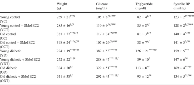
The general characteristics of the rats have been shown
in Table1. OC rats were significantly heavy relative to
YC group (p < 0.01). DM caused a reduction in body weights of YC as well as OC animals. SMe1EC2 treat-ment did not significantly change the weights of YC (p > 0.05) but markedly prevented weight loss in YD and OD animals (0 < 0.05).
In comparison with YC or YCT group, the difference between the mean final blood glucose levels of OC and OCT rats was not statistically significant (p > 0.05). DM led to a persistent hyperglycemia in both young and old rats (p < 0.001). However, SMe1EC2 treatment gave a rise to significant ameliorations in blood glucose levels of both groups of diabetic animals (p < 0.001).
Plasma triglyceride levels did not significantly change in SMe1EC2 treated control groups compared to untreated corresponding rats (p > 0.05). DM in-creased plasma triglyceride levels in both young and old rats (p < 0.01) that were significantly restored by SMe1EC2 treatment (p < 0.05).
Aging of the rats resulted in an increase in systolic blood pressure (p < 0.05), which was not significantly change by SMe1EC2 treatment. Systolic blood pressure was aggravated by DM in both young (p < 0.01) and old rats (p < 0.001) but was significantly prevented by
SMe1EC2 treatment (p < 0.05).
Protein oxidation/nitrosative stress markers and RAGE levels
In OC rats, AGEs levels of aorta were found to be significantly increased and DM caused further
augmen-tation in AGEs levels of old rats (Fig. 1a). DM also
significantly aggravated AGEs levels in young rats (Fig.
1a). OD rats showed the highest value of AGEs levels
when compared to the AGEs levels of YD or OC rats (p < 0.05). AGEs levels was unchanged by SMe1EC2 treatment in YC rats but significantly improved in OCT, YDT (p < 0.01), and ODT (p < 0.001) animals
com-pared to their corresponding controls (Fig.1a).
In OD rats, the increased level of AGEs associated
with a significant decline in aorta RAGE level (Fig.1b).
In comparison with YC rats, DM led to augmentation in aorta RAGE level more significantly in YD than OD rats (p < 0.001). SMe1EC2 treatment did not produce a significant alteration in RAGE level of YC rats
(p > 0.05) but protected the rats against DM-induced
alterations in aorta RAGE level (p < 0.01) (Fig.1b).
In the aorta of OC rats, 4-HNE (Fig.1c) and 3-NT
(Fig.1d) levels were increased statistically less
signifi-cant compared with the aorta of YC animals (p < 0.05); however 4-HNE and 3-NT levels severely exacerbated in the presence of DM and significantly prevented by
SMe1EC2 treatment (p < 0.001) (Fig. 1c, d). The
in-crease in aorta 3-NT levels was the most in YD rats
compared to other groups of rats (Fig.1d).
Constriction and relaxation responses
Cumulative addition of phenylephrine (Phe 10−9 to
10−5M) or potassium chloride (KCl 10–60 mM) to the
organ bath resulted in concentration-dependent
contrac-tions of aortic rings in all the group of animals (Fig.2).
Aging itself did not significantly affect the
vasoconstric-tive response to Phe (Fig.2a). DM caused a significant
increase in the vasoconstrictor effect of Phe in both young and old rats that was reflected by a significant
increase in apparentEmax(p < 0.01). The
vasoconstric-tive response of aorta to Phe was not statistically differ-ent in YCT and OCT rats compared to YC or OC rats
(Emaxp > 0.05). SMe1EC2 treatment led to a significant
downward shift in Phe-induced vasoconstrictions of YD
and OD animals (Emaxp < 0.001). An important feature
of Phe-induced constriction was the lowest level in YDT
and ODT rats compared to other groups of rats (Fig.2a).
The responsiveness of aorta to KCL did not
signifi-cantly change in OC rats compared to YC rats (Emaxp >
0.05) (Fig. 2b). The treatment of rats with SMe1EC2
caused a significant inhibition in response of the aorta to KCL in old but not in young rats compared to their untreated controls. DM led to a significant increase in KCL-induced vasoconstriction only in young rats
(Emaxp < 0.001) but not in old rats when compared with
treatment of both groups of diabetic animals results in significantly downward shift in the vasoconstrictor
re-sponse to KCL (10–60 mM) (Emaxp < 0.05) (Fig.2b).
We observed the lowest contractile response to KCL in
OCT animals compared to other group of animals
(Emaxp < 0.001), and the aorta of YD rats displayed the
most contractions to KCL than other group of rats
(Emaxp < 0.05).
Table 1 The final measurements of body weight, blood glucose, triglyceride, and systolic blood pressure in experimental rats Weight (g) Glucose (mg/dl) Triglyceride (mg/dl) Systolic BP (mmHg) Young control (YC) 269 ± 21††ƒƒ 105 ± 8ƒƒƒ### 82 ± 4ƒƒ# 123 ± 2††ƒƒ###
Young control + SMe1EC2 (YCT)
285 ± 16†ƒƒ 110 ± 9ƒƒƒ### 85 ± 8ƒƒ 128 ± 2†ƒƒ###
Old control (OC)
383 ± 37**ƒƒƒ# 117 ± 34ƒƒƒ### 81 ± 3ƒƒ# 140 ± 4*ƒ##
Old control + SMe1EC2 (OCT)
398 ± 24***ƒƒƒ# 107 ± 26ƒƒƒ### 88 ± 7ƒƒ 141 ± 3*ƒ##
Young diabetic (YD)
224 ± 19**†††## 382 ± 53***††† 126 ± 21**††## 159 ± 5**†
Young diabetic + SMe1EC2 (YDT)
252 ± 22*††# 288 ± 47***†††ƒ 89 ± 10ƒ 147 ± 6*#
Old diabetic (OD)
304 ± 30†ƒ 329 ± 51***††† 113 ± 8*† 169 ± 4***††
Old diabetic + SMe1EC2 (ODT)
311 ± 38†ƒ 292 ± 43***†††ƒ 93 ± 12ƒ# 134 ± 5*†ƒ##
Mean ± SEM,n = 8–10 rats per group. *p < 0.05, **p < 0.01, ***p < 0.001 vs young control;†p < 0.05,††p < 0.01,†††p < 0.001 vs old control;ƒp < 0.05,ƒƒp < 0.01,ƒƒƒp < 0.001 vs young diabetic;#p < 0.05,##p < 0.01,###p < 0.01 vs old diabetic
Fig. 1 The results of measurements of AGE-protein adduct levels (a), RAGE levels (b), 4-HNE-protein adduct levels (c), and 3-NT levels in aorta homogenates of rats grouped as young control (YC), young control treated with SMe1EC2 (YCT), old control (OC), old control treated with SMe1EC2 (OCT), young diabetic (YD), young
diabetic treated with SMe1EC2 (YDT), old diabetic (OD), and old diabetic treated with SMe1EC2 (ODT). Mean ± SEM,n = 8–10 rats per group. *p < 0.05, **p < 0.01, ***p < 0.001 vs YC;†p < 0.05,††p < 0.01,†††p < 0.001 vs OC;ƒp < 0.05,ƒƒp < 0.01,ƒƒƒp < 0.001 vs YD;#p < 0.05,##p < 0.01,###p < 0.001 vs OD
Cumulative addition of ACh (10−9to 10−5M) to the organ bath resulted in concentration-dependent de-creases in the tension of aorta pre-contracted with Phe
(Fig.3a). The responsiveness of aorta to ACh
signifi-cantly decreased by aging (p < 0.01), and DM did not produce a further attenuation in OD rats compared to
OC rats (p > 0.05) (Fig. 3a). DM led to a maximum
decrease in ACh-induced relaxation in YD rats
com-pared to other group of rats (p < 0.01). The cumulative
dose–response curves of ACh were similar in YC and YCT rats, but SMe1EC2 protected aorta against aging-induced ACh hypo-responsiveness, reflected by a
significant alteration in apparentEmaxin OCT rats
com-pared with OC rats (p < 0.01) (Fig.3a). SMe1EC2
treat-ment also significantly prevented the inhibitory effect of
DM on ACh relaxation in YDT rats (p 0.001) (Fig.3a).
The relaxation response to salbutamol was found to be decreased in OC rats compared to YC animals
(Emaxp < 0.001) (Fig.3b). DM inhibited the relaxation
response of aorta to salbutamol significantly (p < 0.001),
and theEmaxfrom the cumulative dose–response curves
of salbutamol was similar in YD and OD rats (Fig.3B).
SMe1EC2 treatment did not significantly change the
dose–response curve of salbutamol in YCT or OCT rats
when compared with untreated control rats (p > 0.05). Salbutamol relaxation was inhibited by DM in YD rats in similar degree that we observed in OC rats and was found to be not different in OC, OD, and YD rats. SMe1EC2 treatment was able to improve salbutamol
relaxation in YDT and ODT rats (Emaxp < 0.001).
On the other hand, neither aging nor DM or SMe1EC2 treatment affected the responsiveness of
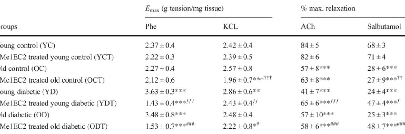
aor-ta to SNP (daaor-ta not shown). The Emax values for the
responses of aorta to Phe, KCL, ACh, and salbutamol
are demonstrated in Table2. Either aging, diabetes, or
SMe1EC2 treatment had no significant effect upon the
pD2pattern of contractions or relaxations in all group,
indicating that there has not been any significant change in the sensitivity of aortic rings in different experimental groups.
Calcium channel regulation of aorta constriction
To elucidate the mechanism of action of aging, DM or SMe1EC2 treatment on calcium mobilization, the tran-sient vasoconstriction to a single dose of Phe was
re-examined in the presence of Ca2+free medium (Krebs
solution without Ca2+, CFM) as well as in the presence
of cyclopiazonic acid, a SERCA inhibitor, plus Ca2+
free medium (Ceylan-Isik et al.2011). Figure4shows
the response of aorta to Phe (3 × 10⁻⁶ M) before and
after CFM (a) and CFM plus cyclopiazonic acid (CYP) (b). Phe constrictions were significantly inhibited in YC but not in OC rats when bath medium was changed by CFM and was further inhibited in the presence of CFM plus CYP (p < 0.01). CFM alone or combined with CYP resulted in a significant decrease in Phe constrictions in
YD and OD rats (p < 0.001). In SMe1EC2 treated rats,
Phe constrictions did not significantly change in the rings exposed to CFM or CFM plus CYP compared to
before exposures (p > 0.05) (Fig.4b).
Fig. 2 Concentration-dependent vasoconstrictions to phenyleph-rine (Phe, 10−9–10−4M) (a) and KCL (10–60 mM) (b) in aorta rings isolated from rats grouped as young control (YC), young control treated with SMe1EC2 (YCT), old control (OC), old control treated with SMe1EC2 (OCT), young diabetic (YD), young diabetic treated with SMe1EC2 (YDT), old diabetic (OD), and old diabetic treated with SMe1EC2 (ODT). Mean ± SEM,n = 8–10 rats per group. *p < 0.05, **p < 0.01, ***p < 0.001 vs YC;
†p < 0.05,††p < 0.01,†††p < 0.001 vs OC;ƒp < 0.05,ƒƒp < 0.01, ƒƒƒp < 0.001 vs YD;#p < 0.05,##p < 0.01,###p < 0.001 vs OD
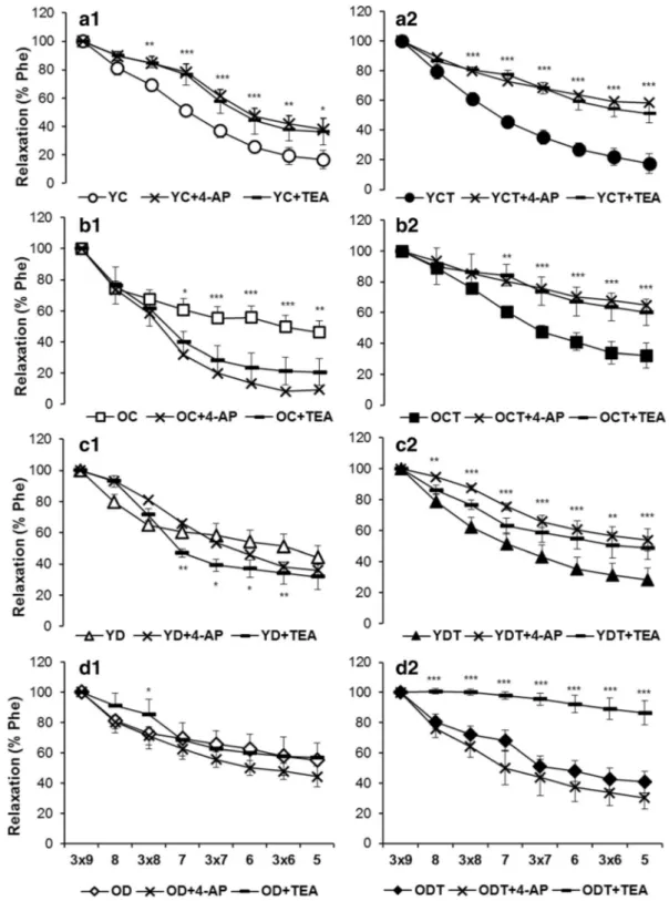
Potassium channel regulation of aorta relaxation
Figure5shows the effects of TEA and 4-AP on
ACh-induced relaxations. In YC rats, preincubation of aortic rings with TEA or 4-AP inhibited the relaxant effect of
ACh in the same degree (p < 0.001) (Fig. 5A1).
SMe1EC2 treatment of YC rats resulted in a significant increase in the inhibitory effect of TEA or 4-AP on ACh-induced relaxation compared to SMe1EC2
un-treated YC rats (p < 0.001) (Fig.5A2). ACh-induced
relaxations have ~ 40% decrease in OC relative to YC rats, displayed completely normalization by TEA or
4-AP (Fig. 5B1). TEA or 4-AP inhibited ACh-induced
relaxation in OCT rats, which showed a significant
improvement with SMe1EC2 treatment (p < 0.01) (Fig.
5B2). We did not achieve further inhibition with TEA or
4-AP in YD or OD rats characterized by ~ 50%
reduc-tion in ACh-induced relaxareduc-tions (Fig. 5C1, D1).
SMe1EC2-induced amelioration in ACh-induced relax-ation by SMe1EC2 treatment was significantly inhibited
with TEA or 4-AP in YD rats (p < 0.001) (Fig. 5C2).
The influence of KCa blockade and Kv inhibition on ACh dose–response curves was considerably different in ODT rats, which showed a better relaxation response to ACh compared to untreated OC rats. Namely, ACh-induced relaxations were markedly inhibited by TEA
(p < 0.001), it did not change significantly by 4-AP in
ODT rats (p > 0.05) (Fig.5D2).
Discussion
This study provides strong evidence of a vaso-protective action of SMe1EC2 in aging and diabetes. The results have shown that SMe1EC2 in old and diabetic rats can serve to maintain physiological blood pressure by main-taining ACh relaxation through mainly smooth muscle hyperpolarization and protection of NO against destruc-tive effects of free radicals. Not only through these mechanisms, but SMe1EC2 also contributes to the
reg-ulation of blood pressure by protecting β-adrenergic
receptor-mediated relaxation and by controlling
α1-adrenergic receptor signaling for contraction. We ob-served that in the old and diabetic vasculature, SMe1EC2 treatment: (1) prevents impairment of endo-thelial function, (2) reduces systemic oxidant genera-tion, and (3) suppresses smooth muscle depolarization and calcium mobilization.
It is well known that, the vascular contraction
in-duced by Phe, a selective α1-receptor agonist, is
bal-anced with ACh-stimulated and NO-mediated
endothe-lial relaxation through the mechanism by which Ca2+
influx is reduced (Bolotina et al.1994; Peng et al.1996).
When this regulatory system is inadequate, the aortic contractions to Phe or other adrenergic agents increase
Fig. 3 Concentration-dependent vasorelaxations in response to acetylcholine (ACh, 10−9–10−5 M) (a) and salbutamol (10−9– 10−4M) (b) in the phenylephrine-preconstricted (Phe, 10−6–3 × 0−6M) aortic rings isolated from rats grouped as young control (YC), young control treated with SMe1EC2 (YCT), old control (OC), old control treated with SMe1EC2 (OCT), young diabetic (YD), young diabetic treated with SMe1EC2 (YDT), old diabetic (OD), and old diabetic treated with SMe1EC2 (ODT). Mean ± SEM,n = 8–10 rats per group. *p < 0.05, **p < 0.01, ***p < 0.001 vs YC;†p < 0.05,††p < 0.01,†††p < 0.001 vs OC;ƒp < 0.05,ƒƒp < 0.01,ƒƒƒp < 0.001 vs YD;#p < 0.05,##p < 0.01,###p < 0.001 vs OD
(Koçak et al.2000; Bauer and Sotníková2010; Karasu
2010), which leads to rise in blood pressure in aging
(Faconti et al. 2015) and diabetes (Cohen and Tong
2010). This relationship seems to be not working in
the aorta of old rats, because OC rats showed no signif-icant increase in Phe contractions despite inhibition of ACh and salbutamol relaxations. In that case, the reason
for the increase in Phe or KCl contractions observed in diabetic rats cannot be explained by the reduced regula-tory effect of endothelium-dependent and/or endothelium-independent relaxation alone since diabe-tes did not produce a further decline in ACh or salbutamol relaxations in YD and OD compared to OC rats. In old age, Phe signaling seems to be not sensitive
Table 2 The maximum contractions to Phe and KCL and maximum relaxations to ACh and salbutamol in experimental groups
Emax(g tension/mg tissue) % max. relaxation
Groups Phe KCL ACh Salbutamol
Young control (YC) 2.37 ± 0.4 2.42 ± 0.4 84 ± 5 68 ± 3
SMe1EC2 treated young control (YCT) 2.22 ± 0.3 2.39 ± 0.5 82 ± 6 71 ± 4
Old control (OC) 2.27 ± 0.4 2.57 ± 0.8 57 ± 8*** 28 ± 6***
SMe1EC2 treated old control (OCT) 2.12 ± 0.6 1.96 ± 0.7***††† 63 ± 8*** 27 ± 9***††
Young diabetic (YD) 3.63 ± 0.3*** 2.86 ± 0.6** 41 ± 7*** 24 ± 4***
SMe1EC2 treated young diabetic (YDT) 1.43 ± 0.4***ƒƒƒ 2.43 ± 0.4ƒƒ 65 ± 6***ƒƒƒ 47 ± 4***ƒ
Old diabetic (OD) 3.48 ± 0.8*** 2.48 ± 0.4 57 ± 10*** 25 ± 3***
SMe1EC2 treated old diabetic (ODT) 1.53 ± 0.7***### 2.22 ± 0.8*# 58 ± 6***### 48 ± 7***### Mean ± SEM,n = 8–10 rats per group. *p < 0.05, **p < 0.01, ***p < 0.001 vs young control;††p < 0.01,†††p < 0.001 vs old control;ƒp < 0.05,ƒƒp < 0.01,ƒƒƒp < 0.001 vs young diabetic;#p < 0.05,###p < 0.01 vs old diabetic
Fig. 4 Vasoconstrictions to a single dose of phenylephrine (Phe, 3 × 10⁻⁶M) in the absence (black bar) or in the presence of Ca+2-free medium (white bar) (a) or Ca+2-free medium +
cyclopiazonic acid (10−6M) (white bar) (b) in aorta rings isolated from rats grouped as young control (YC), young control treated with SMe1EC2 (YCT), old control (OC), old control treated with SMe1EC2 (OCT), young diabetic (YD), young diabetic treated with SMe1EC2 (YDT), old diabetic (OD), and old diabetic treated with SMe1EC2 (ODT). Mean ± SEM,n = 8–10 rats per group. **p < 0.01, ***p < 0.001 vs YC in the presence of Ca+2; $p < 0.01, @p < 0.001 vs. without Ca+2-free medium or Ca+2-free medium + CYP
Fig. 5 Vasorelaxation response to cumulatively increased concen-tration of acetylcholine (ACh, 10−9–10−5M) in the preconstricted (Phe, 10−6–3 × 0−6M) aortic rings before and after incubation with TEA (10−4M) or 4-AP (10−4M). The rats grouped as young control (YC), young control treated with SMe1EC2 (YCT), old
control (OC), old control treated with SMe1EC2 (OCT), young diabetic (YD), young diabetic treated with SMe1EC2 (YDT), old diabetic (OD), and old diabetic treated with SMe1EC2 (ODT). Mean ± SEM, n = 8–10 rats per group. *p < 0.05, **p < 0.01, ***p < 0.001 vs before incubation with TEA or 4-AP
to the reduced endothelial relaxation capacity of vascu-lature, but an activated vasoconstrictor signaling appears to play an excessive role in YD and OD rats. Contrary to this, we and other investigators previously demonstrated in the rat model of aging or diabetes mellitus that endo-thelial control of vascular tonus is shifted in favor of vasoconstriction, and insufficient NO and also some arachidonoic acid metabolites mediate to increase Phe
contractions (Karasu et al. 1997a; Koçak et al. 2000;
Peredo et al.2006; Reyes-Toso et al.2007; Sotníková
et al.2011; Ceylan-Isik et al.2011). The reason for why
aging per se did not have an effect on the vasoconstrictor response of aorta to Phe may be related to aging time; in this current study, we used rats that were 20 months old, others usually used 10-month-old rats. In that case, the comparison of aorta relaxations and contractions in the presence of NOS inhibitor L-NAME would be more
determinative (Novella et al.2013).
The Phe contractions in aorta are mediated by the
release of Ca2+from the intracellular stores and Ca2+
influx through voltage-gated calcium channels (CaV)
and receptor-operated channels (ROCs) (Horowitz
et al. 1996; Hill-Eubanks et al. 2011). KCl also
pro-motes vasoconstriction via smooth muscle membrane
depolarization and then Ca2+influx through CaV
(Hill-Eubanks et al. 2011). Our findings indicate that the
mechanisms involving regulation of CaVto mediating
Phe contractions remain as physiologically active level during aging course, since not only Phe contractions but also KCL contractions stayed as unchanged in OC com-pared to YC rats. However, when diabetes mellitus developed in young stages of rats, the increased activity
of CaVis most likely to be a primary contributing factor
leading to an increase in Phe contraction and hyperten-sion, because KCL contractions only elevated in YD but not in OD compared to YC, OC, and OD rats. Accord-ingly, the expressions or activity of contractile proteins
such as PKC, which leads to Ca2+flow through CaV, has
been shown to increase in diabetic vessels due to
oxi-dative and carbonyl stress (Webb 2003; Wang et al.
2012a; Karasu2010; Chettimada et al.2014). Because
altered Ca2+mobilization and impaired calcium
signal-ing play a crucial role in the development of vascular reactivity abnormalities in aging or diabetes mellitus
(Zhu et al.2001; Karasu2010; Ceylan-Isik et al.2011;
Ma et al. 2008; Wang et al. 2012a); in details, the
function of sarcoplasmic reticulum Ca2+-ATPase
(SERCA) was assessed by treating the aorta rings in
Ca2+-free conditions with or without cyclopiazonic acid
(CYP). We found that CFM or CFM plus CYP inhibited Phe contractions in YC and both groups of diabetic but not OC rats, suggesting that exaggerated vasoconstric-tion seen in diabetic aorta is also related to SERCA and IP3R activation and sensitive to the inhibition with CFM
or CFM plus CYP (Table3).
Phe contractions did not change in YCT and OCT rats but dramatically inhibited in YDT and ODT rats. SMe1EC2 treatment also prevented exaggeration of KCl contractions in YD rats. These are associated with its blood pressure lowering and salbutamol relaxation normalizing effects in YDT and ODT rats. ACh relaxa-tion improving effect of SMe1EC2 was only seen in OCT and YDT animals. In addition, SMe1EC2 treat-ment kept blood pressure at normal levels in ODT rats without any improvement in endothelial relaxation. In-terestingly, Phe contractions in YDT and ODT rats as well as KCl contractions in OCT rats were obtained at the lowest levels compared to other group of rats. These
results suggest that exacerbatedα1-adrenergic receptor
signaling is inhibited by SMe1EC2, and SME1EC2 affects the aorta when aging makes the aorta more
sensitive to calcium mobilization via CaV. In addition,
SMe1EC2 treatment seems to be unable to affect Ca2+
release from IP3-sensitive intracellular stores since nei-ther CFM nor CFM plus CYP exhibited a significant inhibition in the contractions of aorta to a single dose of Phe in YCT, OCT, YDT, and ODT rats. This is in agreement with the reported endothelial function protecting effect of SMe1EC2 and its mother substance stobadine in hyperglycemic rats (Zúrová-Nedelcevová
et al.2006; Sotníková et al.2011). Conversely, there is
also an in vitro study showing that SMe1EC2 does not affect the responses of aortic rings to Phe, KCL, and
ACh (Broskova et al.2013).
The increased K+efflux causes membrane potential
hyperpolarization, leading to vasodilatation that may play a critical compensatory vasodilator role in disease states in which NO-mediated dilation is impaired
(Nelson and Quayle1995). In fact, the vasorelaxation
induced by ACh can be attributed in part to the opening
of voltage-sensitive K+ channels (Kv) and
large-conductance calcium-activated K+ channels (KCa)
(Malakul et al. 2008). Kv and KCachannels are
abun-dantly expressed in vascular smooth muscle cells in aorta and play a crucial role in counteracting of vaso-constriction and high blood pressure (Lísková et al.
2010). Thus, we examined the contribution of K+
Ta b le 3 The table summarizes ho w the ef fects of pharmacological ag ents used in the study change in experimental groups and also in ex perimental conditions Ag onist Medium Response o f aorta rings Alterations in respons e of aor ta rings in ex perimental g roups YC Y C T OC OCT YD Y DT OD O DT Phenylephrine (Phe) (in cumulatively in-cr ea sed co n cent ra tions ) Ca 2+ presence Constrictio n U nchanged* Un changed* Unchanged* Increased* D ecr ea sed* Inc re ase d* D ecr ea sed* Phenylephrine single dose (3 × 1 0 − 6 M) Ca 2+ presence Constrictio n U nchanged* Un changed* Unchanged* Increased* D ecr ea sed* Inc re ase d* D ecr ea sed* Phenylephrine single dose (3 × 1 0 − 6 M) Ca 2+ fr ee De cr eas ed cons tric-ti on # De cr ea se d # Unchanged # Unchanged # D ecr ea sed # U n chang ed # De cr ease d # U n chang ed # Phenylephrine single dose (3 × 1 0 − 6 M) Ca 2+ fr ee + C YP De cr eas ed cons tric-ti on # De cr ea se d # Unchanged # Unchanged # D ecr ea sed # U n chang ed # De cr ease d # U n chang ed # Potassium ch loride (KCL) (in cumulatively in-cr ea sed co n cent ra tions ) Ca 2+ presence Constrictio n U ncha nged* Unchanged* Decreased* ‡ƒ & Incr ea sed* ‡ƒ & U n chang ed* Unc h ange d* D ecr ea sed *‡ ƒ & Acetylcholine (ACh) (in cu mula tiv ely inc re ase d co ncent ra tions ) Ca 2+ presence in phe pr ec ontr ac ted aor ta Relaxation U nchanged* De cr ea sed* De cre ase d* incr ea sed ‡ D ecr ea se d* ‡ & In cr eas ed ƒ de- crea se d* Unchanged ‡ inc rea sed ƒ de cr ea se d* U n chang ed & ‡ in cr ea sed ƒ de cr ease d * Acetylcholine (in cu mula tiv ely inc re ase d co ncent ra tions ) Ca 2+ pr es en ce + tetraeth ylamonium (TEA) in Phe p re contr act ed aor ta De cr eas ed re la xa-ti on § In cr ea sed § In cr ease d § De cre ase d § Unchanged § D ecr ea sed § Unchanged § D ecr ea sed § Acetylcholine (in cu mula tiv ely inc re ase d co ncent ra tions ) Ca 2+ pr es en ce + 4-aminopyridine (4-AP) in Phe p re contr act ed aor ta De cr eas ed re la xa-ti on ɸ In cr ea sed ɸ In cr ease d ɸ De cre ase d ɸ Unchanged ɸ D ecr ea sed ɸ Unchanged ɸ U n chang ed ɸ Salbutamol (in cu mula tiv ely inc re ase d co ncent ra tions ) Ca 2+ pr es en ce in P h e pr ec ontr ac ted aor ta Relaxation U nchanged* De cr ea sed* Unc h ange d ‡ decreased * Unchanged ‡ decr ea sed* In cr eas ed ƒ‡ de- crea se d* Unchanged ƒ‡ de cr ea se d* In cr eas ed ƒ‡ de cr ease d * * C omparison w ith res ponse of aortic rings obtained from the YC rats ‡Comparison with the respon se of aortic ring s ob tain ed from the OC rats ƒ Comparison with response of aortic rings obtained from the YD rats & Comparison with the response of aorta rings obtained from the OD rats # Comparison of resp onses within the study grou p in the presence of Ca 2+ in the m edi u m § Comparison of resp onses within the study grou p in the absence o f T EA in the m edium ɸComparis on of res ponses within the study group in the absence of 4-AP in the m edium
KCa, tetraethylamonium (TEA), and 4-aminopyridine
(4-AP), a selective inhibitor of KV. ACh relaxations
were blunted by pharmacological blockade of K+
chan-nels with TEA and 4-AP in YC rats, suggesting that K+
channels opening contributes to vasodilatation evoked by ACh. When YC rats treated with SMe1EC2, ACh relaxation was inhibited more significantly by TEA or 4-AP than those of SMe1EC2 untreated YC rats, sug-gesting that ACh relaxation is realized independently from endothelial NO release in the presence of SMe1EC2 and the hyperpolarization-mediated relaxa-tion is likely playing a major regulatory role in control-ling of vasoconstrictor tonus by SMe1EC2. Interesting-ly, in OC rats, the presence of TEA or 4-AP led to an increase in ACh-induced relaxation. This response that seems to be counter to the expectation, may occur in the presence of aging-dependent regulation of the
mem-brane potential through changes in K+channel activity,
which changes the activity of voltage-dependent Ca2+
channels. Admittedly, additional experiments that go beyond the scope of the present study will be required
to further identify the specific subtype of K+channels
involved in the vasorelaxations produced by ACh in old rats. On the other hand, pretreatment of the aorta with 4-AP or TEA did not alter the depressed ACh-induced relaxation seen in YD and OD rats. This finding
sup-ports the hypothesis that the involvement of K+channels
in ACh relaxation is inhibited by diabetes. Our results are consistent with those obtained previously in aorta where diabetes was found to inhibit the vasodilation
mediated by KCaor Kv channels (Malakul et al.2008;
Kavak et al. 2009; Wang et al. 2012a). Furthermore,
since diabetes-induced decrease in
β-adrenoceptor-mediated vascular dilatation has been shown to be pri-marily associated with endothelium-independent mech-anisms, our findings, which show a decrease in salbutamol relaxation in YD rats, can be explained by
the dysfunction of KCa or Kv (Karasu et al. 1997b;
Ferro et al. 2004; Chai et al. 2005; Ko et al. 2008).
Current study also showed that aging alone is an impor-tant triggering factor to induce a decrease in salbutamol-induced relaxation, which shows no further inhibition in OD rats. This supports the reduction of the NO-mediated component of salbutamol relaxations due to aging and also overlaps with the finding that diabetes does not cause an additional inhibition in the already inhibited ACh relaxation in old rats. Nonetheless, we do
not totally discard a possible decrease of density ofβ2
-adrenergic receptors by aging (Deisher et al. 1989;
Schutzer et al.2006). In fact, the blunted regulatory role
of KCa as well as NO on Phe-induced contractions in
DM has been well documented (Chai et al. 2005;
Majithiya and Balaraman2006). Previous studies also
indicated a reduced KATP,Kv activity in the cholinergic
relaxation of aortic rings of STZ-diabetic rats (Chai et al.
2005; Porto et al.2010). In the base of our findings, it is
possible to say that the regulating role of KCaand Kv on
Phe contractions is emerging during aging course. This is consistent with a previously published report, which
suggests that the regulating potency of KCa on
NA-induced contractions is similar to that elicited by NO but is more evident in arteries from hypertensive rats
than in those from control rats (Lísková et al. 2010).
Another remarkable finding of this study is that TEA or 4-AP produces a significant inhibition ACh relaxation
in YDT rats; suggesting that the contribution of KCaand
Kv to the relaxant effect of ACh is enhanced in diabetic state by SMe1EC2. Moreover, when old diabetic rats treated with SMe1EC2, the relaxation to ACh was not changed by 4-AP, but was largely inhibited by TEA,
indicates the involvement of the activation by Ca2+of
K+channels in the endothelial-mediated control of
vas-cular tone in old diabetic rats. It has been reported that
the opening of KCa channels in aorta is increased by
diabetes (Ye et al.2004).
In fact, this is the first study indicating the regulatory role of systemic administration of SMe1EC2 on ion channel dynamics, relaxation, and contraction capacity of aorta and blood pressure in YD and OD rats. We inserted a table demonstrating the effects of the test substance on the contractile and dilatative properties of
the aorta from each experimental group (Table2).
Pres-ent findings are in agreemPres-ent with the previous works showing that SMe1EC2 has an anti-dysrhythmic effect
through regulation of cardiac KCa (Félétou 2009;
Broskova and Knezl2011). Accordingly, mother
sub-stance stobadine has been shown to decrease aortic stiffness and arterial blood pressure in diabetic rats
(Broskova et al.2013). In addition, we cannot exclude
the possibility of inhibition of other vasoconstrictors as a potential mechanism of action of SMe1EC2 to preserve aortic relaxation and to protect the increased vasocon-strictive response to phenylephrine. The fact that, SMe1EC2 allows the regulation of active vessel wall components by different pathways, which may also involve the inhibition of other vasoconstrictors like prostanoids or arachidonic acid metabolites which are
In the current study, diabetes led to an increase in AGEs, RAGE, 4-HNE, and 3-NT, but SMe1EC2 treat-ment improved all alterations observed in the stress markers in diabetic rats. This is in accordance with the reported inhibition of endothelial function by carbonyl compounds and oxidonitrosative intermediates in
dia-betic vessels (Zobali et al.2001; Negre-Salvayre et al.
2008; Karasu 2010; Wang et al. 2012b; Sell and
Monnier2012). AGEs exert their cellular effects mainly
through interaction with cell-surface RAGE (Barlovic
et al.2011; Yamagishi et al.2012). We found that when
the AGE level is increased the RAGE level is decreased in aged rats, implying a receptor down regulation to compensate the destructive effects of excessive produc-tion of AGEs and/or possibly other RAGE ligands or an aging-dependent decrease in the synthesis of RAGE protein. In concert of all the aforementioned results, this study revealed that SMe1EC2 is a promising pharma-cological agent to be used in the treatment of vascular disorders including hypertension in case of aging and diabetes.
Acknowledgements This article has been written by Prof. Karasu who is the leader of ADIC study group. We thank Ahmet Cumaoğlu and Elif Aydın for their technical help during measure-ment of some biomarkers. This work originally includes a part of the PhD thesis of Dr. ArzuŞakul and was partly supported by the Research Foundations of Gazi and Ankara Universities (GU-Pro-ject No. 01/2010-126, AU-Pro(GU-Pro-ject No. 10B336002), COST Action BM1203 and the Slovak Academy of Sciences (VEGA grant APVV-51-017905).
Compliance with ethical standards
Conflict of interest The authors declare that they have no con-flicts of interest.
References
Barlovic DP, Soro-Paavonen A, Jandeleit-Dahm KA (2011) RAGE biology, atherosclerosis and diabetes. Clin Sci (Lond) 121:43–55
Baumann M (2012) Role of advanced glycation end products in hypertension and cardiovascular risk: human studies. J Am Soc Hypertens 6:427–435
Bauer V, Sotníková R (2010) Nitric oxide–the endothelium-de-rived relaxing factor and its role in endothelial functions. Gen Physiol Biophys 29:319–340
Bolotina VM, Najibi S, Palacino JJ, Pagano PJ, Cohen RA (1994) Nitric oxide directly activates calcium-dependent potassium
channels in vascular smooth muscle. Nature 368(6474):850– 853
Broskova Z, Knezl V (2011) Protective effect of novel pyridoindole derivatives on ischemia/reperfusion injury of the isolated rat heart. Pharmacol Rep 63:967–974
Broskova Z, Sotnikova R, Nedelcevova J, Bagi Z (2013) Effect of a novel stobadine derivative on isolated rat arteries. Interdiscip Toxicol 6:63–66
Ceylan-Isik AF, Ari N, Stefek M, Sotnikova R, Ozansoy G, Horakova L, Karasu C (2011) Effects of a long-term treat-ment with an antioxidant pyridoindole on vascular respon-siveness in diabetes-induced aging rats. Curr Aging Sci 4: 150–157
Chai Q, Liu Z, Chen L (2005) Effects of streptozotocin-induced diabetes on Kv channels in rat small coronary smooth muscle cells. Chin J Physiol 48:57–63
Chettimada S, Ata H, Rawat DK, Gulati S, Kahn AG, Edwards JG, Gupte SA (2014) Contractile protein expression is upregu-lated by reactive oxygen species in aorta of Goto-Kakizaki rat. Am J Physiol Heart Circ Physiol 306:H214–H224 Cohen RA, Tong X (2010) Vascular oxidative stress: the common
link in hypertensive and diabetic vascular disease. J Cardiovasc Pharmacol 55:308–316
Cumaoglu A, Stefek M, Bauer V, Ari N, Aricioglu A, Karasu C (2010) Glycoxidative and nitrosative stress in kidney of experimental diabetic rats: effects of the prydoindole antiox-idant stobadine. Neuro Endocrinol Lett 31:313–318 Deisher TA, Mankani S, Hoffman BB (1989) Role of cyclic
AMP-dependent protein kinase in the diminished beta adrenergic responsiveness of vascular smooth muscle with increasing age. J Pharmacol Exp Ther 249:812–819
Drummond GR, Selemidis S, Griendling KK, Sobey CG (2011) Combating oxidative stress in vascular disease: NADPH oxidases as therapeutic targets. Nat Rev Drug Discov 10: 453–471
Ergin V, Hariry RE, Karasu C (2013a) Carbonyl stress in aging process: role of vitamins and phytochemicals as redox regu-lators. Aging Dis 4:276–294 Exp Biol Med (Maywood) 241: 343–352
Ergin V, Bali EB, Hariry RE, Karasu C (2013b) Natural products and the aging process. Horm Mol Biol Clin Investig 16:55– 64
Faconti L, Bruno RM, Ghiadoni L, Taddei S, Virdis A (2015) Ventricular and vascular stiffening in aging and hypertension. Curr Hypertens Rev 11:100–109
Félétou M (2009) Calcium-activated potassium channels and en-dothelial dysfunction: therapeutic options? Br J Pharmacol 156:545–562
Ferro A, Coash M, Yamamoto T, Rob J, Ji Y, Queen L (2004) Nitric oxide-dependent beta2-adrenergic dilatation of rat aor-ta is mediated through activation of both protein kinase A and Akt. Br J Pharmacol 143:397–403
Fleming TH, Humpert PM, Nawroth PP, Bierhaus A (2011) Reactive metabolites and AGE/RAGE-mediated cellular dys-function affect the aging process: a mini-review. Gerontology 57:435–443
Gu Q, Wang B, Zhang XF, Ma YP, Liu JD, Wang XZ (2014) Chronic aerobic exercise training attenuates aortic stiffening and endothelial dysfunction through preserving aortic mito-chondrial function in aged rats. Exp Gerontol 6:37–44
Hill-Eubanks DC, Werner ME, Heppner TJ, Nelson MT (2011) Calcium signaling in smooth muscle. Cold Spring Harb Perspect Biol 3:a004549
Horowitz A, Menice CB, Laporte R, Morgan KG (1996) Mechanisms of smooth muscle contraction. Physiol Rev 76:967–1003
Horváth EM, Benko R, Kiss L, Murányi M, Pék T, Fekete K, Bárány T, Somlai A, Csordás A, Szabo C (2009) Rapid 'glycaemic swings' induce nitrosative stress, activate poly(ADP-ribose) polymerase and impair endothelial func-tion in a rat model of diabetes mellitus. Diabetologia 52:952– 961
Juranek I, Horakova L, Rackova L, Stefek M (2010) Antioxidants in treating pathologies involving oxidative damage: an up-date on medicinal chemistry and biological activity of stobadine and related pyridoindoles. Curr Med Chem 17: 552–570
Karasu C (2000) Time course of changes in endothelium-dependent and -inendothelium-dependent relaxation of chronically diabet-ic aorta: role of reactive oxygen species. Eur J Pharmacol 392:163–173
Karasu C (2010) Glycoxidative stress and cardiovascular compli-cations in experimentally-induced diabetes: effects of antiox-idant treatment. Open Cardiovasc Med J 4:240–256 Karasu C, Ozansoy G, Bozkurt O, Erdoğan D, Omeroğlu S
(1997a) Antioxidant and triglyceride-lowering effects of vi-tamin E associated with the prevention of abnormalities in the reactivity and morphology of aorta from streptozotocin-diabetic rats. Antioxida nt s i n D ia bet es-I nduc ed Complications (ADIC) study group. Metabolism 46:872– 879
Karasu C, Ozansoy G, Bozkurt O, Erdoğan D, Omeroğlu S (1997b) Changes in isoprenaline-induced endothelium-de-pendent and -indeendothelium-de-pendent relaxations of aorta in long-term STZ-diabetic rats: reversal effect of dietary vitamin E. Gen Pharmacol 29:561–567
Kavak S, Emre M, Meral I, Unlugenc H, Pelit A, Demirkazik A (2009) Repetitive 50 Hz pulsed electromagnetic field ame-liorates the diabetes-induced impairments in the relaxation response of rat thoracic aorta rings. Int J Radiat Biol 85:672– 679
Ko EA, Han J, Jung ID, Park WS (2008) Physiological roles of K+ channels in vascular smooth muscle cells. J Smooth Muscle Res 44:65–81
Koçak G, Karasu C (2002) Elimination of O2•/H2O2by
alpha-lipoic acid mediates the recovery of basal EDRF/NO avail-ability and the reversal of superoxide dismutase-induced relaxation in diabetic rat aorta. Diabetes Obes Metab 4:69–74 Koçak G, Aktan F, Canbolat O, Ozoğul C, Elbeğ S, Yildizoglu-Ari N, Karasu C (2000) ADIC study group–antioxidants in diabetes-induced complications. Alpha-lipoic acid treatment ameliorates metabolic parameters, blood pressure, vascular reactivity and morphology of vessels already damaged by streptozotocin-diabetes. Diabetes Nutr Metab 13:308–318 Lamoke F, Shaw S, Yuan J, Ananth S, Duncan M, Martin P,
Bartoli M (2015) Increased oxidative and nitrative stress accelerates aging of the retinal vasculature in the diabetic retina. PLoS One 10(10):e0139664
Lísková S, Petrová M, Karen P, Kunes J, Zicha J (2010) Influence of calcium-dependent potassium channel blockade and nitric oxide inhibition on norepinephrine-induced contractions in
two forms of genetic hypertension. J Am Soc Hypertens 4: 128–134
Ma L, Zhu B, Chen X, Liu J, Guan Y, Ren J (2008) Abnormalities of sarcoplasmic reticulum Ca2+mobilization in aortic smooth muscle cells from streptozotocin-induced diabetic rats. Clin Exp Pharmacol Physiol 35:568–573
Majithiya JB, Balaraman R (2006) Metformin reduces blood pressure and restores endothelial function in aorta of streptozotocin-induced diabetic rats. Life Sci 78:2615–2624 Malakul W, Thirawarapan S, Suvitayavat W, Woodman OL (2008) Type 1 diabetes and hypercholesterolaemia reveal the contribution of endothelium-derived hyperpolarizing fac-tor to endothelium-dependent relaxation of the rat aorta. Clin Exp Pharmacol Physiol 35:192–200
Negre-Salvayre A, Coatrieux C, Ingueneau C, Salvayre R (2008) Advanced lipid peroxidation end products in oxidative dam-age to proteins. Potential role in diseases and therapeutic prospects for the inhibitors. Br J Pharmacol 153:6–20 Nelson MT, Quayle JM (1995) Physiological roles and properties
of potassium channels in arterial smooth muscle. Am J Phys 268(4 Pt 1):C799–C822
Novella S, Dantas AP, Segarra G, Vidal-Gómez X, Mompeón A, Garabito M, Hermenegildo C, Medina P (2013) Aging-related endothelial dysfunction in the aorta from female senescence-accelerated mice is associated with decreased nitric oxide synthase expression. Exp Gerontol 48:1329– 1337
Peng W, Hoidal JR, Farrukh IS (1996) Regulation of Ca2+ -acti-vated K+channels in pulmonary vascular smooth muscle cells: role of nitric oxide. J Appl Physiol 81:1264–1272 Peredo HA, Rodríguez R, Susemihl MC, Villarreal I, Filinger E
(2006) Long-term streptozotocin-induced diabetes alters prostanoid production in rat aorta and mesenteric bed. Auton Autacoid Pharmacol 26:355–360
Porto NP, Jucá DM, Lahlou S, Coelho-de-Souza AN, Duarte GP, Magalhães PJ (2010) Effects of K+channels inhibitors on the cholinergic relaxation of the isolated aorta of adult offspring rats exposed to maternal diabetes. Exp Clin Endocrinol Diabetes 118:360–363
Ray PD, Huang BW, Tsuji Y (2012) Reactive oxygen species (ROS) homeostasis and redox regulation in cellular signal-ing. Cell Signal 24(5):981–990
Reyes-Toso CF, Obaya-Naredo D, Ricci CR, Planells FM, Pinto JE, Linares LM, Cardinali DP (2007) Effect of melatonin on vascular responses in aortic rings of aging rats. Exp Gerontol 42:337–342
Schutzer WE, Xue H, Reed JF, Mader SL (2006) Effect of age on vascular beta2-adrenergic receptor desensitization is not me-diated by the receptor coupling to Galphai proteins. J Gerontol A Biol Sci Med Sci 61:899–906
Sell DR, Monnier VM (2012) Molecular basis of arterial stiffen-ing: role of glycation - a mini-review. Gerontology 58:227– 237
Sotníková R, Nedelčevová J, Navarová J, Nosáĺová V, Drábiková K, Szöcs K, Křenek P, Kyseĺová Z, Bezek S, Knezl V, Dřímal J, Brosková Z, Kristová V, Okruhlicová L, Bernátová I, Bauer V (2011) Protection of the vascular endothelium in experimental situations. Interdiscip Toxicol 4:20–26 Wang RX, Shi HF, Chai Q, Wu Y, Sun W, Ji Y, Yao Y, Li KL,
Zhang CY, Zheng J, Guo SX, Li XR, Lu T (2012a) Molecular mechanisms of diabetic coronary dysfunction due to large
conductance Ca2+-activated K+ channel impairment. Chin Med J 125:2548–2555
Wang Z, Jiang Y, Liu N, Ren L, Zhu Y, An Y, Chen D (2012b) Advanced glycation end-product Nε-carboxymethyl-lysine accelerates progression of atherosclerotic calcification in di-abetes. Atherosclerosis 221:387–396
Webb RC (2003) Smooth muscle contraction and relaxation. Adv Physiol Educ 27:201–206
Yamagishi S, Maeda S, Matsui T, Ueda S, Fukami K, Okuda S (2012) Role of advanced glycation end products (AGEs) and oxidative stress in vascular complications in diabetes. Biochim Biophys Acta 1820:663–671
Ye CL, Shen B, Ren XD, Luo RJ, Ding SY, Yan FM, Jiang JH (2004) An increase in opening of BK(Ca) channels in smooth muscle cells in streptozotocin-induced diabetic mice. Acta Pharmacol Sin 25:744–750
Yülek F, Or M, Ozoğul C, Isik AC, Ari N, Stefek M, Bauer V, Karasu C (2007) Effects of stobadine and vitamin E in diabetes-induced retinal abnormalities: involvement of oxi-dative stress. Arch Med Res 38:503–511
Zhu BH, Guan YY, Min J, He H (2001) Contractile re-sponses of diabetic rat aorta to phenylephrine at dif-ferent stages of diabetic duration. Acta Pharmacol Sin 22:445–449
Zobali F, Cakici I, Karasu C (2001) Effects of peroxynitrite on the reactivity of diabetic rat aorta. Pharmacology 63:58–64 Zúrová-Nedelcevová J, Navarová J, Drábiková K, Jancinová V,
Petríková M, Bernátová I, Kristová V, Snirc V, Nosál'ová V, Sotníková R (2006) Participation of reactive oxygen species in diabetes-induced endothelial dysfunction. Neuro Endocrinol Lett Suppl 2:68–71