AFFINITY OF GLYCOPEPTIDE NANOFIBERS TO GROWTH FACTORS AND THEIR EFFECTS ON CELLS
A THESIS SUBMITTED TO
THE GRADUATE SCHOOL OF ENGINEERING AND SCIENCE OF BILKENT UNIVERSITY
IN PARTIAL FULFILLMENT OF THE REQUIREMENTS FOR THE DEGREE OF
MASTER OF SCIENCE IN
MATERIALS SCIENCE AND NANOTECHNOLOGY
By
NURCAN HAŞTAR September, 2017
i
AFFINITY OF GLYCOPEPTIDE NANOFIBERS TO GROWTH FACTORS AND THEIR EFFECTS ON CELLS
By Nurcan Haştar, September, 2017
We certify that we have read this thesis and that in our opinion it is fully adequate, in scope and in quality, as a thesis for the degree of Master of Science.
Michelle Marie Adams (Advisor)
Ayşe Begüm Tekinay (Co-Advisor)
Çağlar Elbüken
Melih Önder Babaoğlu
Approved for the Graduate School of Engineering and Science:
Ezhan Karaşan
ii
ABSTRACT
AFFINITY OF GLYCOPEPTIDE NANOFIBERS TO GROWTH FACTORS AND THEIR EFFECTS ON CELLS
Nurcan HAŞTAR
M.Sc. in Material Science and Nanotechnology Advisor: Michelle Marie Adams
Co-Advisor: Ayşe Begüm Tekinay
September, 2017
The development of scaffolds for growth factor delivery is a promising approach for tissue regeneration applications due to their crucial roles during regeneration. Growth factor secretion and interactions with glycosaminoglycans are essential steps for the regulation of cellular behavior. Therefore, glycosaminoglycan-mimetic scaffolds provide a great opportunity to modulate the effects of growth factor actions on cell fate. In this thesis, sugar-bearing peptide amphiphile molecules were characterized and tested for VEGF, FGF-2 and NGF affinity. ELISA-based affinity analyses revealed that glycopeptide nanofibers had high affinity to NGF; however, glycopeptides alone were not enough to interact efficiently with VEGF and FGF-2. Since VEGF and NGF contain heparin-binding domains, the addition of a sulfonated peptide amphiphile increased the affinity of the nanofiber network to these growth factors. Glycopeptide-sulfonate nanofibers were also found to promote in vitro tube formation through their VEGF and FGF-2 affinity. VEGF release profiles of HUVECs indicated that increasing concentration of VEGF may provide autocrine signaling and enhance tube
iii
formation without any exogenous pro-angiogenic factor addition. In addition, when NGF-responsive PC-12 cells were cultured on glycopeptide nanofibers, they extended their neurites to an extent comparable with a widely-used positive control molecule (poly-L-lysine). These results suggest that glycosaminoglycan-mimetic glycopeptide nanofiber networks can be used as efficient growth factor presentation platforms for tissue regeneration applications to induce angiogenesis or peripheral nerve regeneration.
KEYWORDS: Growth factors, glycosaminoglycans, extracellular matrix, peptide nanofibers, neovascularization, neurite extension
iv
ÖZET
BÜYÜME FAKTÖRLERİNİN ŞEKERLİ NANOFİBERLERE AFİNİTESİ VE BU NANOFİBERLERİN HÜCRELERE ETKİLERİ
Nurcan HAŞTAR
Malzeme Bilimi ve Nanoteknoloji, Yüksek Lisans Tez Danışmanı: Michelle Marie Adams Tez Eşdanışmanı: Ayşe Begüm Tekinay
Eylül, 2017
Büyüme faktörleri, doku yenilenmesi sırasında önemli görevler almaktadırlar. Bu nedenle büyüme faktölerinin etkili bir biçimde hedeflenen bölgeye ulaştırılması doku yenilenmesi çalışmaları için önem taşımaktadır. Büyüme faktörlerinin glikozaminoglikanlarla etkileşimleri, fonksiyonlarını doğru bir şekilde yerine getirmeleri için önemli bir basamaktır. Bu sebeple, glikozaminoglikan benzeri yapıların büyüme faktörlerinin sunulması için tasarlanan yapı iskelelerinde kullanımları doku yenilenmesi için umut vaat eden bir yaklaşımdır. Bu çalışmada, glikozaminoglikanların monomerlerine benzer şekerli peptit nanofiberlerin, vasküler endotelyal büyüme faktörü (VEGF), fibroblast büyüme faktörü (FGF-2) ve sinir büyüme faktörüne (NGF) olan afinitesi ve bu peptit nanofiberlerin hücrelere olan etkileri araştırılmıştır. Şeker içeren nanofiberler NGF’e kuvvetle bağlanma gösterirken, VEGF ve FGF-2 moleküllerine afinitelerinin daha az olduğu
v
gözlemlenmiştir. VEGF ve FGF-2, heparin bağlanma domaini içerdiği ve heparinde yüksek miktarda sülfat molekülü bulunduğu için, bu nanofiberlerin yapısına sülfatlı peptit amfifil molekülünün eklenmesi, bu büyüme faktörlerine olan afinitesini artırmıştır. Büyüme faktörlerinin şekerli nanofiberlere afiniteleri göz önüne alınarak, bu nanofiberlerin endotel ve NGF’e yanıt veren PC-12 hücrelerine olan etkileri incelenmiştir. Sonucunda endotel hücreleri ekstra büyüme faktörüne ihtiyaç duymadan tüp benzeri yapılar oluşturmuş ve PC-12 hücreleri yüksek oranda nörit uzatmışlardır. Bu sonuçlar değerlendirildiğinde, üretilen şekerli yapı iskeleleri, yeni damar oluşumu gereken durumlarda ve periferik sinir yaralanmalarının onarımında, büyüme faktörlerinin etkili sunulması için in vivo çalışmalarda uygun birer platform sağlayabilirler.
ANAHTAR SÖZCÜKLER: Büyüme faktörleri, glikozaminoglikanlar, ekstraselüler matriks, peptit nanofiberler, yeni damar oluşumu, nörit uzatımı
vi
ACKNOWLEDGEMENTS
First, I would like to express my sincere thanks to my advisor Michelle Marie Adams for her invaluable support. I would like to express my gratitude thanks to my co-advisor Ayşe Begüm Tekinay. I am very glad to be a part of her team. In addition, I am very thankful for her guidance, supports and scientific advice throughout my master studies which give me the opportunity to advance myself. Additionally, I would like to thank Prof. Mustafa Özgür Güler for his guidance and scientific advice during my master studies.
I would like to acknowledge the graduate scholarship from BIDEB 2210-C TÜBİTAK (The Scientific and Research Council of Turkey) for financial support.
Throughout my Bilkent journey, I gain many friends who support and motivate me. Besides known ones, I have a chance to get to know various types of person. Firstly, I would like to thank my İYTE family. All of them are unique and special people. I would like to thank İdil Uyan (actually Arıöz now) for being an amazing pal for all the time and be there whenever I need. Also, I would like to thank Zeynep Orhan (actually now Okur) for her valuable friendship and for making Ankara livable place for with her travel plans. I would like to thank Merve Şen (still ŞEN thank god) for giving me a chance to know her colorful personal character better and for her psychological supports. And I would like to thank Gökhan Günay for his chatting about the questioning the life and his Turkish coffee passion. I would like to express my grateful thanks to my boyfriend Fatih Yergöz, without his support, patience, and love, it will be very hard to finish this master studies.
vii
I would like to thank all NBT, BML and UNAM members, especially Ashif Shaikh Özüm Şehnaz Çalışkan, Yasin Tümtaş, Gözde Uzunallı, Seher Üstün Yaylaci, Gülistan Tansık, Elif Arslan, Ruslan Garifullin, M. Arif Khalily, Melike Sever, Melis Şardan Ekiz, Göksu Çınar, Kübra Kara, Begüm Dikeçoğlu, Çağla Eren Çimenci, Hamid Muhammed Syed, Zehra Tatlı Yıldırım, Aslı Ekin Doğan, Faruk Okur, Mustafa Fadlelmullah, Nazan Fadlelmullah for creating such a warm and lively environment in Bilkent University.
I would like to thank Özge Uysal for her cheerful, amazing friendship, Nuray Gündüz for her anti- (aykırı) and amusing friendship, Canelif Yılmaz for her unexpected reactions and helpfulness, Oğuz Tuncay for his careless but actually careful friendship, Mustafa Beter for our little but entertaining chit chats, Ahmet Emin Topal for his special songs for me and for every one and Alper for his wise, thoughtful and charitable friendship.
Least but not last, I would like to thank my family. Especially, I would like to thank my mother Fatma Haştar for her warm-hearted love and support, and my sisters Zehra Şahin and Esin Bozdemir for their encouragement and endless supports. Finally, I would like to thank my deceased dearest father Mehmet Haştar. I know that if he can able to see my graduation, I believe that he will be very proud of me.
viii
Contents
Abbreviations ... xiii
1. Introduction ... 1
1.1. Carbohydrates of Extracellular Matrix ... 1
1.2. Growth Factors and Their Interaction with Glycosaminoglycans ... 6
1.3. Growth Factor Presentation Strategies ... 10
1.4. Self-Assembled Peptide Nanofiber Networks for Regenerative Medicine ... 14
Chapter 2 ... 18
2.1. Introduction ... 18
2.2. Experimental Section ... 20
2.2.1. Materials ... 20
2.2.2. Synthesis of Purification of Amphiphilic Glycopeptides ... 20
2.2.3.1. Scanning Electron Microscopy (SEM) Imaging... 21
2.3. Results and Discussion ... 26
2.3.1. Synthesis and Characterization of Glycopeptide Nanofibers ... 26
34 34 2.3.2. Affinity Analysis of Growth Factors to Glycopeptides ... 35
2.3.3. Viability of Cells on Glycopeptide Nanofibers ... 37
2.3.4. Glycopeptide-Sulfonate Nanofiber Networks Promote in vitro Tube-like Structures ... 40
ix
2.3.6. Time-Dependent VEGF and FGF-2 Secretion Analyses of
HUVECs Cultured on Glycopeptide-Sulfonate Nanofibers………...44
2.4. Conclusions and Future Perspectives ... 47 Bibliography ... 53
x
LIST OF FIGURES
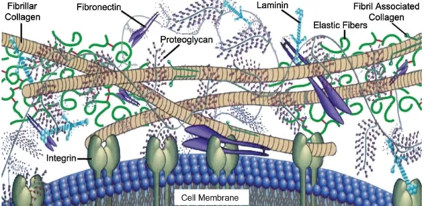
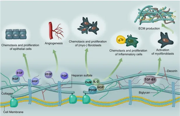
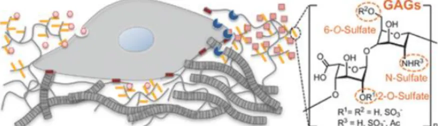
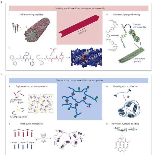
Figure 1: Representative image of the extracellular matrix. Structural proteins such as collagen provide physical support. Adhesive proteins like laminin and fibronectin function by interacting with integrin. Carbohydrate components of ECM, proteoglycans and connected unbranched glycosaminoglycans, have distinct biological functions. Copyrighted from © 2016 Elsevier Ltd. Published by Elsevier. Ltd [2]. ... 1 Figure 2: Chemical structures of glycosaminoglycans. Disaccharide units and their linkage types are provided with their common names. Copyright© 2013, Dovepress [2]. ... 4 Figure 3: Growth factors bearing heparin-binding domains associate with heparan sulfate glycosaminoglycans and exert their effects depending on their type, which could be to mediate the differentiation, migration, proliferation processes of cells. Copyrighted from American Journal of Physiology - Gastrointestinal and Liver Physiology Published 15 May 2015 Vol. 308 no. 10, G807-G830 DOI: 10.1152/ajpgi.00447.2014 [3]. ... 7 Figure 4: Properties of GAG-based, ECM-mimicking materials include the capacity to deliver growth factors, provide a porous environment, create a platform for cell adhesion and biodegrade naturally in the ECM. Copyrighted Advanced Materials, Volume 28, Issue 40, October 26, 2016, DOI: 10.1002/adma.201601908 [1]. ... 12 Figure 5: An overview of supramolecular interactions. a) Stacking motifs create one-dimensional self-assembled structures such as i) peptide amphiphiles and ii) aromatic
xi
group-containing oligopeptides; iii) which gather and self-assemble through hydrogen bonding and hydrophobic interactions. b) Formation of polymeric supramolecular biomaterials through molecular recognition strategies; i) cross-linking by recognition of the docking domain by polymer-conjugated engineered recombinant proteins; ii) Crosslinking through host-guest affinity; iii) elongation of oligomeric precursor through hydrogen-bonding; iv) metal-ligand interaction-facilitated oligomer extension. Copyrighted by Nature Materials 15, 13–26 (2016) doi:10.1038/nmat4474 [50]. ... 16 Figure 6: Chemical structures and LC-MS analyses of a) galactose-PA and b) mannose-PA. ... 28 Figure 7: Chemical structure and LC-MS analyses of a) glucose PA and b) N-AcetylglucosaminePA. ... 28 Figure 8: Chemical structures and LC-MS analyses of a) SO3-PA and b) EE-PA. .. 30 Figure 9: Chemical structure and LC-MS analyses of K-PA. ... 31 Figure 10: CD spectra analyses of peptide amphiphile molecules. ... 31 Figure 11: CD spectra of glycopeptide nanofibers that are formed by the addition of the oppositely charged K-PA. a) Only glycopeptide and K-PA combinations; b) Glycopeptide/sulonfate PA and K-PA combinations and c) Equimolar glycopeptide/K-PA combinations that were analyzed for NGF binding ... 32 Figure 12: SEM imaging reveals the ECM-like nanofibrous, porous network of glycopeptide nanofibers. (Combinations for NGF binding) ... 33 Figure 13: Height maps of glycopeptide nanofibers. ... 33 Figure 14: SEM imaging reveals the ECM-like nanofibrous, porous network of glycopeptide nanofibers. (Combinations for VEGF and FGF 2 binding) ... 34
xii
Figure 15: a) VEGF affinity to glycopeptide and glycopeptide-sulphate nanofiber network. b) FGF-2 affinity to glycopeptide and glycopeptide-sulfonate nanofiber network. Values represent mean ±SEM (****p<0.0001). ... 36 Figure 16: NGF affinity to glycopeptide nanofibers. Values represent mean ± SEM (****p<0.0001). ... 37 Figure 17: Representative images of live-dead assay of HUVECs that are cultured on glycopeptide nanofibers. Green dots indicate live cells (calcein-AM) and red dots indicate dead cells (ethidium homodimer). ... 38 Figure 18: Results of live-dead assay indicate that all glycopeptide nanofibers are biocompatible. ... 39 Figure 19: Representative images of PC-12 cells and quantification of live dead assay. ... 39 Figure 20: Brightfield microscope images of HUVECs cultured on nanofibers. Tube formation assay quantification, average length of formed tubular structures. Values represent mean ±SEM (***p<0.001). ... 42 Figure 21: Brightfield microscope images of PC-12 cells cultured on glycopeptide nanofibers and quantification of average neurite extension. Values represent mean ±SEM (**p<0.01). ... 43 Figure 22: FGF-2 release profiles of HUVECs cultured on glycopeptide-sulfonate nanofibers. ... 45 Figure 23: Time dependent release of VEGF secreted from HUVECs cultured on glycopeptide-sulfonate nanofibers. Values represent mean ±SEM (****p<0.0001). 46
xiii
Abbreviations
AFM Atomic force microscopy
ANOVA Analysis of variance
CD Circular dichroism
DCM Dichloromethane
DIEA N,N-diisopropylethylamine
DMEM Dulbecco's modified Eagle's medium
DMF N, N-Dimethylformamide
ELISA Enzyme-linked immunosorbent assay
ECM Extracellular matrix
FGF-2 Fibroblast growth factor-2
FBS Fetal bovine serum
Fmoc 9-Fluorenylmethoxycarbonyl GAG Glycosaminoglycan GF HBTU Growth Factor N,N,N′,N′-Tetramethyl-O-(1H-benzotriazole-1-yl) uronium hexafluorophosphate
HUVEC Human umbilical vein endothelial cell LC-MS Liquid chromatography-mass spectroscopy
NGF Nerve growth factor
PA Peptide amphiphile
SEM Scanning electron microscopy
TCP Tissue culture plate
TFA Trifluoroacetic acid
1
Chapter 1
1. Introduction
1.1. Carbohydrates of Extracellular Matrix
All cells of the human body are surrounded by an extracellular matrix (ECM) that contributes to their behaviors despite not strictly being a part of the cell environment. Components of ECM demonstrate tremendous variety but mainly consist of proteins, carbohydrates, and complexes formed by the combination of both (Figure 1). Biomechanical characteristics of ECM, as well as the composition and spatiotemporal orientations of its components, may differ depending on tissue type. In addition to providing mechanical support to cells, this network delivers signals to regulate cellular activities such as migration, differentiation, and proliferation through its dynamic properties [4].
Figure 1: Representative image of the extracellular matrix. Structural proteins such as collagen provide physical support. Adhesive proteins like laminin and fibronectin function by interacting with integrin. Carbohydrate components of ECM, proteoglycans and connected unbranched glycosaminoglycans, have distinct biological functions. Copyrighted from © 2016 Elsevier Ltd. Published by Elsevier.
2
Two main types of ECM elements are glycosaminoglycans, which are typically bound to protein cores and called proteoglycans in this form, and fibrous proteins such as collagen, elastin, laminin, and fibronectin that are responsible for structural support by forming the fibrillary structures of the ECM and assisting in cell adhesion [5-6]. Collagens are the most abundant proteins of ECM, and twenty-eight different types of collagen have so far been defined in the literature [5]. These tensile fibers resist deformation during the mechanical stretching of tissues [7]. Collagen deposition is performed by tissue-specific cells in connective tissues, e.g. collagens are secreted from osteoblasts in bone and from chondroblasts in cartilage tissue. In addition, a small percentage of collagen molecules are modified with monosaccharides and disaccharides [8]. In contrast to collagen, elastin mediates stretching in response to mechanical forces and provides elasticity to tissues like elastic cartilage, arterial vessels, and lungs. Elastins form microfibril-coated fibers that contain glycoproteins called fibrillins, which are members of the fibronectin family and are crucial for elastin function. Due to their strongly hydrophobic amino acid composition, elastins are able to recover their original shapes after exposure to mechanical forces [9]. While collagens and elastins provide physical support and are the main structural components of the ECM, glycoproteins are responsible for mediating cellular adhesion. Fibronectin is one of the glycoproteins of the ECM and assists in the adhesion of cells to their ECM [10] or facilitates their migration at some of the crucial events during development [11]. It has several domains that interact with other ECM structures such as heparin, proteoglycans, glycosaminoglycans, and fibrin, as well as transmembrane receptors called integrins [12]. Epithelial cells and fibroblasts are the main cell types that secrete
3
fibronectin. In the ECM, secreted fibronectins form fibrils and can be found in the solid state, while the soluble form of fibrils are found in body fluids such as blood [13]. Laminin is mainly synthesized by neurons, bone marrow cells, muscle cells and the epithelium, and it is a main component of the basal lamina [6]. This glycoprotein is formed by three different elongated polypeptides and has various domains that are recognized by some of the glycosaminoglycans, integrins and type IV collagen. It mediates the interaction between basement membrane, type IV collagen and cells, and establishes a connection between cell adhesion sites and the ECM network [14].
Glycosaminoglycan (GAG) units of proteoglycans are the carbohydrate reservoir of the ECM. Proteoglycans can be thought of as a storage unit containing GAGs that are covalently attached to a core protein. Proteoglycans can be found in intracellular sites, cell membranes, and the ECM environment. Their interactions with ECM molecules, growth factors, chemokines or receptors are mediated by their core proteins or attached GAG sites. These interactions result in the organization of the ECM and behaviors such as adhesion, proliferation, differentiation or migration [15]. Unbranched polysaccharides that either reside freely in the ECM or connect to proteoglycans through one or more binding sites are called glycosaminoglycans. GAGs are composed of disaccharide units. One saccharide unit is an amino sugar (galactosamine or D-glucosamine) and the second unit is uronic acid (D-glucuronic acid or L-iduronic acid) [16]. Anionic characteristics of GAGs arise from the presence of carboxyl and sulfate groups on some of the sugar subunits. Due to negatively-charged subunits, GAGs have the capacity to absorb high amounts of water and form a gel-like environment for cells. These hydrated gels help to absorb mechanical forces and provide transportation of molecules from the ECM to cells. Their sugar subunits can be sulfated at carbon 4 or
4
6 or non-acetylated nitrogen atoms, increasing the diversity of GAG chains. This sulfation arrangement leads to the formation of over 1,000,000 different patterns [17].
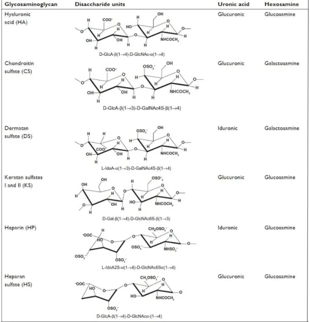
Figure 2: Chemical structures of glycosaminoglycans. Disaccharide units and their linkage types are provided with their common names. Copyright© 2013, Dovepress [2].
5
Six types of GAGs are present in ECM and are classified depending on the type of their saccharide unit and its glycosidic linkage (Figure 2). Only one type does not contain sulfated saccharide units and is called hyaluronic acid (HA, hyaluronate, hyaluronan). Hyaluronic acid is composed of D-glucuronate (GlcA) and N-acetylglucosamine (GlcNAc), which is linked through β (1, 3) glycosidic bonds. HA is non-epimerized and does not have any connection with core proteins of the ECM. It is mostly found in the synovial fluid, articular cartilage, vitreous humor, skin and loose connective tissue [18]. Other types of GAGs are chondroitin sulfate (CS), dermatan sulfate (DS), keratan sulfate (KS), heparin (HP) and heparan sulfate (HS). CS is the most abundant GAG and consists of GlcA and GalNAc-4 or 6 sulfate with a β (1, 3) linkage. CS is attached to core proteins of proteoglycans called lecticans. Versican, aggrecan, and decorin are members of this family. CS participates in developmental stages of the central nervous system, the wound healing process and various signaling events resulting in cell division and morphogenesis. It is a major component of the tendons, aorta, cornea, bone, cartilage and intervertebral discs [19]. DS is related to CS and was previously classified as chondroitin sulfate B. The amino sugar component of DS is N-acetylgalactosamine (GalNAc), which is sulfated at position 4 and are linked by a β (1,3) bond to GlcA or an α (1,3) bond to L-iduronate (IdoA). It is found in skin, lung, heart valves and blood vessels and plays a role in clot formation, wound healing, fibrous tissue production and infection [20]. Disaccharide units of keratan sulfate consist of β (1,4) linked galactose and GlcNAc, which is sulfated at the sixth carbon. This GAG appears in the brain, cornea and cartilage tissues, and binds to
6
keratocan, lumican, and aggrecan proteoglycans to assist in the cell movement, axon guidance and ligand recognition [21]. Heparin (HP) and heparan sulfates share identical disaccharide units, which are IdoA (typically sulfated at the 2 position) or GlcA (also typically sulfated at the 2 position) linked to N-sulfo-D-glucosamine-6-sulfate with α (1,4) (if IdoA) or β (1,4) (if GlcA) bonds, but heparin has a much higher sulfate content than heparan sulfate. Due to its highly negative charge, HP forms strong electrostatic interactions with thrombin and serves as an anticoagulative agent. Mast cells store heparin in their intracellular compartments, and the GAG is also present in the arteries of the lungs, liver, and skin [22]. Heparan sulfate is a fundamental type of GAG for the ECM and basement membranes of cells. HS contains more acetylated glucosamine and interacts with syndecans and glycosylphosphatidylinositol (GPI)-linked glypicans. In addition, it has an affinity to growth factors such as fibroblast growth factor (FGF), vascular endothelial growth factor (VEGF), hepatocyte growth factor (HGF) and chemokines [23].
1.2. Growth Factors and Their Interaction with Glycosaminoglycans
A wide range of growth factors is required to orchestrate and regulate the physiological functions of tissues. Growth factors are secreted polypeptides that participate in crucial steps of development, regeneration, and morphogenesis. They exert their effects by interacting with specific receptors on the cell membrane. Receptor-growth factor interactions result in the activation of associated pathways by phosphorylating the internal domain of receptors. Pathway activation regulates the expression or inhibition of related genes, which typically modulate the survival, proliferation, differentiation or migration of cells (Figure 3) [3] [24].
7
Oxygen is an indispensable requirement of cells. Transportation of oxygen to cells and tissues is crucial for survival and facilitated by blood vessels. Therefore, the formation of new blood vessels from pre-existing ones (angiogenesis) is an essential event for wound healing and the regeneration of tissues. In addition, angiogenesis is observed in pathological processes such as metastasis and rheumatoid arthritis.
Figure 3: Growth factors bearing heparin-binding domains associate with heparan sulfate glycosaminoglycans and exert their effects depending on their type, which could be to mediate the differentiation, migration, proliferation processes of cells. Copyrighted from American Journal of Physiology - Gastrointestinal and Liver Physiology Published 15 May 2015 Vol. 308 no. 10, G807-G830 DOI: 10.1152/ajpgi.00447.2014 [3].
8
Endothelial cells undergo a differentiation during new blood vessel formation. After their division, some regions of the basement membrane and extracellular matrix near endothelial cells are partially degraded to allow their migration, which culminates in the formation of tubular structures [25]. Proangiogenic and antiangiogenic factors stay in balance under normal physiological conditions, resulting in the steady renewal of blood vessels. Vascular endothelial growth factor (VEGF) is one of the major proangiogenic signals. Interaction of VEGF with its receptor triggers a signaling pathway that results in the survival, growth, and migration of endothelial cells. Seven different types of VEGF have been described in the literature, called VEGF-A through VEGF-F, and placental growth factor (PlGF). Among them, VEGF-A is the major factor that is responsible for neovascularization. Alternative splicing of the VEGF-A transcript leads to seven isoforms that consist of distinct amino acid numbers; 121, 145, 148, 165, 183, 189, or 206 [26]. VEGF165 is the most predominant form of VEGF-A and includes a C-terminal heparin binding domain. It can either be found free or bound to the ECM. In the ECM, VEGF interacts with the heparan sulfate (HS) GAG and/or heparin and neuropilin co-receptors through its heparin binding domain. In addition, HS and heparin interact with the VEGF receptor and mediate the stabilization of VEGF, neuropilin and its associated receptor complex. This complex increases the stability and lifetime of VEGF and provides controlled release and effective presentation of the growth factor by enhancing its affinity to its receptor. 6-O sulfation of the HS chain was previously indicated as an essential motif for the affinity of VEGF to HS [27].
Fibroblast growth factors have been identified at the beginning of the seventies and investigated extensively since then. Acidic and basic FGF were first described
9
according to their contribution to fibroblast growth and isoelectric points [28]. Following research characterized 20 sub-types of FGF. Although the name of FGF implies that its only function is related to fibroblast growth, FGF pathway actually regulates the development of embryonic tissues, maintains the homeostasis of tissues, and enhances regeneration and angiogenesis [29]. Acidic fibroblast growth factor (aFGF, FGF-1) and basic fibroblast growth factor (bFGF, FGF-2) are not by themselves sufficient to activate. Therefore, secreted FGF-1 and FGF-2 are bound to heparin sulfate proteoglycan (HSPG) and internalized directly into cells [30]. HS and HP can then interact with FGF and its receptor (FGFR) and stabilize this ternary complex by enhancing their interaction. HS triggers the dimerization of FGF and directs its interaction with the extracellular domain of FGFR. FGF binding leads to the dimerization of FGFRs, tyrosine kinase domains of which phosphorylate each other to start a signaling cascade [31]. FGF-HS interactions and ternary complex formation are regulated by the length and total charge density of sulfates, regardless of HS sulfation pattern [32]. As such, proper signaling of FGF requires HS since HS acts as a reservoir of FGF, protects it from degradation, and regulates its diffusion area.
Nerve growth factor (NGF) is one of the neurotrophic factors that are responsible for the proliferation, growth, and survival of the neurons of the peripheral nervous system. Expression of the NGF gene produces three proteins, which are alpha-, beta- and gamma-NGF. The functional form of NGF is formed when gamma subunit acts as a serine protease and separates the N-terminal of the beta subunit [33]. NGF has two known receptors; TrkA and p75NTR. TrkA is the principal receptor and mediates fundamental responses of cells by interacting with NGF. Since TrkA is a kind of tyrosine kinase receptor, its interaction with NGF triggers its dimerization and
10
phosphorylation, which results in the activation of differentiation pathways [34]. Some neurotrophins, including NGF, exhibit affinity to the 4- and 6-O sulfated GalNAc epitopes of chondroitin sulfate. It has been shown that NGF association with CS leads to the assembly of a signaling complex and modulates related pathways [35]. However, exogenous presentation of CS to the culture medium of cells prevents the activation of NGF-TrkA by sequestering NGF and keeping it away from the cell membrane [36].
GAGs and proteoglycans are able to modulate the growth factor activity in the following ways: They can sequester growth factors to act as a natural reservoir of signaling molecules while delaying their immediate activity; they can preserve growth factors from proteolytic degradation and therefore extend their action time, and they can bring them to spatiotemporally appropriate positions to efficiently present them to cell surface receptors [32].
1.3. Growth Factor Presentation Strategies
Tissue engineering applications are improving day by day through an enhanced understanding of the biological, chemical and physical requirements of damaged tissue and the corresponding development of appropriate strategies for each situation. Growth factors are vital elements of tissue regeneration. Therefore, their convenient, effective and active forms should be delivered by appropriate scaffolds for an optimal recovery process. Since a broad range of growth factors exhibit an affinity towards GAGs, developing GAG-mimetic scaffolds or incorporating GAGs to the structure of scaffolds could be an effective approach for regulating and enhancing the regeneration process through their proper representation.
11
Direct delivery of growth factors has failed in clinical trials since the concentration of the growth factors needs to be above a threshold in order to show their effects on target cells. In addition, directly administrated growth factors are more prone to degradation and may not reach the target area if injected intravenously or intramuscularly. As such, excessive doses of growth factors and several injections may be required for therapeutic effect. Increasing concentration of delivered growth factors may lead to undesirable consequences in case of angiogenic growth factors such as VEGF and FGF2, which can promote the development of pathological vessels and unbalance cellular dynamics to facilitate cancer formation [37, 38]. Therefore, unique and smart administration strategies need to be developed (Figure 4).
Natural and synthetic polymeric hydrogels are widely tested biomaterials due to their high hydration capacity and structural resemblance to the ECM. Physical entrapment or covalent placement of growth factors inside polymers may delay their denaturation and control their release profile thanks to the enhanced degradation kinetics of polymeric scaffolds [39]. Chemical immobilization of growth factors to polymers can be performed through covalent or non-covalent interactions. Non-covalent incorporation of growth factors into scaffolds can be performed through charged and hydrophobic group interactions or the addition of mediator molecules such as fibronectin, heparin or small oligopeptide sequences within the matrix structure. Fibrin networks are especially useful for this purpose, as they enhance clot formation and stop bleeding, in addition to easily encapsulating growth factors such as NGF or VEGF through their mesh-like structures and ECM-mimicking capabilities. Proteolytic degradation of fibrin by plasmin releases growth factors in a controllable manner and without losing their activity. As such, fibrin scaffolds have previously been used with
12
considerable success to enhance nerve regeneration (NGF) [40] and induce angiogenesis (VEGF) [41].
Figure 4: Properties of GAG-based, ECM-mimicking materials include the capacity to deliver growth factors, provide a porous environment, create a platform for cell adhesion and biodegrade naturally in the ECM. Copyrighted Advanced Materials, Volume 28, Issue 40, October 26, 2016, DOI: 10.1002/adma.201601908 [1].
13
Combining ECM mimetic molecules and microporous polymers may also result in better regeneration. Conjugation of heparin to fibrin matrices has been investigated for the non-covalent loading of growth factors, and although NGF has a moderate affinity to heparin, heparin-loaded fibrin matrices were observed to facilitate the slow, controlled release of NGF and promote neurite extension [42]. In a similar way, loading of VEGF to alginate in conjunction with a covalently-attached cell adhesion peptide sequence enhanced the survival rate of endothelial cells and provided blood transmission to ischemic tissue [43]. In addition to natural polymers, synthetic polymers can also be used for growth factor release and exhibit some advantages such as ease of fabrication and chemical modification. For instance, encapsulation of VEGF to poly (d, l-lactide-co-glycolide) (PLG) polymer provides the sustained release of GF and enhances angiogenesis [44].
However, covalent interaction or encapsulation of growth factors to scaffolds may disrupt the active sites of GFs and interfere with their therapeutic effects. Interaction of growth factors with ECM components makes them spatiotemporally available and their association protects growth factors from degradation while providing their sustained release. Therefore, utilizing ECM components during the design of biomaterials could enhance the outcomes of growth factors delivery. It has been shown that heparin incorporation to poly (ethylene glycol) (PEG) with photo-crosslinkable vinyl epitopes promotes human mesenchymal stem cell (hMSCs) proliferation due to the sustained release of FGF-2 [45] and, additionally, three dimensional culturing could be used to induce the osteogenic differentiation of hMSCs through the use of the heparin binding molecules BMP-2 and fibronectin [46]. These systems were also able to facilitate the slow release of their contents, as an FGF-2 release from gels consisting
14
of heparin/hyaluronan and heparin/CS combinations (which were conjugated to PEG diacrylate through the thiol modification of GAGs) was significantly slower than gels without heparin. In addition, subcutaneous injection of FGF-2-loaded heparin and GAG combined hydrogels promoted blood vessel formation due to the sustained release of FGF-2 [47]. Another strategy for heparin mobilization to hydrogels was developed by Mizushima et al., who used an ethylenediamine linker to crosslink carboxylates of heparin to carboxylates of alginate. The heparin-alginate hydrogel decreased the release rate of FGF-2 in vitro and FGF-2 loaded hydrogel implantation to the dorsum of rats significantly enhanced angiogenesis [48]. Overall, both natural and synthetic ECM-mimetic scaffolds are highly promising for tissue regeneration studies due to their ability to enhance the sustained release of growth factors while protecting them from degradation.
1.4. Self-Assembled Peptide Nanofiber Networks for Regenerative Medicine Regeneration of tissues requires dedicated bioactive signaling networks to form fully functional and desired the type of tissue. Supramolecular materials are generated to interact with each other through non-covalent intermolecular bonds depending on environmental stimuli. These biomaterials satisfy the strict requirements of tissue regeneration processes by providing appropriate signals in a controllable manner, exhibiting the ability to adjust their mechanical properties and being degraded without toxicity after completing their mission. Self-assembling peptide hydrogels are next-generation biomaterials for tissue engineering applications and offer biocompatible, biodegradable and bioactive epitope bearing networks that mimic the ECM of cells and thereby support their proliferation, adhesion, and migration [49].
15
Self-assembled systems are produced by two main approaches: (a) the extension of oligomeric chains and crosslinking of the resulting polymeric molecules and (b) self-assembly of monomeric molecules such as peptide amphiphiles and aromatic group-edited oligopeptides (Figure 5). Self-assembly is driven by noncovalent interactions such as electrostatic interactions, hydrogen bonding or hydrophobic interactions [50]. Bioactive sequence-edited polymers supply ECM mimetic porous, fibrous environments to cells while directing their responses by providing the proper signals for healing. As an example of self-assembled peptide and polymer hydrogels, Stupp and co-workers designed starPEG-peptide-HEP (heparin) hydrogels that present sequences derived from the bioactive molecules of ECM, such as IKVAV ( laminin-1- derived) and RGD (derived from fibronectin). These hydrogels were demonstrated to effectively facilitate the spreading and elongation of HUVECs, as well as the neurite outgrowth of dorsal root ganglia [51]. Peptide nanofiber systems provide an opportunity to easily present a precise set of bioactive epitopes depending on the type of damaged tissue. For example, the induction angiogenesis was facilitated by a heparin-binding sequence (LRKKLGKA) containing peptide amphiphile (heparin-PA) and heparin nanofibers in a rat corneal assay [52]. Further, heparin-PA nanofibers have been found to promote extensive revascularization in a chronic rat ischemic hind limb model [53]. The addition of growth factor-binding sequences to self-assembled nanofibers can also enable the sustained and efficient delivery of growth factors and encourage the regeneration of damaged tissues. Stupp and co-workers, for example, designed and synthesized peptide amphiphiles (PA) containing a BMP-2 binding sequence (NH2–TSPHVPYGGGS–COOH) and demonstrated that the injection of this PA molecule with low doses of BMP-2 (100 ng) led to spinal fusion in rats [54].
16
Figure 5: An overview of supramolecular interactions. a) Stacking motifs create
one-dimensional self-assembled structures such as i) peptide amphiphiles and ii) aromatic group-containing oligopeptides; iii) which gather and self-assemble through hydrogen bonding and hydrophobic interactions. b) Formation of polymeric supramolecular biomaterials through molecular recognition strategies; i) cross-linking by recognition of the docking domain by polymer-conjugated engineered recombinant proteins; ii) Crosslinking through host-guest affinity; iii) elongation of oligomeric precursor through hydrogen-bonding; iv) metal-ligand interaction-facilitated oligomer extension. Copyrighted by Nature Materials 15, 13–26 (2016) doi:10.1038/nmat4474 [50].
17
Since many of the growth factors have heparin-binding domains, the design of heparin mimetic peptide nanofibers could be highly effective for a range of regenerative medicine applications. Previously, our group designed and synthesized heparin-mimetic (HM) peptide nanofibers (lauryl-VVAGEGDK(p-benzosulfonate)S-Am) that exhibited the ability to induce angiogenesis in vitro and in vivo [55]. Further investigation of the angiogenesis induction mechanisms of HM peptide nanofibers revealed that HM peptide nanofibers interacted with VEGF and FGF-2 molecules and promoted angiogenesis even in the absence of external growth factor addition by concentrating and presenting endogenously produced growth factors. In addition, although NGF has a moderate affinity towards heparin, HM-peptide nanofibers supported neurite outgrowth of PC12 cells, which suggests that NGF attached to HM peptide nanofibers and exerted its effects without losing its bioactivity [56]. The capacity of HM peptide nanofibers to induce angiogenesis was also confirmed through in vivo applications, and is peptide system was able to enhance the healing of acute wounds [57], shorten recovery times in burn wounds [58], support and increase the functionality of pancreatic islet transplantation [59], and assist in the repair of cardiac tissue /degeneration after myocardial infarction [60]. Additionally, chondrogenesis-promoting potential of HM peptide nanofibers was indicated by the growth and differentiation of prechondrogenic cells [61]. Furthermore, HM-peptide nanofibers supported the mineralization of osteoblast-like cells [62] and led bone regeneration and mineralization in a rabbit tibial bone defect model [63]. Thus, mimicking glycosaminoglycans by self-assembled peptide nanofiber networks is a promising approach in many areas of tissue engineering.
18
Chapter 2
2. AFFINITY OF GLYCOPEPTIDE NANOFIBERS TO
GROWTH FACTORS AND THEIR EFFECTS ON
CELLS
2.1. Introduction
Regeneration of tissues requires the coordinated action of a complex network of signaling molecules between intracellular, intercellular and extracellular environments. Growth factors are one of the essential components of the regeneration process, and their activity patterns depend on the type of tissue [64]. Appropriate presentation of growth factors at the site of injury has been shown to enhance the regeneration of skin, [47, 65] bone [66] and cartilage [67] tissues. Non-covalent growth factor localization strategies, such as glycosaminoglycan and proteoglycan integration to scaffolds [52, 68] or covalent immobilization of growth factors to biomaterials [69, 70], have been considerably successful in directing and manipulating the fate of cells and recently emerged as a preferred approach for tissue regeneration. However, complications associated with the direct introduction to growth factors to delivery sites, such as burst release, rapid degradation of growth factors [64] within scaffolds, steric hindrance of active sites during receptor recognition, loss of native growth factor conformation, and limited internalization of growth factor-receptor complexes due to improper covalent interactions, complicate the use of growth factors for mediating wound recovery [71]. Therefore, smart and controllable growth factor release platforms that mimic growth factor-affine structures would be promising candidates for regeneration.
19
The present chapter describes the design and synthesis of a glycopeptide nanofiber array containing four monosaccharide epitopes: glucose, N-acetylglucosamine, galactose, and mannose. These GAG-inspired, growth factor-affine nanostructures were tested for their ability to present growth factors for tissue regeneration through binding assays for the heparin-binding growth factors VEGF and FGF-2, as well as the non-heparin-binding growth factor NGF.
20
2.2. Experimental Section
2.2.1. Materials
9-Fluorenylmethoxycarbonyl (Fmoc) and tert-butoxycarbonyl (Boc) protected amino acids, lauric acid, 2-(1H-benzotriazol-1-yl)-1,1,3,3-tetramethyluronium hexafluorophosphate (HBTU), 4-(2′,4′-dimethoxymethyl-Fmoc-aminomethyl)-phenoxyacetamido-norleucyl-MBHA resin (Rink amide MBHA resin) and diisopropylethylamine (DIEA) were supplied from Merck and all other chemicals were purchased from Sigma-Aldrich, Thermo Scientific, Invitrogen, and Fisher.
2.2.2. Synthesis of Purification of Amphiphilic Glycopeptides
Glucose-, galactose-, mannose- and N-acetylglucosamine-bearing PA molecules were synthesized by solid phase peptide synthesis method. All carbohydrate residues have been functionalized into allyl glycosides and then converted to carboxylic acid derivatives, which could be easily attached to the side chain amine group on the resin-bound PA. Allyl glycoside can be synthesized by various glycosidation methods depending on the carbohydrate; in this work, BF3.Et2O was used for glucose, mannose and N-acetylglucosamine PAs while the Koenigs-Knorr reaction silver carbonate (Ag2Co3) was used for galactose PA. We have attached the above-mentioned carbohydrate carboxylic acid to the glutamic acid-containing PA using the standard amino acid coupling agent HBTU/DIPEA on a solid phase. C12-VVAGEK (Mtt) was synthesized on peptide synthesizer and then Mtt group was cleaved to produce free amines for coupling the carbohydrate carboxylic acid derivative. After successful coupling of the carbohydrate, deprotection of the acetate group was also performed on solid phase using 10% hydrazine in DMF under overnight shaking. Glycopeptide was cleaved from the solid support using peptide cleavage cocktail ( TFA:TIS:H2O,
21
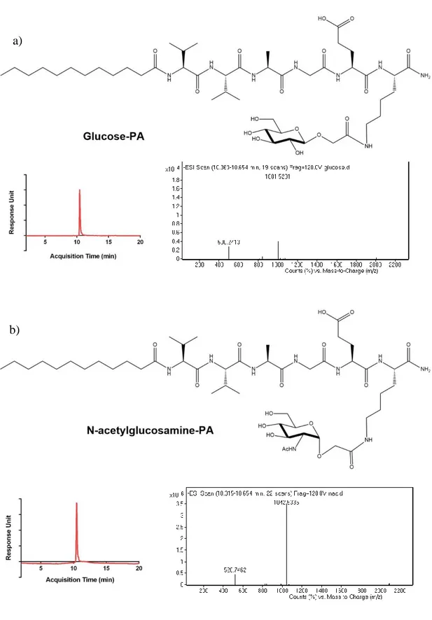
95:2.5:2.5). A similar synthetic procedure is applied for the synthesis of all other Glyco-PAs. Characterization of PAs was performed by using an Agilent liquid chromatography-mass spectrometry (LC-MS). The crude Glyco-PAs were purified by using An Agilent 1200 preparative reverse-phase HPLC 0.1% (NH4OH) in acetonitrile and water as mobile phase gradient. After the HPLC, acetonitrile was removed on rotary evaporator and then samples were lyophilized to get pure Glyco-PAs.
2.2.3. Physical and Chemical Characterization of Peptide Amphiphiles
2.2.3.1. Scanning Electron Microscopy (SEM) Imaging
Glycopeptide nanofibers were prepared by mixing monosaccharide PA and K-PA (at a 1:0.33 and 1:1 molar ratios) and, for sulfated systems, monosaccharide PA, SO3-PA and K-PA (at a 1:1:0.65 molar ratio). Mixed glycopeptide nanofibers (1 wt%) on silicon wafers were incubated at 37 ºC for 30 min and gradually dehydrated with 20, 40, 60, 80 and 100% ethanol. Samples were critical point-dried using an Autosandri-815B CPD equipment (Tousimis). Critical point-dried samples were sputter-coated with 9 nm of gold-palladium and imaged under a beam energy of 30 keV using an FEI Quanta 200 FEG SEM system.
2.2.3.2. Atomic Force Microscopy (AFM) Imaging
Glycopeptide nanofibers (under the same combinations for SEM imaging) were air-dried on glass slides and imaged under contact made using SiNi cantilevers using an Asylum Research MFP-3D atomic force microscope. A scan rate of 0.5 Hz was used for all images. 5 μm x 5 μm topographs were acquired for each sample at a pixel resolution of 512 x 512.
22
2.2.3.3. Circular Dichroism (CD) Analyses
A J-815 Jasco CD spectrophotometer was utilized to determine the secondary structures of peptide nanofibers at room temperature. Stock solutions of each PA were dissolved inside water at 1 mM concentration and mixed with oppositely charged K-PA. For CD spectroscopy measurements, peptide nanofibers were diluted to 0.25 µM. Measurements were carried out in the range of 300 nm to 190 nm with 100 nm/min scanning speed. Bandwidth was adjusted to 1 nm while data pitch and data interval were set as 0.1 nm. Digital integration time was chosen as 1 s at standard sensitivity. The molar ellipticity of each nanofiber system was calculated using three replicate measurements.
2.2.4. Affinity of Growth Factors to Peptide Nanofibers via ELISA- Binding Assay
The affinity of VEGF, FGF-2, and NGF to peptide nanofibers was analyzed using ELISA. Each glycopeptide amphiphile was mixed with positively-charged K-PA (C12 -VVAGK) (1:0.33 molar ratio of VEGF and FGF2 molecules and 1:1 molar ratio for NGF molecule) inside wells (Maxisorp, NUNC-Immuno plate, Thermo Scientific). Additionaly, glycopeptide – sulfonate peptide amphiphile (C12-VVAGE (K-p-sulfobenzoate) - K PA (1: 1: 0.65 molar ratio) combinations were prepared and tested against VEGF and FGF2. Wells were coated with peptide nanofibers (3 wells per each group) and dried overnight inside a laminar flow hood. Dried peptide nanofibers were washed thrice with wash buffer and tapped for drying. Assay buffer was added to each well for blocking. After the washing and drying of wells, growth factor was added to wells and incubated overnight at +4 ºC. On the next day, ELISA plate was incubated
23
at room temperature for 30 min and washed five times with wash buffer. After tapping and drying steps, a biotinylated antibody of growth factor was added to wells. Detection of biotinylated antibody was carried out with horseradish peroxide (HRP) conjugated streptavidin molecule. 3, 3’ 5, 5’– tetramethylbenzidine (TMB), the substrate of HRP, was added and incubated for 15-20 min. To finalize the reaction, stop solution (sulfuric acid) was added and the absorbance at 450 nm wavelength was measured by a microplate reader (Spectramax M5). Absorbance at 600 nm wavelength was taken as the reference point for calculations.
2.2.5. Live-Dead Assay- Cellular Viability Analyses
Biocompatibility of human umbilical vein endothelial (HUVEC) and PC-12 (ATCC® CRL-1721™) cells cultured on peptide nanofibers was tested by Live/Dead Assay. Peptide nanofibers were coated on 96 well plates (n=3). After 24 h and 48 h of incubation, plates were centrifuged at 2500 rpm for 5 min, washed with PBS and centrifuged at 2500 rpm for 1 min. Calcein-AM (which stains live cells) and ethidium homodimer-1 (which stains dead cells) were added to wells at concentrations recommended by the manufacturer. Five photos from each well for each group were taken by fluorescence microscopy (ZEISS Axio Observer A1) and counted using the Image J cell counter software.
2.2.6. Tube Formation Assay
In vitro tube formation assay was carried out by coating each glycopeptide - K-PA (1:0.33 molar ratio) and glycopeptide-sulfonate - K-PA (1:1:0.65 molar ratio) nanofibers on 96 well plates and drying peptide combinations overnight in a laminar flow hood. On the next day, HUVECs were trypsinized and counted with a
24
hemocytometer. 5 x 10⁴ cells/well were seeded inside 1% fetal bovine serum (FBS) containing low glucose DMEM and incubated for 36 h. Five photos from each well for each group were taken by ZEISS Axio Observer A1 microscope and the length of tubular structures was quantified by Image J software.
2.2.7. Neurite Extension Assay
Glycopeptide/K-PA (1:1 molar ratio) nanofibers were coated to well plates (3 wells for each group). After overnight drying, PC12 cells were seeded in expansion medium (RPMI with 10% horse serum (HS), 5% FBS and 1% penicillin/streptomycin (P/S)). On the next day, medium was changed with differentiation medium (MEM with 2% HS, 1% FBS, 1% P/S) with additional NGF (20 ng/mL). Five photos from each well for each group were taken by ZEISS Axio Observer A1 microscope at the end of the six-day incubation. Lengths of extended neurites were quantified by Image J software.
2.2.8. VEGF and FGF-2 Secretion Analysis
Secretion of VEGF and FGF-2 from HUVECs cultured on glycopeptide/K-PA (1: 0.33 molar ratio) and glycopeptide/sulfonate/K-PA (1: 1: 0.65 molar ratio) nanofiber systems were analyzed with ELISA at four-time points; 12 h, 24 h, 36 h and 48 h. VEGF and FGF-2 coating antibodies, AHG9119D (Invitrogen) and MAB233 (R&D Systems) respectively, were coated to Maxisorp ELISA plates and incubated overnight at +4 ºC. On the next day, blocking of wells was performed with assay buffer. For each time point, supernatants of each group were collected and added to antibody-coated wells and incubated overnight at +4 ºC. On the next day, biotinylated antibodies, streptavidin-HRP, TMB and stop solution were added to wells one by one.
25
Absorbances at 450 nm wavelength were read by taking the 600 nm wavelength as a reference point using a microplate reader (Spectramax M5).
2.2.9. Statistical Analysis
One-way analysis of variance (ANOVA) with Tukey’s post-tests was carried out for each experimental setting using GraphPad Prism 6 software. Standard errors of the means were represented in graphics. p < 0.05 was accepted as statistically significant.
26
2.3. Results and Discussion
2.3.1. Synthesis and Characterization of Glycopeptide Nanofibers
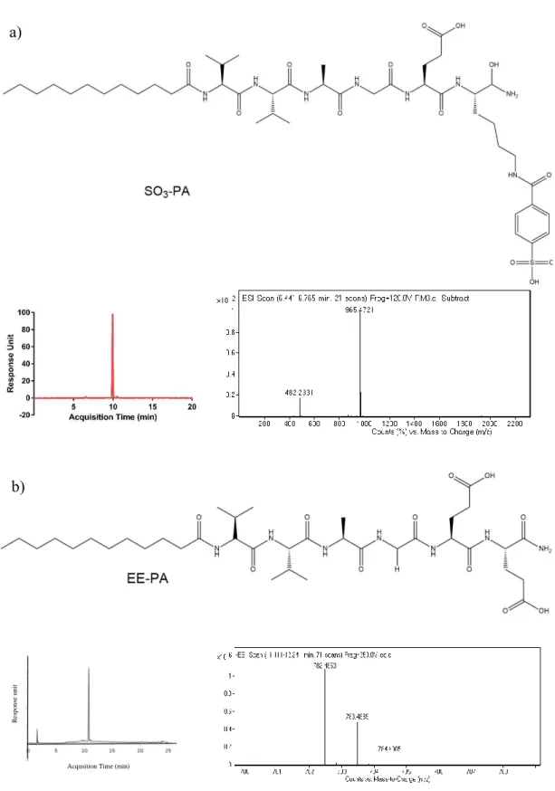
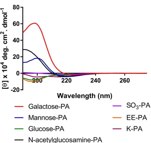
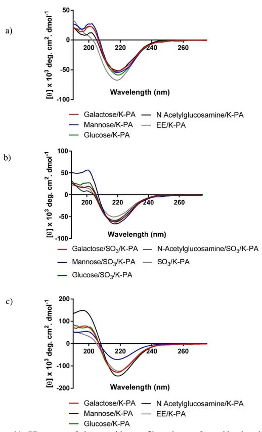
Glycopeptide amphiphiles (glucose, galactose, mannose and N-acetylglucosamine PAs), sulfonate-PA, E-PA, and K-PA were synthesized by solid phase peptide synthesis method and their purities were validated by LC-MS (Figure 6, 7, 8, 9). Each glycopeptide had different monosaccharide units and N-acetyl-glucosamine-PA was synthesized because one of the saccharide units of GAGs is an acetylated amino sugar. Additionally, sulfonate-PA-including combinations were designed in order to obtain much more similar structures to GAGs. E-PA was used as the non-bioactive PA control group since it does not have any saccharide group. Nanofiber networks were formed by mixing with oppositely-charged K-PA. Each peptide pH was adjusted to physiological pH (≈ 7.4) before the formation of the nanofiber networks. GAG-mimetic nanofibers were adjusted to have a highly negative charge to better imitate natural GAG networks through the addition of K-PA at a lower molarity. This system was used for VEGF and FGF-2 interaction studies. Since NGF does not have a heparin binding domain, the NGF affinity of glycopeptide and K-PA nanofiber systems was determined using peptide mixtures containing each component at the same molarity. All individual PA molecules were analyzed by CD spectroscopy in order to confirm that β-sheet structure is only observed following interaction with the oppositely charged K-PA, and we did not observe a β-sheet signal in this case (Figure 10). For each condition, the constituted nanofiber systems were analyzed by CD spectroscopy in order to determine their secondary structures. A positive peak around 200 nm and a corresponding negative peak around 220 nm are indicators of β-sheet formation, which is also a sign of nanofiber formation in each system (Figure 11). Self-assembled nanofibers were further visualized by AFM imaging, and height maps of each system
27
were found to indicate nanofiber formation (Figure 12). SEM imaging of glycopeptide nanofiber systems demonstrated an ECM-like porous fibrous network formation for each combination (Figure 13, 14).
28
Figure 6: Chemical structures and LC-MS analyses of a) galactose-PA and b) mannose-PA.
a)
29
Figure 7: Chemical structures and LC-MS analyses of a) glucose-PA and b) N-acetylglucosamine-PA
a)
30 b) a) R e spo n se uni t
Acqusition Time (min)
31 R e spo n se uni t
Acqusition Time (min)
Figure 9: Chemical structure and LC-MS analyses of K-PA.
32 b)
c) a)
Figure 11: CD spectra of glycopeptide nanofibers that are formed by the addition of the oppositely charged K-PA. a) Only glycopeptide and K-PA combinations; b) Glycopeptide/sulonfate PA and K-PA combinations and c) Equimolar glycopeptide/K-PA combinations that were analyzed for NGF binding
33
Galactose/K-PA Mannose/K-PA Glucose/K-PA
N-Acetylglucosamine/K-PA EE/K-PA
Galactose/K-PA Mannose/K-PA Glucose/K-PA
N-Acetylglucosamine/K-PA EE/K-PA SO3/K-PA
Figure 13: SEM imaging reveals the ECM-like nanofibrous, porous network of glycopeptide nanofibers. (Combinations for NGF binding)
34
Galactose/K-PA Mannose/K-PA Glucose/K-PA
N-Acetylglucosamine/K-PA EE/K-PA
Galactose/SO3/K-PA Mannose/SO3/K-PA Glucose/SO3/K-PA
N-Acetylglucosamine/SO3/K-PA SO3/K-PA
Figure 14: SEM imaging reveals the ECM-like nanofibrous, porous network of glycopeptide nanofibers. (Combinations for VEGF and FGF 2 binding)
35
2.3.2. Affinity Analysis of Growth Factors to Glycopeptides
The Affinity of glycopeptide nanofibers to VEGF, FGF-2 and NGF molecules were analyzed by ELISA method. Galactose and mannose nanofibers showed higher absorbance than glucose and N-acetylglucosamine nanofibers. However, EE/K-PA nanofiber (glutamic acid-bearing nanofiber) had a higher affinity to VEGF and FGF than non-mixed glycopeptide nanofibers. Since the EE/K-PA nanofıber has two carboxyl groups in its chemical structure, it is likely to be exhibit more electrostatic activity inside the buffer environment and effectively bear a greater negative charge than glycopeptides, although total charges of the nanofiber systems were equal. Since glycosaminoglycans consist of sulfated forms of sugars, SO3-PA was also incorporated into the nanofiber systems. The addition of sulfonate PA to glycopeptides significantly enhanced VEGF and FGF-2 binding to nanofibers. In particular, sulfonate-added galactose, mannose and glucose nanofibers bound to VEGF significantly higher than the N-acetylglucosamine- SO3 nanofiber (Figure 14a). It has been shown that VEGF interaction with HS is mediated through carboxylate groups and the 2-O, 6-O and N-sulfation of the HS chain [83]. Therefore, the lower affinity of N-acetylglucosamine-sulfonate nanofibers could result from the incompatibility of the spatiotemporal positions of sulfonate and N-acetylglucosamine groups on the surface of nanofibers for VEGF binding.
The addition of SO3-PA to glycopeptides also increased FGF-2 binding to mannose and glucose glycopeptides. However, SO3 PA-added galactose and N-acetylglucosamine glycopeptides showed the lower binding capacity to FGF-2 (Figure 14 b). Since total negative charge and length of HS is important for FGF-2 binding,
36
the assembly of mannose-sulfonate, glucose-sulfonate and sulfonate nanofiber combinations may present more negative epitopes on the surface of nanofibers.
Figure 15: a) VEGF affinity to glycopeptide and glycopeptide-sulphate nanofiber network. b) FGF-2 affinity to glycopeptide and glycopeptide-sulfonate nanofiber network. Values represent mean ±SEM (****p<0.0001).
37
The entire glycopeptide array was also investigated to determine whether monosaccharides had any affinity to NGF. Since NGF has moderate affinity against sulfated GAGs, only saccharide-bearing peptide nanofibers were investigated for the charge-neutralized combinations of glycopeptides and K-PA. ELISA results indicated that all monosaccharide bearing peptide nanofibers had high affinity to NGF (Figure 15). Therefore, glycopeptide nanofibers could be used as sustained NGF-secreting scaffolds for peripheral nerve regeneration studies.
2.3.3. Viability of Cells on Glycopeptide Nanofibers
Biocompatibility is among essential features of designed and synthesized scaffolds for tissue engineering studies. Besides their bioactive epitopes, constituents of peptide nanofibers are natural components of cells, as alkyl tails are composed of carbon atoms and small amino acid chains are naturally found in the ECM environment. All glycopeptide scaffolds including sulfonate-including ones provide a proper environment for the viability of HUVECs and PC-12 cells (Figure 17, 18, 19).
Figure 16: NGF affinity to glycopeptide nanofibers. Values represent mean ± SEM (****p<0.0001).
38
Galactose/K-PA Mannose/K-PA Glucose/K-PA
N-Acetylglucosamine/K-PA EE/K-PA
Galactose/SO3/K-PA Mannose/SO3/K-PA Glucose/SO3/K-PA
N-Acetylglucosamine/SO3/K-PA SO3/K-PA
Figure 17: Representative images of live-dead assay of HUVECs that are cultured on glycopeptide nanofibers. Green dots indicate live cells (calcein-AM) and red dots indicate dead cells (ethidium homodimer).
39
Galactose/K-PA Mannose/K-PA Glucose/K-PA
N-Acetylglucosamine/K-PA EE/K-PA PLL
Figure 19: Representative images of PC-12 cells and quantification of live dead assay.
Figure 18: Results of live-dead assay indicate that all glycopeptide nanofibers are biocompatible.
40
2.3.4. Glycopeptide-Sulfonate Nanofiber Networks Promote in vitro Tube-like Structures
Angiogenesis induction potential of glycopeptide nanofibers and glycopeptide-sulfonate nanofibers was further investigated via in vitro tube formation assay. HUVECs have the ability to form tube-like structures when they are stimulated with pro-angiogenic factors such as VEGF and FGF-2 [84]. As previously stated, GAGs consist of repeated disaccharide units modified with sulfonated or N-acetylated sugars. The glycopeptide nanofibers had less affinity to VEGF and FGF-2 than SO3-PA-added ones (Figure 15) and they did not form any tube-like structures (Figure 20 a). Although SO3-PA has a significantly higher affinity for VEGF and FGF-2, SO3 presence alone was not sufficient for tube formation. However, the combinations of glycopeptide-PAs and SO3-PA were able to induce the formation of tube-like structures without additional pro-angiogenic factors (Figure 20 b). Despite N-acetylated amino sugars and the oxidated form of glucose (a hydroxyl group that is converted into a carboxyl group) that are present in the structures of GAGs, galactose/SO3-PA and mannose/SO3-PA nanofibers demonstrated significantly higher tube length induction and form ordered tube-like structures to a greater extent than glucose/ SO3-PA and N-acetylglucosamine/ SO3-PA nanofibers. The presence of carboxyl groups in the structure of nanofiber networks is crucial for tube formation, as very few tube-like structures were observed in HUVECs cultured on EE/K-PA nanofibers, despite the higher affinity of this combination to VEGF and FGF-2. Therefore, we conclude that the existence of sulfate and carboxyl groups together with sugar units is indispensable for the coordinated formation of tubular structures in vitro.
41 a)
b)
Galactose/K-PA Mannose/K-PA Glucose/K-PA
N-Acetylglucosamine/K-PA SO3/K-PA
Galactose/SO3/K-PA Mannose/SO3/K-PA Glucose/SO3/K-PA
N-Acetylglucosamine/SO3/K-PA EE/K-PA c)
42
2.3.5. Glycopeptide Nanofibers Enhance Neurite Extension
Patterns of the NGF affinity of glycopeptides led us to investigate their effects on PC-12 cells, which are neuron-like and NGF-responsive. Previously, heparin-conjugated fibrin matrices were used to control the slow and sustained release of NGF. In these studies, heparin was assumed to immobilize NGF while fibrin matrices provided a porous, ECM-mimetic network for the encapsulation of NGF [42, 85]. In here, we demonstrated that glucose and N-acetylglucosamine bearing nanofibers significantly enhanced and supported neurite outgrowth without NGF encapsulation inside these nanofiber systems (Figure 20). The NGF-binding potential of glycopeptides could be used to support nerve regeneration and the additional encapsulation of NGF to these nanofibers could create a favorable environment for the sustained and slow release of NGF.
Figure 20: Brightfield microscope images of HUVECs cultured on nanofibers. Tube formation assay quantification, average length of formed tubular structures. Values represent mean ±SEM (***p<0.001).
43
N-Acetylglucosamine/K-PA EE/K-PA PLL Galactose/K-PA Mannose/K-PA Glucose/K-PA
Figure 21: Brightfield microscope images of PC-12 cells cultured on glycopeptide nanofibers and quantification of average neurite extension. Values represent mean ±SEM (**p<0.01).
44
2.3.6. Time-Dependent VEGF and FGF-2 Secretion Analyses of HUVECs Cultured on Glycopeptide-Sulfonate Nanofibers
Glycopeptide-sulfonate nanofibers promoted tube-like structures without any exogenous angiogenic factor addition to the environment. Therefore, we sought to determine whether the glycopeptide-sulfonate nanofibers were able to store endogenously secreted VEGF and present this growth factor to cells in a convenient manner. Time-dependent VEGF secretion from HUVECs was analyzed using an ELISA based method. Secreted VEGF concentration gradually increased in all glycopeptide-sulfonate groups until 36 h (Figure 23). This release pattern could be related to the autocrine signaling of VEGF, as tube-like structures were observed at the end of 36 hours. Galactose-sulfonate and mannose-sulfonate nanofibers, which formed more ordered tube-like structures and had higher average tube lengths at the end of 36 h, secreted the highest concentration of VEGF even compared to other glycopeptide-sulfonate nanofibers. At 48 h, VEGF concentration of galactose-sulfonate and mannose-galactose-sulfonate nanofibers decreased and came to a similar level with glucose-sulfonate and N-acetylglucosamine-sulfonate nanofibers. However, sulfonate and EE/K-PA control nanofibers exhibited a lower level of VEGF secretion throughout 48 h compared to glycopeptide-sulfonate nanofibers. Low levels of VEGF secretion could be due to a strong affinity between secreted VEGF from HUVECs and sulfonate and EE/K-PA nanofibers, and this concentrated binding may have inhibited the autocrine signaling of VEGF and induced the formation of few to no tube-like structures. Additionally, despite the presence of carboxyl and sulfonate groups, this combination does not resemble GAGs as much as the glycopeptide-sulfonate
45
nanofibers. Thus galactose-sulfonate and mannose-sulfonate nanofibers created much more GAG-mimetic nanostructures and efficiently enhanced in vitro tube formation. We also analyzed the FGF-2 release profiles of HUVECs cultured on glycopeptide-sulfonate nanofıbers (Figure 22). At each time point, there were no significant differences between all groups except FGF-2 release from HUVECs cultured on mannose-sulfonate nanofibers. These results suggest that VEGF binding to and release from glycopeptide-sulfonate nanofibers is the principal factor for directing tube formation.
Figure 22: FGF-2 release profiles of HUVECs cultured on glycopeptide-sulfonate nanofibers.
46
Figure 23: Time dependent release of VEGF secreted from HUVECs cultured on glycopeptide-sulfonate nanofibers. Values represent mean ±SEM (****p<0.0001).