Insulin Resistance and Serum Leptin Levels
in Men with Obstructive Sleep Apnea
Syndrome
ABSTRACT
Aim: The aim of this study was to assess the insulin resistance and serum leptin levels in patients with obstructive sleep apnea syndrome (OSAS), and to compare body mass indexes (BMI) of OSAS patients with matched controls without OSAS.
Method: Twenty eight patients having apnea-hypopnea index (AHI)≥5 included in the study. Thirty two healthy subjects assumed as a con-trol group. Venous blood was obtained in the fasting state for the measurement of glucose, insulin and leptin levels. Insulin resistance index was based on the homeostasis model assesment method (HOMA-IR).
Result: There was no significant difference in the serum leptin lev-els (control group, 32.88±24.22 ng/ml, OSAS group, 24.93±25.84 ng/ ml) and HOMA-IR (control group, 3.01±1.81, OSAS group, 2.58±1.21) between control group and OSAS patients. Insulin resistance and cir-culating plasma leptin concentrations in OSAS patients were indepen-dent of the AHI and were not different from the control group. Conclusion: We concluded that insulin resistance and plasma leptin concentrations are mostly associated with the degree of obesity and BMI. Those parameters seem not to be related with the AHI in OSAS patients.
Key words: Apnea hypopnea index, insulin resistance, leptin, OSAS Ankara Education and Research Hospital,
Department of Gastroenterology1 and
Nephrology2, Ankara, Turkey
Zonguldak Karaelmas University, Faculty of Medicine, Department of Pulmonology3,
Biochemistry6 and Endocrinology7,
Zongul-dak, Turkey.
Ankara Ataturk Education and Research Hospital, Department of Pulmonology4,
Ankara, Turkey.
19 Mayıs University, Faculty of Medicine, Department of Endocrinology5, Samsun,
Turkey.
Eur J Gen Med 2011;8(4):273-279 Received: 07.09.2011
Accepted: 03.11.2011
Correspondence: Tacettin Örnek, Zonguldak Karaelmas University. Faculty of Medicine. Department of Pulmonology. 67600, Zonguldak, Turkey
Tel: 0905363387324
E-mail: [email protected]
Erdem Koçak
1, Gülay Koçak
2, Tacettin Örnek
3, Hatice Bakırtaş
4, Hulusi Atmaca
5, Murat Can
6, Taner
INTRODUCTION
Obstructive sleep apnea syndrome (OSAS) is character-ized by repeated collapse of the pharynx during sleep, which leads to oxygen desaturation, fragmentation of sleep, and often daytime sleepiness (1). A high preva-lence of the condition, affecting approximately 2 to 4% of middle-aged adults, was found in an epidemiologic study (2). OSAS is well defined syndrome that includes one or two of the following symptoms: severe snoring, nocturnal respiratory arrest, repeated nocturnal awaken-ing, non-recuperative sleep, diurnal fatigue, and altered concentration. The respiratory responces are related to the extent of hypoxemia and hypercapnia that develop as a result of the disordered breathing (3). The diagnosis of this syndrome should be suspected on clinical evaluation and is confirmed by polysomnography. Extensive research related with obesity has shown that the location of body fat deposits rather than their size is more important in determining the risk of developing of obesity-linked dis-orders (4). It is well known that excess weight in adults is associated with increased incidence of hypertension, car-diovascular disease, stroke, insulin resistance and type 2 diabetes mellitus (5,6). Leptin is a circulating hormone that is expressed abundantly throughout the body specifi-cally in adipose tissue (7-9), although it is also secreted from other tissues including human placenta and stom-ach (10,11). Plasma leptin concentrations are increased in people who are obese in direct proportion to body fat mass (12). It has been reported that circulating plasma leptin levels are raised in men with newly diagnosed un-treated OSAS (13-14).
We have therefore aimed to evaluate the insülin resis-tance and serum leptin levels in patients with OSAS.
MATERIALS AND METHODS Subjects
Sixty male patients who had been referred to our hospi-tal, for suspected OSAS were evaluated. After an over-night sleep study 28 patients having apnea-hypopnea index (AHI)≥5 included in the study as obstructive sleep apnea group. Thirty two healthy subjects assumed as a control group. Exclusion criteria included the follow-ings: cardiopulmonary or vascular disease, hypertension (140/90 mmHg or on medication), chronic renal disease and diabetes mellitus. Subjects who smoked or had sys-temic infections at the time of the study were also ex-cluded. The following parameters were evaluated; age, body mass index (BMI), leptin, fasting glucose, insulin, insulin resistance and AHI. The average of two weight and height measurements were used to calculate BMI as weight (kg)/[height (m)]2. Leptin was determined in plasma using the DSL-10–23100 ACTIVE Human Leptin ELISA-kit (Diagnostic System Laboratories; Webster, TX). The sensitivity of this assay was 0.5ng/mL, and the terassay coefficient of variation was 4.6%. HOMA of in-sulin resistance; plasma glucose was measured by the glucose-oxidase method on a Beckman glucose analyzer (Beckman Instruments, Fullerton, CA). Serum insulin was measured by a double-antibody radioimmunoassay. The estimate of insulin resistance by HOMA ( HOMA-IR) was calculated with the formula fasting serum insulin (µU/ml) fasting plasma glucose (mmol/l)/22.5.
Study Procedures
All patients gave written informed consent to participate in the trial. The study protocol was approved by the uni-versity ethics committee, and the study was performed in accordance with the guidelines of the Declaration
Obstrüktif Uyku Apne Sendromu Olan Erkek Hastalarda İnsülin Rezistansı ve Serum Leptin Seviyeleri
Amaç: Bu çalışmanın amacı obstrüktif uyku apne sendromu (OUAS) olan hastalarda insülin rezistansı ve serum leptin seviyelerini incelemek ve OUAS olan hastaların vücut kitle indeksini (VCİ) kontrol grubuyla karşılaştırmaktır.
Metod: Apne hipopne indeksi (AHİ)≥5 olan 28 hasta çalışmaya dahil edildi. Sağlıklı 32 birey kontrol grubu olarak kullanıldı. Çalışmaya katılan bireylerden glukoz, insülin ve leptin seviyelerini ölçmek için açlık durumunda venöz kan elde edildi. İnsülin rezistans indeksinde, hemostaz modeli analiz methodu (HOMA-IR) temel alındı.
Bulgular: OUAS grubu ile kontrol grubu arasında serum leptin seviyeleri (kontrol grubu, 32.88 ± 24.22 ng/ml, OUAS grubu, 24.93 ± 25.84 ng/ml) ve HOMA-IR (kontrol grubu, 3.01 ± 1.81, OUAS grubu, 2.58 ± 1.21) açısından anlamlı bir fark yoktu. OUAS olan hastalarda insülin rezistansı ve plazma leptin konsantrasyonları AHİ’den bağımsızdı ve kontrol grubundan farklı değildi.
Sonuç: İnsülin rezistansı ve plazma leptin konsantrasyonları sıklıkla VKİ ve obesitenin derecesiyle ilişkili olduğunu düşünüyoruz. Bu parametreler OUAS olan hastalarda AHİ ile ilişkili olmadığı gözükmektedr.
of Helsinki and its current revision.An overnight poly-somnography in the sleep laboratory was performed. Blood was drawn at 8 am after an overnight fast for the determination of multiple clinical chemistry param-eters. Polysomnography (Somnostar alpha) was started at 21.00 hours and ended at 06.30 hours. Surface elec-trodes were applied using standard techniques to obtain an electroencephalogram, an electromyogram of the chin, an electrocardiogram, and an electrooculogram. Ventilation was monitored by inductive plethysmogra-phy. Airflow was monitored by thermistors placed at the nose and mouth, while arterial oxygen saturation (SaO2) was monitored continuously with a pulse oximeter . A polygraph was run continuously at 10 mm/s to record all of the above physiological data simultaneously through-out the course of the experiment. All parameters were stored in a data recorder for subsequent analysis. Apnea was defined as the cessation of airflow at the nose and mouth lasting for more than 10 seconds. Hypopnoea was defined as a decrease of 50% or more in thoracoabdomi-nal motion associated with a fall in the baseline oxygen saturation of 4% or more. All AHI values were calculated to express the number of episodes of apnoea and hypop-noea per hour of total sleep time. AHI<5 were excluded from study. Patients with sleep disorders, except OSAS, such as upper airway resistance syndrome, periodic leg movements or narcolepsy were excluded. OSAS was de-fined as the combination of an AHI of 5 or more events/h with daytime sleepiness.
Statistical Analysis
Results are expressed as mean ± standard error. We used an unpaired two-sided Student's t-test to analyse any differences in demographic and haemodynamic char-acteristics between these two groups. In correlation analysis Pearson correlation was used. All statical analy-sis performed with SPSS 11.0 programe. Statistical sig-nificance was defined as p < 0.05.
RESULTS
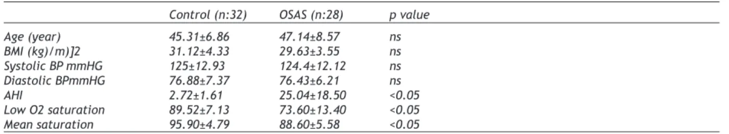
Table 1 shows the main clinical characteristics and poli-somnographic data of all subjects. There was no signifi-cant differences between control and OSAS group with respect to age, BMI, blood pressure. Fasting glucose, insulin, leptin levels and HOMA-IR were examined in whole groups. There was no significant differences be-tween control and OSAS group with respect to fasting glucose, insulin, leptin levels and HOMA-IR (Table 2). To examine the relationship between apne-hipopnea in-dex, serum leptin levels and HOMA-IR, OSAS group were diveded into two subgroups; AHI 5-20, AHI ≥ 20. Table 3 shows the comparision of fasting glucose, insulin, leptin levels, HOMA-IR and the main clinical characteristics in those control and OSAS subgoups. There was no sig-nificant differences between OSAS subgroup1 and OSAS subgroup 2 with respect to HOMA-IR, serum leptin, insu-lin and fasting glucose expect AHI index.
Table 1. Comparison of sample characteristics in those control and OSAS group Control (n:32) OSAS (n:28) p value
Age (year) 45.31±6.86 47.14±8.57 ns BMI (kg)/m)]2 31.12±4.33 29.63±3.55 ns Systolic BP mmHG 125±12.93 124.4±12.12 ns Diastolic BPmmHG 76.88±7.37 76.43±6.21 ns AHI 2.72±1.61 25.04±18.50 <0.05 Low O2 saturation 89.52±7.13 73.60±13.40 <0.05 Mean saturation 95.90±4.79 88.60±5.58 <0.05 ns: nonsignificant
Table 2. Comparison of fasting glucose, insulin, leptin levels and HOMA-IR in those control and OSAS group Control (n:32) OSAS (n:28) p value
Fasting glucose (mg/dl) 101.75±11.92 96.72±14.25 ns
Insulin (uIU/ml) 10.36±4.35 12.59±7.56 ns
Leptin (ng/ml) 24.93±25.84 32.88±24.22 ns
To determine whether the degree of BMI was related to serum leptin levels and HOMA-IR, the subjects were seperated into two groups with respect to BMI; BMI < 30 (kg/m)2 ( group 1), BMI ≥ 30 (kg/m)2 (group 2). Serum leptin levels and HOMA-IR were higher in group 2. Table 4 shows the comparision of fasting glucose, insulin, leptin levels, HOMA-IR and the main clinical character-istics in those group 1 and group 2.
DISCUSSION
In the present study we found that serum leptin lev-els and insulin resistance in male patients with OSAS are not associated with AHI. Furthermore, as expected serum leptin levels and insulin resistance were showed significant correlation with obesity. The results of pu-plished data on the relationship between OSAS, insulin resistance and leptin levels are conflicting.
Leptin is a hormone that reduce food receiving and raise consumption of energy by inhibiting hypothalamic NPY synthesis. Serum leptin levels were increased because of the leptin resistance in obese individuals (15,16). And also previous reports had shown that serum leptin
lev-els are higher in patients with OSAS than simple obese paitents (17,18). TNF-α and IL-6 levels in patients with OSAS were significantly higher because of high levels of serum leptin and it has been suggested that high levels of these inflammatory markers were contributed to in-flammation in the upper airway (19). Ozturk et al was found that there was a significant realtionship between serum leptin levels and degree of OSAS. And they also showed that this relationship was independent from age and BMI (20). In patients with OSAS serum leptin lev-els are decreasing distinctly after the CPAP treatment with a correlation of improvement in AHI levels (21,22). Nonetheless Schafer et al was showed that leptin con-centrations when controlled for body fat are not related to the degree of OSAS (23). In Barcelo’s study serum leptin levels in patients with OSAS was related to obe-sity (24). Recently Kapsimalis et al suggested that cen-tral obesity, which reflects visceral obesity has a major effect on leptin levels and the effect of apne –related hypoxemia maybe smaller (25). In our study we found no correlation between serum leptin levels and AHI. Obesity, particularly abdominal obesity, is associated with resistance to the effects of insulin on peripheral
Table 3. Comparison of fasting glucose, insulin, leptin levels, HOMA-IR and clinical characteristics in those con-trol and OSAS subgroups.
Control (n :32) Subgroup 1 Subgroup 2 p value AHI 5-20 (n:15) AHI ≥20 (n:13) Age (year) 45.31±6.86 48.0±7.99 46.0±8.23 ns BMI (kg)/m)]2 31.12±4.33 30.48±4.42 29.85±2.74 ns AHI 2.72±1.61 12.4±5.48 41.83±21.07 <0.05 Insulin (uIU/ml) 12.59±1.33 9.04±2.70 12.11±5.76 ns Fasting glucose (mg/dl) 98.72±14.25 101.88±11.50 101.58±12.15 ns Leptin ( ng/ml) 32.88±24.22 21.34±18.91 29.7±33.88 ns HOMA-IR 3.01±1.81 2.21±0.67 3.07±0.47 ns
Table 4. Comparison of fasting glucose, insulin, leptin levels, HOMA-IR and clinical characteristics in those group 1 and group 2.
BMI<30kg/m² BMI ≥30kg/m² p value
group 1 (n:28 ) group 2 (n:32) İnsulin (uIU/ml) 9.1±4.96 13.6±6.69 <0.05 Leptin ( ng/ml) 21.95±19.83 35.49±27.71 <0.05 HOMA-IR 2.28±1.31 3.27±1.64 <0.05 Fasting glucose (mg/dl) 102.25±15.87 96,28±10.12 ns Age (year) 44.5±8.0 4 7.5±7.24 ns BMI (kg/m²) 27.31±1.80 33.16±3.40 <0.05 AHI 13.25±20.14 13.03±15.83 ns
glucose and fatty acid utilization, often leading to type 2 diabetes mellitus. The syndromes of insulin resistance actually make up a broad clinical spectrum, which in-cludes obesity, glucose intolerance, diabetes, and the metabolic syndrome, as well as an extreme insulin-resistant state. In obese patients, down regulation of insulin receptor leads to insulin resistance and hyperin-sulinemi (26). And previous reports have shown a linked relationship between OSAS and obesity (27). Conflicting results have been reported on the potential link be-tween OSAS and insulin resistance. According to some studies including obese patients with OSAS, significant insulin resistance was determined (28,29). And also sig-nificant relationship between nocturnal hypoxemia, AHI and insulin resistance regardless of age and BMI have been reported in OSAS patients (29,30).
In patients with OSAS, it has been suggested that the presence of other mechanisms might be lead to insulin resistance with regardless of BMI and body fat. A lot of mechanisms that support the relationship between OSAS and insulin resistance have been considered but the in-vestigators were focused on three important reasons. Firstly, in OSAS patients, hypoxia continued throughout the night was lead to elevated blood catecholamine lev-els and activated sympathetic system (31). Second, hy-poxia occured during the night as a stres factor was lead to an increase of glucocorticoid secretion (32), Third, it has been shown that hypoxia alone contributes insulin resistance in patients with chronic pulmonary disease (33).
In Rajala’s study increasing per unit of BMI leads four times respiratory impairment (34). In many studies, the prevalence of respiratory impairment reached about 40% in exceedingly obese patients (35,36). In a study which performed by Stoohs and collegues, there was a significant relationship with insulin resistance and respi-ratory impairment in sleep but they also suggested that this relationship was entirely dependent on body mass (36). A similar study, Somers and collegues didn’t found any relationship between insulin resistance and sleep disorders (37). On the contrary in another study which enrolled 261 male patient, was showed a significant re-lationship beetween sleep apnea and insulin levels with regardless of BMI (38).
Up to date, reviewing the literature, conflicting results have been reported between leptin levels, insulin re-sistance and OSAS. Some deficiencies and limitations
are observed in these studies which include followings; small number of patients, no gender discrimination, the patients which had systemic disease such as DM, hyper-tension were comprised the study, no assesment of ratio of body fat. For this reason in the present study we en-rolled only male individuals to standardize the differ-ences of results and also we excluded the patients who have systemic diseases.
In the present study, we evaluated the association be-tween serum leptin levels, insulin resistance and apnea-hypopnea index; however no correlation was found. So that in order to prove the accuracy of serum leptin lev-els and insulin resistance, the subjects were seperated into two groups with respect to BMI. As expected insulin resistance and plasma leptin concentrations are mostly associated with the degree of obesity.
In the literature different results had been reported. What is the potential mechanism which lead to differ-ent results between OSAS, leptin levels and insülin re-sistance? The distrubition of fat to the upper body does seem to be more specific marker of the health hazards of obesity than overweight alone. Lara et al showed that different fat patterns for each sex, changes with age in body fat distribution, and different usefulness of external anthropometric measures in males and fe-males to predict fat deposits and their distribution at the abdominal level (39). Furthermore Jensen et al con-cludede that a single-slice CT scan (or other imaging technique) with or without DXA is required for accurate predictions of intraabdominal fat (40). These conflicting results appear to suggest that BMI alone is a poor marker of body fat deposits and it seems that BMI alone did not correctly reflect the amount of viseral obesity.
In conclusion, in this study we found that in OSAS pa-tients serum leptin levels and insulin resistance was as-sociated with BMI but rather not asas-sociated with apnea-hipopnea index. To analyse the relationship with OSAS, insulin resistance and serum leptin levels, further inves-tigations are needed. Future studies should be included larger population and the factors which affect the body fat deposits should be evaluated both invasive and non-invasive methods.
REFERENCES
1. Guilleminault C. Clinical features and evaluation of ob-structive sleep apnea, In: Kryger MH, Roth T, Dement WC. (Eds.). Principles and practice of sleep medicine, W.B. Saunders, Philadelphia, 1994
2. Young T, Palta M, Dempsey J, Skatrud J, Weber S., Badr S. The occurrence of sleep-disordered breathing among middle-aged adults. N Engl J Med 1993;328:1230-5 3. Peled N, Greenberg A, Pillar G Zinder O, Levi N, Lavie
P. Contributions of hypoxia and respiratory disturbance index to sympathetic activation and blood pressure in ob-structive sleep apnea syndrome. Am J Hypertens 1998; 11:1284–9
4. Bjorntorp P. Body fat distribution, insulin resistance, and metabolic diseases. Nutrition 1997;13:795–803
5. Must A, Spadano J, Coakley EH, Field AE, Colditz G, Dietz WH. The disease burden associated with overweight and obesity. JAMA 1999;282:1523-9
6. Pi-Sunyer FX. Medical hazards of obesity. Ann Intern Med 1993;119:655-60
7. Auwerx J, Staels B. Leptin. Lancet 1998;351:737–42 8. Considine RV, Sinha MK, Heiman ML, et al. Serum
immu-noreactive-leptin concentrations in normal-weight and obese humans. N Engl J Med 1996;334:292–5
9. Masuzaki H, Ogawa Y, Isse N, et al. Human obese gene expression, Adipocyte-specific expression and regional differences in the adipose tissue. Diabetes 1995;44:855–8 10. Masuzaki H, Ogawa Y, Sagawa N, et al. Nonadipose tissue
production of leptin: leptin as a novel placenta-derived hormone in humans. Nature Med 1997;100:1029–33 11. Sobhani I, Bado A, Vissuzaine C, et al. Leptin secretion
and leptin receptor in the human stomach. Gut 2000;47: 178–83
12. Rosenbaum M, Nicolson M, Hirsch J, et al. Effects of gender, body composition and menopause on plasma con-centrations of leptin. J Clin Endocrinol Metab 1996;81: 3424-7
13. Ip MS, Lam KS, Ho C, Tsang KW, Lam W. Serum leptin and vascular risk factors in obstructive sleep apnea. Chest 2000;118:580–6
14. Phillips BG, Kato M, Narkiewiczk K, Choe I, Somers WK. Increases in leptin levels, sympathetic drive, and weight gain in obstructive sleep apnea. Am J Physiol Heart Circ Physiol 2000;279:H234–7
15. Walder K, Lewandowski P, Morton G, et al. Leptin resis-tance in a polygenic, hyperleptinemic animal model of obesity and NIDDM: Psammomys Obesus. Int J Obes 1999; 23:83-9
16. Fitzpatrick M. Leptin end the obesity hypoventilation syndrome: A leap of faith? Thorax 2002;57:1–2
17. Zirlik S, Hauck T, Fuchs FS, et al. Leptin, Obestatin and Apelin levels in patients with obstructive sleep apnoea syndrome. Med Sci Monit 2011; 17: CR159-64
18. Tokuda F, Sando Y, Matsui H, Koike H, Yokoyama T. Serum
levels of adipocytokines, adiponectin and leptin, in pa-tients with obstructive sleep apnea syndrome. Inter Med 2008;47:1843-9
19. Vgontzas AN, Papanicolaou DA, Bixler EO et al. Sleep ap-nea and daytime sleepiness and fatigue: Relation to vis-ceral obesity, insulin resistance, and hypercytokinemia. J Clin Endocrinol Metab 2000;85:1151–8
20. Ozturk L, Unal M, Tamer L, Celikoglu F. The association of the severity of obstructive sleep apnea with plasma leptin levels. Arch Otolaryngol Head Neck Surg 2003;129: 538-40
21. Sanner BM, Kollhosser P, Buechner N, Zidek W, Tepel M. Influence of treatment on leptin levels in patients with obstructive sleep apnea. Eur Respir J 2004;23:601-4 22. Harsch IA, Konturek PC, Koebnick C, et al. Leptin and
ghrelin levels in patients with obstructive sleep apnoea: effect of CPAP treatment. Eur Respir J 2003;22:251-7 23. Schafer H, Pauleit D, Sudhop T, Gouni-Berthold I, Ewig
S, Berthold HK. Body fat distribution, serum Leptin, and cardiovascular risk factors in men with obstructive sleep apne. Chest 2002;122:829-39
24. Barceló A, Barbé F, LIompart E, et al. Neuropeptide Y and Leptin in Patients with Obstructive Sleep Apnea Syndrome: Role of Obesity. Am J Respir Crit Care Med 2004;171:183-7
25. Kapsimalis F, Varouchakis G, Manousaki A, et al. Association of sleep apnea severity and obesity with in-sulin resistance, C-Reactive Protein, and Leptin levels in male patients with obstructive sleep apnea. Lung 2008; 186: 209-17
26. Yki-Järvinen H. Glucose toxicity. Endocrine Reviews 1992; 3:415-31
27. Banno K, Walld R, Kryger MH. Increasing obesity trends in patients with sleep-disordered breathing referred to a sleep disorders center. J Clin Sleep Med 2005;3:364-6 28. Tassone F, Lanfranco F, Gianotti L, et al. Obstructive
sleep apnoea syndrome impairs insulin sensitivity inde-pendently of anthropometric variables. Clin Endocrinol 2003;59:374-6
29. Punjabi NM, Sorkin JD, Katzel LI, Goldberg AP, Schwartz AR, Smith PL. Sleep-disordered breathing and insulin re-sistance in middle-aged and overweight men. Am J Respir Crit Care Med 2002;165:677-82
30. Ip MS, Lam B, Ng MM, Lam WK, Tsang KW, Lam KS. Obstructive sleep apnea is independently associated with insulin resistance. Am J Respir Crit Care Med 2002;165: 670-6
31. Leproult R, Copinschi G, Buxton O, Van Cauter E. Sleep loss results in an elavation of cortisol levels the next eve-ning. Sleep 1997;20:865-70
32. Spiegel K, Leproult R, Van Cauter E. Impact of sleep debt on matabolic and endocrine function. Lancet 1999;354: 1435-9
33. Hjalmarsen A, Aasebø U, Birkeland K, Sager G, Jorde R. Impaired glucose tolerance in patients with chronic hipoxic pulmonary disease. Diabetes Metab 1996;22:37-42
34. Rajala R, Partinen M, Sane T, Pelkonen R, Huikuri K. Seppäläinen AM. Obstructive sleep apnoea syndrome in morbidly obese patients. J Intern Med 1991;230:125-9 35. Richman RM, Elliott LM, Burns CM, Bearpark HM,
Steinbeck KS, Caterson ID. The prevalence of obstructive sleep apnea in an obese female population. Int J Obes Relat Metab Disord 1994;18:173-7
36. Stoohs RA, Facchini F, Guilleminault C. Insulin resistance and sleep-disordered breathing in healthy humans. Am J Respir Crit Care Med 1996;154:170-4
37. Somers VK, Dyken ME, Clary MP, Abbound FM. Sympathetic neural mechanism in obstructive sleep apnea. J Clin Invest 1995;96:1897-904
38. Sharma SK, Kumpawat S, Coel A, Banga A, Ramakrishnan L, Chaturvedi P. Obesity, and not obstructive sleep apnea, is responsible for metabolic abnormalities in a cohort with sleepdisordered breathing. Sleep Med 2008;8:12–7 39. Lara Fernández A, Escolar Castellón JL, Aguilar Cuevas R
Fernández Ruiz A, Lara Fernández AL, González Santos P. Obesity and distribution of body fat. Correlation between anthropometric and tomographic data on areas at the ab-dominal level. Rev Clin Esp 1996;196:437-45
40. Jensen MD, Kanaley JA, Reed JE, Sheedy PF. Measurement of abdominal and visceral fat with computed tomogra-phy and dual-energy x-ray absorptiometry. Am J Clin Nutr 1995;61:274-8