Abstract
The central giant cell granuloma (CGCG) is benign, nonodontogenic, and intraosseous lesion of the jaw. Aggressive subtypes of CGCG have a tendency to recur after excision and require wide resection that leads to major defects in the jaw. In this case report a patient who had severe mandibular bony deficiency as a result of excision of aggressive CGCG, orthodontic, and prosthetic treatment was described. The defect was reconstructed with iliac bone graft. Four years later vertical distraction osteogenesis was performed on the grafted mandible in order to obtain a satisfactory bony height of mandibular ridge. After healing period three endosseous dental implants were placed to grafted region. Because of pubertal growth stage, a hybrid removable denture was constructed. The construction of a hybrid removable denture markedly improved the patient’s speech, mastication, and appearance. After pubertal growth stage, a fixed partial denture construction was planned and future parts of treatment procedures were described to the patient. Distraction osteogenesis and endosseous dental implants can be a good alternative method for the unsatisfactory reconstructions of mandibular deficiencies.
Key words: Dental implant, giant cell granuloma, hybrid prosthesis Date of Acceptance: 16‑Sep‑2013
Address for correspondence: Dr. Emir Yüzbaşıoğlu,
İstanbul Medipol University School of Dentistry, Department of Prosthodontics, Atatürk Bulvarı No: 27, Unkapanı, 34083, Fatih‑İstanbul, Turkey.
E‑mail: eyuzbasioglu@medipol. edu.tr
Introduction
The giant cell granuloma was identified as a clinical entity by Jaffe in 1953. This lesion is characterized by proliferation of fibroblasts and multinucleated giant cells, in a densely packed stroma.[1] Central giant cell granuloma (CGCG) (e.g., giant cell
granuloma of the mandible and maxilla bones) is less common than peripheral giant cell granuloma of the extremities.[2]
CGCG is considered to be a benign intraosseous jaw lesion. The World Health Organization (WHO) has defined it as an intraosseous lesion consisting of cellular fibrous tissue that contains multiple foci of hemorrhage, aggregations of multinucleated giant cells, and occasionally, trabeculae of woven bone.[3]
CGCG is an uncommon, benign, and proliferative lesion accounting for less than 7% of all benign jaw lesions whose etiology is not defined.[4‑6] But the occurrence of CGCG
in the jaws of patients with known genetic diseases, such as neurofibromatosis type 1, cherubism, and Noonan syndrome, indicates that a genetic‑related etiology might be possible.[7] Several have hypothesized that the onset
of the abnormalities in cherubism and CGCG and their location‑restricted presentation may be related to dental developmental processes in children.[8,9] The relation
between neurofibromatosis type 1 and giant cell lesions is not clear. There are only a few reports in the literature describing a patient with neurofibromatosis type 1 and CGCG.[10‑12]
Multidisciplinary approach for the rehabilitation of
central giant cell granuloma: A clinical report
Emir Yüzbaşıoğlu, Alper Alkan1, Mete Özer2, Mehmet Bayram3
Department of Prosthodontics, School of Dentistry, İstanbul Medipol University, İstanbul, 1Department of Oral and
Maxillofacial Surgery, Faculty of Dentistry, Erciyes University, Kayseri,
2Department of Orthodontics, Faculty of Dentistry, Ondokuz Mayıs University,
Samsun, 3Karadeniz Technical University, Trabzon, Turkey
Access this article online Quick Response Code:
Website: www.njcponline.com DOI: 10.4103/1119-3077.134060 PMID: *******
529
CGCG only rarely present in patients with a Noonan genotype (mutation in PTPN11), an neurofibromatosis type 1 genotype, or an neurofibromatosis‑Noonan syndrome (NFNS) phenotype. It is suggested that for the development of CGCG in patients with the Noonan‑like/ multiple giant‑cell lesion syndrome a second hit might be needed either in the same gene or in other genes involved in the same pathway.[13] Manor et al., also investigated
cytogenetic characteristics of CGCG and reported that some chromosomal changes.[14]
CGCG usually occurs predominantly in children and young adults, with more than 60% of all cases occurring in the first 3 decades of life and has a distinct sex predilection, with a female to male ratio of 2:1.[15‑17] Although most
CGCGs of the jaws are slow growing, circumscribed lesions that respond well to simple curettage, a smaller number demonstrate aggressive clinical behavior.
The lesion has frequently been reported to be confined to the tooth‑bearing areas of the jaws. Lesions occur more frequently in the mandible than in the maxilla is more common in the anterior region of the jaws, and mandibular lesions frequently extend across the midline.[18,19]
The radiologic features of the CGCG have not been clearly defined, and conflicting descriptions appear in the literature. Radiographically, CGCG present as radiolucent defects, which may be unilocular or multilocular with well‑defined or ill‑defined margins and varying degrees of expansion of the cortical plates.[20] In some cases teeth displacement can
be found.
The radiographic findings are not specifically diagnostic. It is important to remember that the radiologic appearance of the lesion is not pathognomonic and may be confused with that of many other lesions of the jaws.[21,22] Small unilocular lesions
can be confused with periapical cysts, and multilocular giant cell lesions cannot be distinguished radiographically from ameloblastomas or other multilocular lesions.[17,23]
Aggressive local curettage is regarded as the treatment choice for traditional treatment of the CGCG.[23‑25]
Resection is performed for recurrent or more aggressive variants, which leads to major defects and loss of teeth. However, the extent of tissue removal ranges from simple curettage to en bloc resection. This is particularly mutilating in a growing child or young adult. CGCG can prove difficult to treat either because of its large size, the difficulty of extensive curettage in the fragile facial bones, or because of the risk to facial growth centers and developing teeth.[26]
In such cases, extensive reconstructive procedures are required for anatomic restoration and rehabilitation to achieve satisfactory form and function. Curettage has also been supplemented with cryosurgery[27] and peripheral
ostectomy.[24]
In the past few years a number of alternative nonsurgical therapies have been described. CGCG has also been treated by nonsurgical methods such as radiotherapy,[24] daily
systemic administration of calcitonin,[26,28‑30] intralesional
injection with corticosteroids,[31,32] subcutaneous interferon
injections[33] and intralesional injection with combined
bisphosphonates and corticosteroids.[34]
In this case report, it was described that a patient who had a severe mandibular bony deficiency as a result of excision of aggressive CGCG in which distraction osteogenesis techniques were used to improve the alveolar ridge vertical dimension and dental implants and orthodontic treatment used for to achieve satisfactory results for an esthetic and functional rehabilitation. The reason of preferability of distraction osteogenesis is achieving sufficient bone volume for further restorative procedures. Guided bone regeneration techniques are not predictable for this case to achieve optimal esthetic and functional results. A hybrid removable denture was selected for restorative option, because of pubertal growth stage, the restorative phase of treatment will be modified in several steps.
Case Report
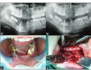
A 9‑year‑old female patient was referred for treatment at the Department of Oral and Maxillofacial Surgery, Faculty of Dentistry, Ondokuz Mayıs University, Samsun, Turkey, with a complaint of mobility of the mandibular incisors and swelling of the adjacent vestibular mucosa [Figure 1]. Radiological examination showed that a well‑defined radiolucent area at the mandibular symphysis extending from tooth no. 33 to tooth no. 46 [Figure 4a]. The lesion was associated with permanent tooth germs and the affected tooth roots were partially resorbed. There was only a thin layer of mandibular basal bone [Figure 2a]. The diagnosis of CGCG was made on clinical and radiological results. Intraoral examination revealed swelling of the right buccal cortical plate, overlaying mucosa was normal in appearance. The patient was in mixed dentition and there were some carious lesions in both permanent and decideous dentition. There was crowding in teeth in association with the lesion. There was no paresthesia of the lower lip, and all teeth in the area of expansion tested vital. Extraoral examination revealed palpation of the expanded bone.
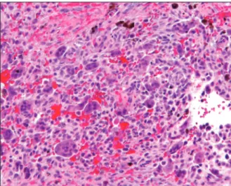
An incisional biopsy confirmed a CGCG which has a typical histological appearance [Figure 3]. Hematological tests were performed for evaluation of possibilities for hyperparathyroidism (serum calcium, phosphate, parathormone, and alkaline phosphatase levels), and brown tumor. The differential diagnosis included fibrous dysplasia, cherubism, aneurasmal bone cyst, giant cell tumor of the long bones, neurofibromatosis type I, cherubism, and
Noonan syndrome. These conditions ruled out by clinical, radiological, and laboratory tests of calcium and PTH levels. There are no predisposing factors in medical history which could generate a genetic disorder in her family.
Surgical treatment was preferred as treatment option and the lesion was aggressively currettaged under general anesthesia via an intraoral approach. A thin layer of cortical bone was left at the lower border of the mandible. There was no intact bone at the vestibule and lingual cortex. The affected region of mandible was resected protecting the inferior alveolar nerve though to mandibular basis. For reconstruction of lesion region an anterior iliac corticocancellous bone graft was harvested and fixed to the basal bone with a reconstruction plate [Figure 2b]. The patient was monitored by regular follow‑up until healing was complete. The reconstruction plate was removed 1 year later [Figure 4b] and the patient monitored both clinically and radiographically for 4 years. There were no sensory deficiencies during follow‑up period. Significant resorption of the grafted bone was observed 4 years later [Figure 4c and d].
Morphology and bone height of the anterior mandible was unable to perform a sufficient functional and esthetic
rehabilitation for patient. To obtain satisfactory bony height of the mandibular ridge vertical distraction osteogenesis was planned. Before vertical distraction operation fixed orthodontic treatment applied to the patient for leveling and alignment of both dental arches for satisfactory prosthetic rehabilitation, proper function, and esthetics.
After the orthodontic treatment, an intraoral bidirectional vertical distraction device (Modus ARS 1.5; Medartis, Basel, Switzerland) was placed under general anesthesia for vertical alveolar augmentation [Figure 5]. The distraction protocol included 7 days of latency after surgery and a distraction rate of 1 mm per day. The bone was distracted by about 13 mm [Figure 6a and b]. The patient followed‑up for 3 months by clinical evaluation, panoramic, and lateral cephalometric radiographs. After 3 months later, the distractor was removed under local anesthesia [Figure 6c and d]. During surgical procedures and healing period, orthodontic treatment was performed both mixed and permanent dentitions to obtain satisfactory alignments and inclinations.
Three dental implants (SwissPlus Tapered 4.1 10 mm implant, Zimmer Dental, USA) were placed to mandibular anterior edentulous region during distractor removal operation. The dental implants followed for 3 months by clinical evaluation and periapical radiographs. After
Figure 1: Pretreatment extraoral and intraoral situation
Figure 2: Pretreatment panoramic radiograph and intraoperative photograph of the grafted bone after curettage of the lesion
b a
Figure 3: Histopathological appearance of lesion
Figure 4: Posttreatment panoramic radiograph after first and second surgery. Radiologic and clinical views of the bone
remodeling after 4 years
d c
b a
531
6 months and completing osseointegration of the three implants [Figure 6a‑b], the definitive impressions of implants were made with an individual tray with polyether impression material (Impregum, 3M Espe, USA) by using open tray technique. A centric relation record was obtained with record using an interocclusal registration material (OccluFast, Zermack, Italy).
Because of patients pubertal growth period, implant supported fixed partial denture reconstruction was not preferred. A hybrid removable denture was planned during this period [Figure 7a‑c]. Three titanium milling abutments connected to implant replicas and milled in surveyor to obtain parallelism and single path of insertion of hybrid removable denture. Two secondary copings were casted for two abutments. The removable dentures were constructed in traditional techniques and the copings connected to acrylic denture base.
Because of inclination of the third implant which is adjacent to mandibular left canine tooth, secondary coping was not constructed [Figure 7d]. The buccal flange of denture will be very thick and the grayish color is markedly visible. For an esthetic appearance and comfortable function removable, denture was constructed with two copings. After pubertal growth stage, a fixed partial denture construction was planned and future parts of treatment procedures were described to the patient [Figure 8 and 9].
Discussion
CGCGs are benign but occasionally aggressive lesions.[16]
Aggressive form is characterized by pain, rapid growth, expansion, and/or perforation of the cortical bone, radicular resorption, and a high tendency to recur.[24,26,29,32‑35]
Management of CGCG traditionally has been accomplished via surgical removal of the lesion. Some authors recommend en bloc resection including uninvolved bone; others,
however, prefer conservative surgical treatment via simple curettage or curettage with peripheral ostectomy,[24] because
these lesions lack characteristics of malignant tumors. Radiotherapy,[24] intralesional steroid injections,[31,32]
calcitonin,[36,28‑30] and systemic alpha interferon therapy[33]
preferred to avoid the need for surgery. These procedures are advantageous for large aggressive lesions in order to cure or reduce the size of the lesion, and thus minimize the need for extensive surgical resection that could result in functional and esthetic deficit.
Calcitonin treatment has been advocated; since it was first suggested in 1993, a number of reports exist in the literature.[26,29,30] This treatment method also avoids the
need for radiotherapy in growing children. However, calcitonin therapy is complicated owing to the great amount of discomfort and the relatively long duration of treatment, which can be intolerable by some patients, especially children.[6] In the present case; the patient refused calcitonin
therapy because of its prolonged treatment period.
Before the surgery the intralesional injections of steroids was prescribed for about 4 months, but there was no evidence of regression of the lesion, so surgical treatment option was performed. In the present case, there were radicular resorption at the anterior teeth, lingual and vestibule cortex perforation, and only a thin layer of basal bone intact. Two alternative treatments were planned: 1) Aggressive curettage and reconstruction with otogen graft and 2) anterior segmental resection. But this form of surgical treatment can be particularly disfiguring for a young patient, thus the first alternative was selected.
Resection of CGCG results in a major defect to the patients. This is of great concern, especially in children and young adults with developing dentition and jaws. In the present
Figure 6: Before and after completion of the distraction. Evidence of new bone formation at device removal. Healing situation at 6
months after distraction
d c
b a
Figure 5: Intraoperative photographs of the vertical alveolar distraction operation
d c
b a
case, surgery may lead to extensive resection and there was no intact bone cortex. For reconstruction, an iliac corticocancellous bone graft was harvested from the iliac crest and fixed to the basal bone with a reconstruction plate. Au t o g e n o u s b o n e g r a f t i n g a n d g u i d e d b o n e regeneration (GBR) with allogenic and alloplastic materials were currently used for reconstructing alveolar bone in
patients with severe mandibular defects. Relatively, small alveolar defects can be augmented by GBR,[36] but it is not
appropriate for larger defects such as in the present case. The usage of allogenic bone has potential risks of disease transmission, rejection, and resorption is most commonly used alternative method to the autogenous harvesting.[37]
When autogenous bone graft is used, bone resorption must be expected. Free bone graft at initial operation was used. There was a great amount of resorption as expected and no sufficient bone to support the dental implants after 1 year from the grafting procedure. For optimum results vertical distraction osteogenesis planned to restore mandibular height. The other treatment option was regrafting with autogenous bone. Because of tendency to resorption and morbidity of a second donor site, the second option was rejected.
Nocini et al.,[38] performed vertical distraction osteogenesis
on the grafted mandible in order to obtain a satisfactory bony height of the mandibular ridge. The authors performed the distraction osteogenesis 3 months after the grafting procedure. In the present case before the distraction osteogenesis, to watch the resorption and remodeling period at least for 12 months was preferred. But this period prolonged to 4 years for the individual problems of the patient.
After dental implant placement and healing period a hybrid removable implant‑supported denture constructed. The age of the patient presented in this case is a primary concern due to future expected growth. The implants in the presented patient were placed when she was 14‑years‑old. Because of patient’s pubertal growth period, this type of treatment option was preferred and after pubertal growth stage, a fixed partial denture construction was planned and future parts of treatment procedures were described to the patient. A removable hybrid implant‑supported denture is relatively easy to fabricate and to adjust compared to a metal‑ceramic prosthesis, considering the expected changes in the patient’s occlusion over time. In the future part of treatment inclination of mandibular right first molar teeth will be altered by fixed orthodontic therapy and the resultant space will restore with a dental implant. At the end of pubertal growth period anterior segment of mandible will be rehabilitated with implant supported metal‑ceramic fixed partial dentures.
Recall visits performed up to 9 months and there were no complications and changes in occlusion. Long‑term follow‑ups at 3 month intervals is required to identify changes in oral structures along with the patient’s pubertal growth period and make adequate adjustments as necessary. After 1 year of prosthesis insertion, there were inflammations around the dental implants due to poor oral hygiene. After proper periodontal treatment and oral
Figure 9: Extraoral photographs of the patient
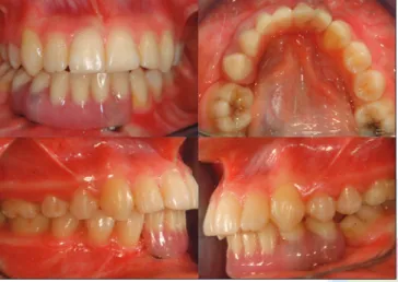
Figure 8: Intraoral photographs of hybrid prosthesis and dental tissues
Figure 7: Dental implant and prepared titanium abutments. Hybrid prosthesis and intaglio surface of the denture
d c
b a
533
hygiene procedures periimplanter tissues healed. Periodic radiographic examination at 6‑month intervals for the 1st year, then yearly, should be performed to observe for
changes of the bone surrounding the implants. There was no radiologic pathology around the dental implants after 1 year of prosthesis insertion.
This clinical report demonstrated one of the treatment modalities of CGCG of mandible in clinical dentistry. The techniques used in this case are associated to the treatment plan and prognosis of the case. Different treatment approaches should be preferred according to the case such as excessive surgical techniques, tooth‑tissue supported prosthesis, etc., Treatment strategy of the case should be changed by the patient’s socioeconomic status and motivation to the long period of treatment sequences.
Summary
This clinical report described the oral rehabilitation of a patient who was diagnosed with a CGCG of the mandible. She underwent mandibular resection, iliac graft reconstruction, distraction osteogenesis, implant placement, and fabrication of a removable hybrid implant partial denture. A multidisciplinary approach was crucial for optimum results. Since changes in her oral structures are expected in the future due to pubertal growth period, close follow‑up is required and is still in progress.
References
1. Jaffe HL. Giant‑cell reparative granuloma, traumatic bone cyst and fibrous dysplasia of the jaw bones. Oral Surg Oral Med Oral Pathol 1953;6:159‑75. 2. Khafif A, Krempl G, Medina JE. Treatment of giant cell granuloma of the maxilla
with intralesional injection of steroids. Head Neck 2000;22:822‑5. 3. Kramer IR, Pindborg JJ, Shear M. Histological typing of odontogenic tumors.
2nd ed.. Berlin: Springer‑Verlag; 1991. p. 31.
4. Potter BJ, Tiner BD. Central giant cell granuloma. Report of a case. Oral Surg Oral Med Oral Pathol 1993;75:286‑9.
5. Spraggs PD, Roth J, Young‑Ramsaran J, Goodwin WJ. Giant cell reparative granuloma of the maxilla. Ear Nose Throat J 1997;76:445‑9.
6. Austin LT Jr, Dahlin DC, Royer RQ. Giant‑cell reparative granuloma and related conditions affecting the jawbones. Oral Surg Oral Med Oral Pathol 1959;12:1285‑95.
7. de Lange J, van den Akker HP, van den Berg H. Central giant cell granuloma of the jaw: A review of the literature with emphasis on therapy options. Oral Surg Oral Med Oral Pathol Oral Radiol Endod 2007;104:603‑15.
8. Ueki Y, Tiziani V, Santanna C, Fukai N, Maulik C, Garfinkle J, et al. Mutations in the gene encoding c‑Abl‑binding protein SH3BP2 cause cherubism. Nat Genet 2001;28:125‑6.
9. Miah SM, Hatani T, Qu X, Yamamura H, Sada K. Point mutations of 3BP2 identified in human‑inherited disease cherubism result in the loss of function. Genes Cells 2004;9:993‑1004. 10. Ardekian L, Manor R, Peled M, Laufer D. Bilateral central giant cell granulomas in a patient with neurofibromatosis: Report of a case and review of the literature. J Oral Maxillofac Surg 1999;57:869‑72. 11. Krammer U, Wimmer K, Wiesbauer P, Rasse M, Lang S, Müllner‑Eidenböck A, et al. Neurofibromatosis 1: A novel NF1 mutation in an 11‑year‑old girl with a giant cell granuloma. J Child Neurol 2003;18:371‑3. 12. Edwards PC, Fantasia JE, Saini T, Rosenberg TJ, Sachs SA, Ruggiero S. Clinically aggressive central giant cell granulomas in two patients with neurofibromatosis 1. Oral Surg Oral Med Oral Pathol Oral Radiol Endod 2006;102:765‑72.
13. Lee JS, Tartaglia M, Gelb BD, Fridrich K, Sachs S, Stratakis CA, et al. Phenotypic and genotypic characterization of noonan‑like/multiple giant cell lesion syndrome. J Med Genet 2005;42:e11.
14. Manor E, Kachko L, Brennan PA, Bodner L. Cytogenetics of central giant cell granuloma of the mandible. J Oral Maxillofac Surg 2013;71:1541‑4. 15. Auclair PL, Cuenin P, Kratochvil FJ, Slater LJ, Ellis GL. A clinical and
histomorphologic comparison of the central giant cell granuloma and the giant cell tumor. Oral Surg Oral Med Oral Pathol 1988;66:197‑208.
16. Stimson PG, McDaniel RK. Traumatic bone cyst, aneurysmal bone cyst, and central giant cell granuloma‑pathogenetically related lesions. J Endod 1989;15:164‑7. 17. Regezi JA, Sciubba JJ. Oral pathology: Clinical‑pathologic correlations. 2nd ed. Philadelphia: Saunders; 1989. p. 379‑81. 18. Waldron CA, Shafer WG. The central giant cell reparative granuloma of the jaws: An analysis of 38 cases. Am J Clin Pathol 1966;45:437‑47. 19. Whitaker SB, Waldron CA. Central giant cell lesions of the jaws. Oral Surg Oral Med Oral Pathol 1993;75:199‑208.
20. Chuong R, Kaban LB, Kozakewich H, Perez‑Atayde A. Central giant cell lesions of the jaws: A clinicopathologic study. J Oral Maxillofac Surg 1986;44:708‑13. 21. Stafne EC, Gibilisco JA. Oral Roentgenographic Diagnosis. 4th ed. Philadelphia:
Saunders; 1975. p. 200.
22. Shafer WG, Hine MK, Levy BM. A textbook of oral pathology. 4th ed. Philadelphia: Saunders; 1983. p. 146. 23. Neville BW, Damm DD, Allen CM, Bouquot JE. Oral maxillofacial pathology. Philadelphia: Saunders; 1995. p. 453. 24. Eisenbud L, Stern M, Rothberg M, Sachs SA. Central giant cell granuloma of the jaws: Experiences in the management of thirty‑seven cases. J Oral Maxillofac Surg 1988;46:376‑84. 25. Harrison D, Lund VJ. Tumours of the upper jaw. Edinburgh: Churchill Livingstone; 1993. p. 232‑5. 26. O’Regan EM, Gibb DH, Odell EW. Rapid growth of giant cell granuloma in pregnancy treated with calcitonin. Oral Surg Oral Med Oral Pathol Oral Radiol Endo 2001;92:532‑8. 27. Webb DJ, Brockbank J. Combined curettage and cryosurgical treatment for the aggressive “giant cell lesion” of the mandible. Int J Oral Maxillofac Surg 1986;15:780‑5. 28. Harris M. Central giant cell granulomas of the jaws regress with calcitonin therapy. Br J Oral Maxillofac Surg 1993;31:89‑94. 29. de Lange J, Rosenberg AJ, van den Akker HP, Koole R, Wirds JJ, van den Berg H. Treatment of central giant cell granuloma of the jaw with calcitonin. Int J Oral Maxillofac Surg 1999;28:372‑6. 30. Pogrel MA. Calcitonin therapy for central giant cell granuloma. J Oral Maxillofac Surg 2003;61:649‑53. 31. Kermer C, Millesi W, Watzke IM. Local injection of corticosteroids for central giant cell granuloma: A case report. Int J Oral Maxillofac Surg 1994;23:366‑8. 32. Carlos R, Sedano HO. Intralesional corticosteroids as an alternative treatment for central giant cell granuloma. Oral Surg Oral Med Oral Pathol Oral Radiol Endod 2002;93:161‑6.
33. Shohat I, Shoshani Y, Taicher S. Medical treatment of central giant cell granuloma of the jaws. Refuat Hapeh Vehashinayim 2002;19:37‑44.
34. da Silva NG, Carreira AS, Pedreira EN, Tuji FM, Ortega KL, de Jesus Viana Pinheiro J. Treatment of central giant cell lesions using bisphosphonates with intralesional corticosteroid injections. Head Face Med 2012;8:23.
35. Bataineh AB, Al‑Khateeb T, Rawashdeh MA. The surgical treatment of central giant cell granuloma of the mandible. J Oral Maxillofac Surg 2002;60:756‑61. 36. Caplanis N, Sigurdsson TJ, Rohrer MD, Wikesjo UM. Effect of allogeneic,
freeze‑dried, demineralised bone matrix on guided bone regeneration in supraalveolar peri‑implant defects in dogs. Int J Oral Maxillofac Implants 1997;12:634‑42.
37. Moghadam HG, Sandor GK, Holmes HH, Clokie CM. Histomorphometric evaluation of bone regeneration using allogenic and alloplastic bone substitues J Oral Maxillofac Surg 2004;62:202‑13.
38. Nocini PF, Albanese M, Buttura da PE, D’Agostino A. Vertical distraction osteogenesis of the mandible applied to an iliac crest graft: Report of a case. Clin Oral Impl Res 2004;15:366‑70.
How to cite this article: Yuzbasioglu E, Alkan A, Ozer M, Bayram M. Multidisciplinary approach for the rehabilitation of central giant cell granuloma: A clinical report. Niger J Clin Pract 2014;17:528‑33.