43/
Acta Chir. orthop. Traum. čech., 83, 2016, No. 1p. 43–46
ORIGINAL PAPER
PŮVODNÍ PRÁCE
Therefore treatment of ligamentous instability needs special attention in management of tibial plateau fractures.
Disruption of posterior tibial slope which is necessary for normal range of motion and ligament function is an-other ignored topic in tibial plateau fractures (15, 21). Changes in normal posterior tibial slope may affect tibial scissoring forces, anterior tibial translation and the force on anterior cruciate ligament (ACL). Therefore increment of posterior tibial slope may lead to ACL injuries (4, 14, 15).
The objective of this study is to evaluate the effect of posterior tibial slope after fracture healing on antero-posterior knee laxity, functional outcome and patient satisfaction.
INTRODUCTION
Fractures of proximal tibia affect function and stability of knee joint. The objective of surgical treatment in proximal tibial fractures is to regain and preserve normal knee functions (23). Anatomical restoration of tibial joint surface and obtaining ligamentous stability are necessary for a painless functional knee joint with a pre-served mechanical axis (2, 6, 10, 11, 12, 20).
Vast majority of tibial plateau fractures result from high energy accidents such as traffic accidents and fall from height (8, 13). Varus and valgus scissoring forces ac-companying axial compression results in ligament and meniscal injuries in up to 50% of the cases (8, 16).
The Effect of Sagittal Plane Deformities after Tibial
Plateau Fractures to Functions and Instability
of Knee Joint
Význam deformit v sagitální rovině u zlomenin tibiálního plató na funkci a stabilitu
kolenního kloubu
M. ERDIL1, F. YILDIZ2, E. KUYUCU1, Ş. SAYAR2, G. POLAT3, H. H. CEYLAN4, F. KOÇYIĞIT5
1Istanbul Medipol University, Orthopaedics & Traumatology, Istanbul, Turkey 2Bezmi Alem University, Orthopaedics & Traumatology, Istanbul, Turkey 3Istanbul University, Orthopaedics & Traumatology, Istanbul, Turkey
4LNB Sultançiftliği Government Hospital, Orthopaedics & Traumatology, Istanbul, Turkey 5Pamukkale University, Physical Therapy and Rehabilitation School, Denizli, Turkey
ABSTRACT
PURPOSE OF THE STUDY
The objective of this study is to evaluate the effect of posterior tibial slope after fracture healing on antero-posterior knee laxity, functional outcome and patient satisfaction.
MATERIAL AND METHODS
126 patients who were treated for tibial plateau fractures between 2008–2013 in the orthopedics and traumatology department of our institution were evaluated for the study. Patients were treated with open reduction and internal fixation, arthroscopy assisted minimally invasive osteosynthesis or conservative treatment.
RESULTS
Mean posterior tibial slope after the treatment was 6.91 ± 5.11 and there was no significant difference when compared to the uninvolved side 6.42 ± 4,21 (p = 0.794). Knee laxity in anterior-posterior plane was 6.14 ± 2.11 and 5.95 ± 2.25 respectively on healthy and injured side. The difference of mean laxity in anterior-posterior plane between two sides was statistically significant.
DISCUSSION
In this study we found no difference in laxity between the injured and healthy knees. However Tegner score decreased significantly in patients who had greater laxity difference between the knees. We did not find significant difference between fracture type and laxity, IKDC functional scores independent of the ligamentous injury.
CONCLUSION
In conclusion despite coronal alignment is taken into consideration in treatment of tibial plateau fractures, sagittal alignment is reasonably important for stability and should not be ignored.
Key words: knee, tibial fracture, fracture fixation.
recom mended to all patients for two months. Patients were requested to attend follow-up visits on second week, sixth week, third month, sixth month and first year in the post-operative period.
The knee flexion was statistically significantly decreased on the affected side compared to the healthy side (100.16 ± 16.14° versus 110.16 ± 13.11, p < 0.001). Mean IKDC and Tegner scores were 67.83 ± 19.98 and 87.22 ± 14.48 respectively. IKDC (p = 0.001) and Tegner scores (p = 0.003) tend to increase as the amount of knee flexion increased.
When the relation between fracture type and radiological and functional parameters is evaluated; there was no re-lation between fracture type and slope, laxity, IKDC Score. However increase of fracture type was correlated with significant decrease in Tegner score (p = 0.045), and flexion angles (p = 0.021).
Mean posterior tibial slope after the treatment was 6.91 ± 5.11 and there was no significant difference when compared to the uninvolved side 6.42 ± 4.21 (p = 0.794). Mean posterior tibial slope was significantly higher in patients with higher posterior tibial slope on the uninvolved side (p = 0.001). Posterior tibial slope after the treatment and posterior tibial slope difference did not affect functional scores (IKDC score p = 0.903, Tegner score p = 0.523, range of motion p = 0.994). (Table 3).
Knee laxity in anterior-posterior plane was 6.14 ± 2.11 and 5.95 ± 2.25 respectively on healthy and injured side. The difference of mean laxity in anterior posterior plane between two sides was statistically significant (1.89 ± 1.53, p = 0.036). There was no correlation between laxity and age, fracture type, range of motion, functional knee scores, posterior tibial slope. However Tegner scores were significantly lower in patients with higher difference between laxity of two knees (p = 0.03). (Table 3). DISCUSSION
The main goal in treatment of tibial plateau fractures is to obtain painless, stabile knee joint with a functional range of motion (23). Anatomical restoration of tibial joint surface is necessary to achieve treatment goal (10, 20). Proximal sagittal plane is as important as coronal plane deformities when evaluating anatomical reduction radiologically.
It is documented in many previous biomechanical and clinical trials that anatomical posterior slope of pro -ximal tibia in sagittal plane is very important in transfer of weight, sagittal plane stability and normal range of motion (1, 2, 8, 9, 13, 15, 16, 21). However up to our knowledge there are no studies in literature investigating
44/
Acta Chir. orthop. Traum. čech., 83, 2016, No. 1ORIGINAL PAPER
PŮVODNÍ PRÁCE
MATERIAL AND METHODS
Hospital records of 126 patients who were treated for tibial plateau fractures between 2008–2013 in the or-thopedics and traumatology department of our institution were evaluated for the study. Of these 32 (16 females, 16 males) patients who attended follow-up visits regularly for at least 12 months were analyzed retrospectively on electronical charts. Patients with trauma of the contralateral knee joint, knee osteoarthritis, inflammatory artropathy, previous knee surgery were excluded. Patients gave written informed consent and were informed about the treatment and post-operative rehabilitation before the treatment.
Patients were treated with open reduction and internal fixation, arthroscopy assisted minimally invasive os-teosynthesis (18) or conservative treatment (Table 1). Patients who were surgically treated were recommended a hinged knee brace and range of motion exercises for knee was started on the second post-operative day. Fracture healing and posterior tibial slope were evaluated on roentgenograms. One and only orthopedist evaluated posterior tibial slope according to technique described by Utzschneider et al. using NovaPACS Diagnostic Viewer 7.5.5.14 (NovaRad Corporation, USA), (22). Tibial anterior translation was evaluated by another or-thopedist that is blinded to roentgenogram findings using digital arhtrometer (Kneelax MR Systems, Haarlem, The Netherlands). International Knee Documentation Committee (IKDC) knee evaluation form and Tegner knee score were used for clinical and functional evaluation (5, 7). Fracture classification was made according to Schatzker classification (17). SPSS version 16.0 (SPSS, Chicago, USA) was used for statistical analyses. The relation between fracture type, anterior translation, pos-terior tibial slope, range of motion, IKDC and Tenger knee scores were analyzed using Spearman non-parametric correlation test.
RESULTS
Mean age of the study patients were 50 (ranged between 29–72) and mean follow-up duration was 34.1 moths (ranged between 12–58 months). Tibial fractures were on the right knee in 16 (50%) patients. Most of the patients had Schatzker type-3 fractures (n:12, 37.5% ). There were accompanying fractures in 5 patients.
The most applied treatment was open reduction, internal fixation and grafting with autogenously iliac bone (n:15, 46.8%). (Table 2). Immobilization was
Schatzker classification Type I – Type II 9 (28.1%) Type III 12 (37.5%) Type IV – Type V 6 (18.7%) Type VI 5 (15.6%)
Table 1. Distribution of fracture types according to Schatzker classification
Table 2. Distribution of applied treatment procedures Treatment alternative
Open reduction-internal fixation 15(46.8%) Arthroscopy assisted minimal
invasive procedure 9 (28.1%) – cannulated screw 4 (12.5%) – screw and plate Conservative treatment (cast) 4 (12.5%)
45/
Acta Chir. orthop. Traum. čech., 83, 2016, No. 1ORIGINAL PAPER
PŮVODNÍ PRÁCE
the effect of post-traumatic sagittal plane deformities on anterior posterior stability of knee and functional results.
Streubel et al. demonstrated change of posterior tibial slope reaching 20° in a series of 74 patients with tibial plateau fractures (21). Barei et al. reported normal posterior tibial slope in 72.4% of 83 patients with complex tibial plateau fractures (2). We achieved posterior tibial slope similar to uninvolved knee (p = 0.794) and no functional differences between two knees. This implies the functional success of anatomical plane reposition and involved knee behaves similar to the healthy one.
Jansen et al. reported tendency towards instability in pivot shift test after tibial plateau fractures (10). Rade-makers et al. reported anteroposterior laxity more than 14% of the patients who were followed for 14 years (12). In this study we found no difference in laxity between the injured and healthy knees. However Tegner score decreased significantly in patients who had greater laxity difference between the knees. Despite absence of correlation between laxity and posterior tibial slope, minimization of instability via anatomical reposition of posterior tibial slope is necessary for a functional knee. There are studies reporting increased ligamentous and meniscal injury as the Shatzker degree of fracture increases (19). On the other hand there are also studies reporting absence of correlation between fracture type and ligamentous, meniscal injury (3, 24). We did not
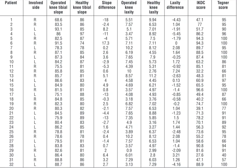
Table 3. Measured posterior tibial slopes, laxity ratios and functional knee scores
Patient Involved Operated Healthy Slope Operated Healthy Laxity IKDC Tegner side knee tibial knee tibial difference knee knee difference score score
slope slope laxity laxity
1 2 3 4 5 6 7 8 9 10 11 12 13 14 15 16 17 18 19 20 21 22 23 24 25 26 27 28 29 30 31 32 R R L L R R L R L L R R R L L R L R R R L L L L R R L L R L R L 68.6 83.5 93.1 86 82.5 91.5 78.3 87.1 87.3 84.2 75.5 85.8 85.7 86.6 84.9 81.5 75.1 84.8 82.3 80.3 84.5 75.9 80.4 86.2 78.8 78.6 79.5 83.8 82.6 86.4 88.8 88.7 86 86 85 97 87 74 78 85 84 87 81 85 81 83 80 81 88 85 80 82 89 89 83 85 81 78 81 83 81 80 86 86 -18 -2.4 8.2 -11 -4 17.3 0.2 2.6 3.6 -2.9 -5.3 0.6 5.1 4 4.9 0.8 -13 -0.3 2.5 -2.1 -4.4 -13 -2.7 1.6 -2.4 0.4 -1.4 0.7 2 6.4 3.2 2.7 5.51 7.57 5.1 3.47 5.71 7.11 10.2 6.19 7.65 7.45 4.39 10 8.57 4.58 4.69 3.57 4.08 3.18 6.82 7.57 7.65 7.35 4.9 4.71 3.89 10.2 7.57 3.57 0.9 6.01 7.29 3.13 9.94 6.53 7.01 8.92 7.5 7.11 8.12 4.55 7.9 5.73 5.31 2.76 11.2 4.45 6.21 4.97 4.93 3.76 7.02 6.53 8.88 5.85 3.16 3.27 6.37 8.12 6.53 4.97 2.99 2.8 6.03 7.29 -4.43 1.04 -1.91 -5.45 -1.79 0 2.08 1.64 -0.25 1.72 -0.92 7.24 -2.63 0.13 -1.52 -1.4 -0.85 -0.58 -0.2 1.04 -1.23 1.5 1.74 1.44 -2.48 2.08 1.04 -1.4 -2.09 3.21 1.26 -4.16 47.1 77 51.7 86.2 94.3 94.3 89.7 88.5 87.4 63.2 85.1 32.2 48.3 60.9 65.5 96.6 49.4 40.2 74.7 39.1 71.3 78.2 70.1 56.3 73.6 55.2 58.6 90.8 81.6 27.6 47.1 88.9 95 95 90 96 100 100 95 100 100 86 81 66 81 97 95 100 87 66 100 77 91 91 89 92 95 78 66 94 91 40 57 100 find significant difference between fracture type and laxity, IKDC functional scores independent of the liga-mentous injury.
Range of motion is an indicator of functional recovery after tibial plateau fractures (6, 20). We found higher IKDC and Tegner scores in patients with higher range of motion. However this situation is affected by fracture type and increase in Tegner scores and range of motion is significantly related to increase in fracture type.
The small number of patient group is one of the limi-tations of this study. Another limitation is inclusion of different fracture types. Although patients with previous ligamentous injury, knee instability, previous knee surgery, and documented ligamentous injury were excluded from the study, accompanying soft tissue injuries were not evaluated with magnetic resonance imaging before the operation because of availability and financial burden.
CONCLUSION
Despite coronal alignment is taken into consideration in treatment of tibial plateau fractures, sagittal alignment is reasonably important for stability and should not be ignored. Further studies with homogenous fracture types and including more patients to evaluate effect of change in posterior tibial slope to knee stability after tibial plateau fractures are recommended.
46/
Acta Chir. orthop. Traum. čech., 83, 2016, No. 1ORIGINAL PAPER
PŮVODNÍ PRÁCE
Conflict of interest: All authors declared that they have no conflict of interest.
References
1. AHEARN, N., OPPY, A., HALLIDAY, R., ROWETT-HARRIS, J., MORRIS, S. A., CHESSER, T. J., LIVINGSTONE, J. A.: The outcome following fixation of bicondylar tibial plateau fractures. J. Bone Jt Surg., 96-B: 956–962, 2014.
2. BAREI, D. P., NORK, S. E., MILLS, W. J., COLES, C. P., HEN-LEY, M. B., BENIRSCHKE, S. K.: Functional outcomes of severe bicondylar tibial plateau fractures treated with dual incisions and medial and lateral plates. J. Bone Jt Surg., 88-A: 1713–1721, 2006. 3. BENNETT, W. F., BROWNER, B.: Tibial plateau fractures: a study of associated soft tissue injuries. J. Orthop. Trauma, 8: 183–188, 1994.
4. BRANDON, M. L., HAYNES, P. T., BONAMO, J. R., FLYNN, M. I., BARRETT, G. R., SHERMAN, M. F.: The association between posterior-inferior tibial slope and anterior cruciate ligament insufficiency. Arthroscopy, 22: 894–899, 2006. 5. BRIGGS, K. K., LYSHOLM, J., TEGNER, Y., Rodkey, W.G., KOCHER, M. S., STEADMAN, J. R.: The reliability, validity, and responsiveness of the Lysholm score and Tegner activity scale for anterior cruciate ligament injuries of the knee: 25 years later. Am. J. Sports Med., 37: 890–897, 2009.
6. CHOO, K. J., MORSHED, S.: Postoperative complications after repair of tibial plateau fractures. J. Knee Surg., 27: 11–19, 2014. 7. GREVNERTS, H. T., TERWEE, C. B., KVİST, J.: The measurement properties of the IKDC-subjective knee form. Knee Surg. Sports Traumatol. Arthrosc., 2014 Sep 6. [Epub ahead of print] 8. HONKONEN, S. E., JARVINEN, M. J.: Classification of fractures
of the tibial condyles. J. Bone Jt Surg., 74-B: 840–847, 1992. 9. HOUBEN, P. F., VAN DER LINDEN, E. S., VAN DEN
WILDEN-BERG, F. A., STAPERT, J. W.: Functional and radiological outcome after intra-articular tibial plateau fractures. Injury, 28: 459–462, 1997.
10. JANSEN, H., FREY, S. P., DOHT, S., FEHSKE, K., MEFFERT, R.H.: Medium-term results after complex intra-articular fractures of the tibial plateau. J. Orthop. Sci., 18: 569–577, 2013. 11. MATTIASSICH, G., FOLTIN, E., SCHEURECKER, G.,
SCHNEIDERBAUER, A., KROPFL, A., FISCHMEISTER, M.: Radio -graphic and clinical results after surgically treated tibial plateau fractures at three and twenty two years postsurgery. Int. Orthop., 38: 587–594, 2014.
12. RADEMAKERS, M. V., KERKHOFFS, G. M., SIEREVELT, I. N., RAAYMAKERS, E. L., MARTI, R.K.: Operative treatment of 109 tibial plateau fractures: five- to 27-year follow-up results. J. Orthop. Trauma, 21: 5–10, 2007.
13. SEGAL, D., MALLIK, A. R., WETZLER, M. J., FRANCHI, A. V., WHITELAW, G. P.: Early weight bearing of lateral plateau fractures. Clin. Ortop., 294: 232–237, 1993.
14. SENISIK, S., OZGURBUZ, C., ERGUN, M., YUKSEL, O., TASKIRAN, E., ISLEGEN, C., ERTAT, A.: Posterior tibial slope as a risk factor for anterior cruciate ligament rupture in soccer players. J. Sports Sci. Med., 10: 763–766, 2011.
15. SHELBURNE, K. B., KIM, H. J., STERETT, W. I., PANDY, M. G.: Effect of posterior tibial slope on knee biomechanics during functional activity. J. Orthop. Res., 29: 223–231, 2011.
16. SHEPHERD, L., ABDOLLAHI, K., LEE, J., VANGSNESS, C. T. Jr.: The prevelence of soft tissue injuries in nonoperative tibial plateau fractures as determined by magnetic resonance imaging. J. Orthop. Trauma, 16: 628–631, 2002.
17. SCHATZKER, J., MCBROOM, R., BRUCE, D.: The tibial plateau fracture; the Toronto experience 1968–1975. Clin. Orthop., 138: 94–104, 1979.
18. SIEGLER, J., GALISSIER, B., MARCHEIX, P. S., CHARISSOUX, J. L., MABIT, C., ARNAUD, J. P.: Percutaneous fixation of tibial plateau fractures under arthroscopy: a medium term perspective. Orthop. Trauma Surg. Res., 97: 44–50, 2011.
19. STANNARD, J. P., LOPEZ, R., VOLGAS, D.: Soft tissue injury of the knee after tibial plateau fractures. J. Knee Surg., 23: 187–192, 2010.
20. STEVENS, D.G., BEHARRY, R., MCKEE, M.D., WADDELL, J. P., SCHEMITSCH, E. H.: The long-term functional outcome of operatively treated tibial plateau fractures. J. Orthop. Trauma, 15: 312–320, 2001.
21. STREUBEL, P. N., GLASGOW, D., WONG, A., BAREI, D. P., RICCI, W. M., GARDNER, M. J.: Sagittal plane deformity in bi-condylar tibial plateau fractures. J. Orthop. Trauma, 25: 560–56, 2011.
22. UTZSCHNEIDER, S., GOETTINGER, M., WEBER, P., HORNG, A., GLASER, C., JANSSON, V., MÜLLER, P. E.: De-velopment and validation of a new method for the radiologic meas-urement of the tibial slope. Knee Surg., 19: 1643–1648, 2011. 23. WATSON, J. T.: High energy fractures of tibial plateau. Orthop.
Clin. North Am., 25: 723–752, 1994.
24. ZAKRZEWSKI, P., ORŁOWSKI, J.: Meniscuses and ligaments injuries in tibial plateau fractures in comparative evaluation of clinical, intraoperative and MR examination. Chir. Narzadow Ruchu Ortop. Pol., 70: 109–113, 2005.
Corresponding author: Ersin Kuyucu
Medipol University-Istanbul
TEM Avrupa Göztepe çıkışı, Bağcılar, Turkey E-mail: [email protected]