BIOACTIVE PEPTIDE NANOFIBERS FOR ACCELERATION OF
BURN WOUND HEALING
A THESIS SUBMITTED TO
THE GRADUATE SCHOOL OF ENGINEERING AND SCIENCE OF BILKENT UNIVERSITY
IN PARTIAL FULFILLMENT OF THE REQUIREMENTS FOR THE DEGREE OF
MASTER OF SCIENCE IN
MATERIALS SCIENCE AND NANOTECHNOLOGY
By
FATİH YERGÖZ May, 2017
i
BIOACTIVE PEPTIDE NANOFIBERS FOR ACCELERATION OF BURN WOUND HEALING
By Fatih Yergöz, May, 2017
We certify that we have read this thesis and that in our opinion it is fully adequate, in scope and in quality, as a dissertation for the degree of Master of Science.
Ayşe Begüm Tekinay (Advisor)
Aykutlu Dâna
Reyhan Neslihan Gürsoy
Approved for the Graduate School of Engineering and Science:
Ezhan Karaşan
ii
ABSTRACT
BIOACTIVE PEPTIDE NANOFIBERS FOR ACCELERATION OF
BURN WOUND HEALING
Fatih Yergöz
M.Sc. in Materials Science and Nanotechnology Supervisor: Ayşe Begüm Tekinay
May, 2017
Burn injuries are one of the most typical types of trauma worldwide, and the unique physiology of burn injuries requires the use of specialized therapeutic materials for treatment and makes the development of such materials especially challenging. Here, we report the use of synthetic, functional and biodegradable peptide nanofiber gels for improved healing of burn wounds to alleviate the progressive loss of tissue function at the post-burn wound site. These bioactive nanofiber gels form scaffolds which recapitulate the morphology and function of the natural extracellular matrix through peptide epitopes, which can trigger angiogenesis through significant affinity to basic growth factors. In this study, the angiogenesis-promoting properties of the bioactive scaffolds were utilized for the treatment of thermal burn model. Following the excision of necrotic tissue, bioactive gels and control solutions were applied topically onto the wound area. The wound healing process was evaluated at 7, 14 and 21 days following injury through histological observations, immunostaining and marker RNA / protein analysis. Bioactive peptide nanofiber treated burn wounds formed well-organized and collagen-rich granulation tissue layers, developed a greater density of
iii
newly formed blood vessels, and exhibited increased re-epithelialization and skin appendage formation with minimal crust formation. Overall, the heparin-mimetic peptide nanofiber gels increased the rate of repair of burn injuries and can be used as effective means of facilitating wound healing.
Keywords: Peptide Nanofiber, Burn Injury, Heparin, Hydrogel, Self-Assembly,
iv
ÖZET
YANIK YARA İYİLEŞMESİNİN HIZLANDIRILMASINDA
BİYOAKTİF PEPTİT NANOFİBERLERİN KULLANIMI
Fatih Yergöz
Malzeme Bilimi ve Nanoteknoloji, Yüksek Lisans Danışman: Ayşe Begüm Tekinay
May, 2017
Yanık yaralanmaları dünya genelinde en yaygın travma tiplerinden biri olmakla birlikte yanık yaralanmalarının kendine özgü fizyolojisi, tedavi için özel terapötik materyallerin kullanılmasını gerektirmekte ve bu tür malzemelerin geliştirilmesini zorlaştırmaktadır. Bu tez kapsamında, yanık yaralanmaları sonucu yara alanında oluşan ilerleyici doku fonksiyonu kaybının hafifletilerek iyileştirilmesi için sentetik, fonksiyonel ve biyolojik olarak parçalanabilir peptit nanofiber jellerin kullanımı anlatılmaktadır. Bu biyoaktif nanofiber jelleri bazik büyüme faktörlerine kuvvetli afiniteyle bağlanarak anjiyogenezi tetikleyebildigi gibi, hücrelere sinyal verebilen peptit epitopları vasıtasıyla hücreler arası ortamın yapısını ve fonksiyonunu taklit edebilme yetisine sahip iskeleler oluşturabilmektedir. Bu çalışmada, biyoaktif iskelelerin anjiyogenezi teşvik edici özellikleri termal yanık yara modelinin tedavisinde kullanılmıştır. Nekrotik dokuların eksizyonunu takiben, biyoaktif jel ve kontrol solusyonları yara alanı üzerine topikal olarak uygulanmıştır.Yaranın iyileşme süreci hasar sonrası 7., 14. ve 21. günlerde histolojik gözlemler, immüno boyama ve markör RNA / protein analizi ile değerlendirilmiştir. Biyoaktif peptit nanofiber ile
v
tedavi edilen yanık yaralarında, iyi organize olmuş ve kollajen bakımından zengin granülasyon dokusu, yara alanında yeni oluşan kan damarlarının yoğunluğunda, minimal kabuk oluşumu ile yeni oluşturulan epitelizasyon ve deri apendiksiyonu seviyesinde artma gözlemlenmiştir. Sonuç olarak, heparin-mimetik peptit nanofiber jelleri yanık yaralanmalarının onarımını arttırmasıyla, yara iyileşmesini kolaylaştırmada etkili bir tedavi yöntemi olarak kullanılabilir.
Anahtar sözcükler: Peptit Nanofiber, Yanık Yarası, Heparin, Hidrojel, Kendindiliğinden Bir Araya Gelme, Neo-vaskülarizasyon, Ekstraselüler matris
vi
ACKNOWLEDGEMENT
There are numerous people to acknowledge for their cooperation, guidance and support during the last two years. So many have made my stay in Ankara a lot easier that I thought it was going to be. This thesis would not have been possible without the help and support of the kind people around me, to only some of whom it is possible to give particular mention here.
It would not have been possible to write this master thesis without continuous motivation and patience of my primary advisor Prof. Ayşe Begüm Tekinay. I appreciate her vast knowledge and skill in many areas not to mention her guidance, advice, and constant, horrifying academic pressure that were responsible for the completion of my thesis in a timely manner, and resulted in the preparation of a high-quality manuscript in the process. The door of Prof. Tekinay office was always open and she was always interested whenever I had a question about my research and writing or encountered trouble related with my experiments. It was a great opportunity to be advised by someone communicative, diligent, enthusiastic and friendly for my master study.
I would like to thank my thesis committee, Dr. Dâna and Dr. Gürsoy for the assistance they provided to make this thesis with high-standards by improving it from various perspectives and for taking time out from their busy schedule to serve as my external reader. I would also like to thank Dr. Mustafa Özgür Güler, who was gracious enough to read and comment on much of my work and gave access to his laboratory and research facilities.
With a special mention, I am thankful to my lifetime friends from IYTE, Nurcan Hastar, İdil Uyan, Merve Şen, Zeynep Orhan, Gökhan Günay for the stimulating
vii
discussions, for the sleepless nights we were working together, and for all the fun we have had in the two years. It was fantastic to have the opportunity to work majority of my master research beside you!
I would like to thank all NBT, BML and other UNAM members especially Cağla Eren, Begüm Dikeçoğlu, Faruk Okur, Mustafa Fadlelmula, Özge Uysal, Sehmus Tohumeken, Burak Demircan, İslam Oğuz Tuncay, Canelif Yılmaz, İbrahim Çelik, Mustafa Beter, Abdurrahman Türksoy and Deniz Yıldız for creating such a warm and entertaining working environment.
The assistance, cooperation and experience of my fellow doctoral students and pos-doc’s were essential for the completion of the field as well as the model deck. I’d like to thank Gülistan Tansık, Elif Arslan, Nuray Gündüz, Melike Sever, Özlem Tufanlı,
Dr. Begüm Kocatürk, Dr. Muhammed Aref Khaliy, Ahmet Emin Topal. In particular, I am grateful to Dr. Gözde Uzunallı for enlightening me the first glance
of research.
I would also like to thank Alper Devrim Özkan as the first reader and reviewer of this thesis, and I am very grateful to him for his very valuable comments and revision on this thesis.
My forever interested, encouraging and always enthusiastic grandmother Ülker Tozlu and grandfather Sabri Yergöz: they were always keen to know what I was doing, although it is likely that they never grasped what it was all about! I will miss your screams of joy whenever a significant momentous was reached.
I would like to acknowledge the financial and technical support of National Nanotechnology Research Center (UNAM). Much of the experimental work would
viii
not have been completed without the equipment and technical assistance of technicians.
I would like to acknowledge the financial assistance of The Scientific and Technological Research Council of Turkey (TÜBİTAK) under the BİDEB 2210/C Master Scholarship.
Finally, I would like to express my very profound appreciation to my father Mehmet Emin & my mother Neslihan and my brothers Enes & Arif Emre and to my girlfriend Nurcan for providing me with unfailing support, for their continuous encouragement, personal support, great patience and endless love throughout my master studies and through the process of researching and writing this thesis. These accomplishments wouldn’t have been possible without them. Thank you.
ix Table of Contents
ÖZET ... iv
ACKNOWLEDGEMENT ... vi
Abbreviations ... xi
List of Figures ... xiv
List of Tables ... xviii
CHAPTER 1 ... 1
INTRODUCTION... 1
1.1. Skin and Burns - Overview ... 2
1.1.1. Socio-Economic Impact ... 3
1.1.2. Etiology of burn wounds ... 3
1.1.3. Burn Wound Progression ... 4
1.2. Anatomy of the Skin ... 5
1.2.1. Skin Embryology ... 5
1.2.2. Epidermis ... 5
1.2.3. Dermis ... 8
1.3. Important Elements Regulating Wound Repair Process ... 11
1.3.1. Growth factors ... 11
1.3.2. The Extracellular Matrix (ECM) ... 12
1.4. Scaffolds ... 15
1.4.1. Properties of scaffolds... 18
1.4.2. Types of Scaffolds Used for Regenerative Medicine ... 19
1.5. Angiogenesis, its Pathways, and its Mechanisms-of-Action ... 25
1.5.1. Role of Angiogenesis in Wound Healing ... 29
1.5.2. Biomaterial-Based Therapeutics to Target the Angiogenesis Process .. 30
1.6. Self-assembled peptides in wound repair and regeneration ... 32
CHAPTER 2 ... 38
A HEPARIN-MIMETIC PEPTIDE AMPHIPHILE ENHANCES THE RECOVERY OF FULL-THICKNESS BURN INJURIES ... 38
x
2.1. Introduction ... 39
2.2. Materials and Methods ... 41
2.2.1. Materials ... 41
2.2.2. Synthesis and Purification of Molecules and Gel Formation ... 41
2.2.3. Physical and Mechanical Characterization of Self-Assembled Peptide Nanofiber Networks ... 43
2.2.4. In vivo Burn Model ... 44
2.2.5. Wound Area Measurement ... 46
2.2.6. Histological Analysis ... 46
2.2.7. Immunohistochemical Staining ... 47
2.2.8. Protein and RNA Isolation from tissue... 48
2.2.9. Quantitative Reverse Transcription Polymerase Chain Reaction ... 49
2.2.10. Western Blot Analysis ... 50
2.2.11. Statistical Analysis ... 51
2.3. Results ... 51
2.3.1. Synthesis and characterization of PA molecules ... 51
2.3.2. Gel treatment and dorsal burn injury model ... 54
2.3.3. Histological analysis ... 59
2.3.4. Modulation of angiogenesis by bioactive peptide nanofiber scaffolds ... 62
2.3.5. Collagen orientation ... 66
2.4. Discussion ... 67
2.5. Conclusions ... 70
CHAPTER 3 ... 72
CONCLUSIONS AND FUTURE DIRECTIONS ... 72
REFERENCES ... 75
xi
Abbreviations
AFM Atomic force microscopy
ANOVA Analysis of variance
Boc Tert-butoxycarbonyl
BSA Bovine serum albumin
CD Circular dichroism
CS Chondroitin sulfate
Col-I Collagen type I
DAB 3,3'-diaminobenzidine
DCM Dichloromethane
DMEM Dulbecco's modified Eagle's medium
DMF N,N-Dimethylformamide
DMSO Dimethyl sulfoxide
ECM Extracellular matrix
EDTA Ethylenediaminetetraacetic acid
E-PA Lauryl-VVAGE-OH
ESC Embryonic stem cells
FBS Fetal bovine serum
FDA U.S. Food and Drug Administration
FGF-2 Fibroblast growth factor-2
Fmoc 9-Fluorenylmethoxycarbonyl
GAG Glycosaminoglycan
xii H&E Hematoxylin and eosin
HBTU N,N,N′,N′-Tetramethyl-O-(1H-benzotriazole-1-yl)
uronium hexafluorophosphate
HM-PA Heparin mimetic peptide amphiphile
HPLC High pressure liquid chromatography
HRP Horseradish peroxidase
HKT Human keratocyte
HUVEC Human umbilical vein endothelial cell
IHC Immunohistochemistry
IPGTT Intraperitoneal glucose tolerance test
iPSC Induced pluripotent stem cell
K-PA Lauryl-VVAGK- NH2
LC-MS Liquid chromatography-Mass spectroscopy
P/S Penicillin/Streptomycin
PA Peptide amphiphile
PBS Phosphate buffered saline
PDGF Platelet-derived growth factor
PEG Poly ethylene glycol
RT Room temperature
SEM Standard error of mean
SDS Sodium dodecyl sulfate
T1D Type 1 Diabetes
TBS Tris buffered saline
TCP Tissue culture plate
xiii
TIS Triisopropylsilane
xiv
List of Figures
Figure 1. Thermal injury zones. The three zones of burn injury is illustraed as
three-dimensional. (Reused from Ref. 6 with permission from Wolters Kluwer Group.) ... 4
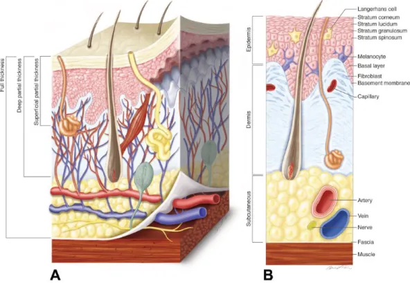
Figure 2. Skin Anatomy. A, Three-dimensional demonstration of the skin. B,
Location of different cell types and important histological zones. (Reused from Ref. 6 with permission from Wolters Kluwer Group.) ... 7
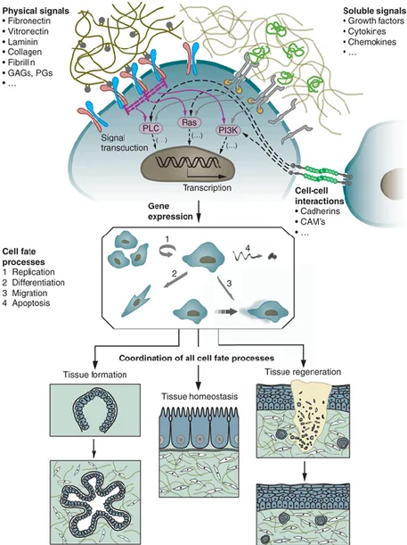
Figure 3. Dynamic interactions of cells with the surrounding ECM. Physical signals,
soluble signals, cell fate processes and cell-cell interactions regulate processes such as tissue formation, tissue homeostasis and tissue regeneration. CAMs, cell adhesion molecules; PGs, proteoglycans; GAGs, glycosaminoglycans; PLC, phospholipase C (Reused from Ref. 19 with permission from Nature Publishing Group). ... 13
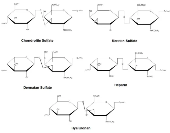
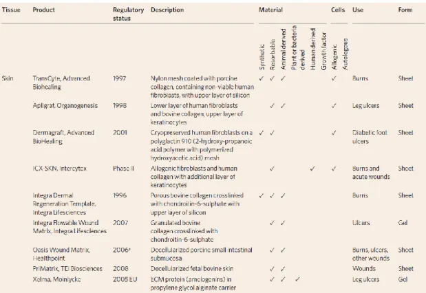
Figure 4. Glycosaminoglycans and their structural units. ... 15 Figure 5. Commercially available tissue engineering products for injuries and
diseases of the skin. (Adapted from Ref. 34 with permission from Nature Publishing Group.) ... 17
Figure 6. General structure and schematic representations of peptide amphiphile
molecules and nanofibers. (A) Chemical structure, (B) different regions of structure, (C) nanofibrous structure of peptide amphiphiles (Reused from Ref. 108 with
permission from Royal Society of Chemistry). ... 25
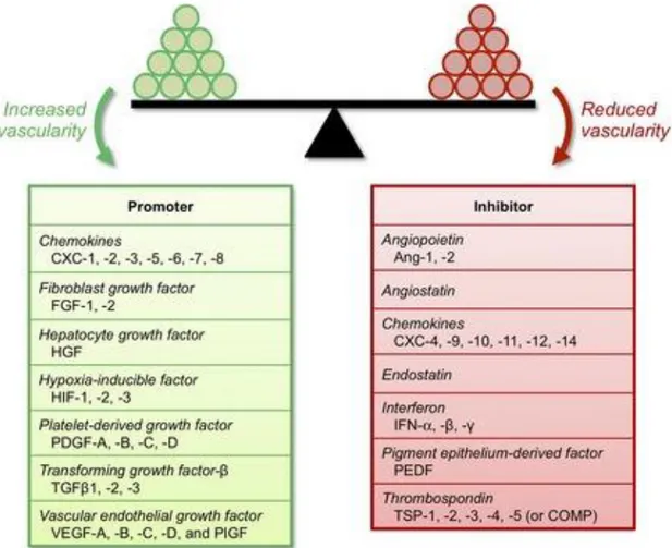
Figure 7. Regulation of angiogenesis process trough pro-angiogenic factors and
anti-angiogenic factors (Reproduced from Ref. 1 with permission from Journal of Cellular and Molecular Medicine 109). ... 28
Figure 8. Vascularization of tissue-engineering scaffolds. (Reused from Ref. 19 with
xv
Figure 9. Chemical structures of K-PA, HM-PA and E-PA. (A) Characterization of
peptide amphiphile molecules and their nanofıbers with circular dichroism. (B) Structure of Heparin. (C) Characterization of peptide nanofibers at pH 7.4 by using SEM. (D,E) General appearance of peptide nanofiber gels (F). Rheology measurements of scaffolds (G). Scale bars are 5 µm. ... 53
Figure 10. (A) RP-HPLC chromatogram of HM-PA , the change of response units
with respect to time at 220 nm. (B) Mass spectrometry of HM-PA. [M-H] -(calculated): 1225.59, [M-H]- (observed): 1224.59, [M-2H]-2/2 (calculated): 611.79, [M-2H]-2/2 (observed): 611.76 [M-3H]-3/3 (calculated): 407.53, [M-3H]-3/3 (observed): 407.49. ... 53
Figure 11. (A) RP-HPLC chromatogram of K-PA , the change of response units with
respect to time at 220 nm. (B) Mass spectrometry of K-PA. [M+H]+ (calculated):653.48, [M+H]+ (observed): 654.49, [2M+H]+(calculated): 1307.96 [2M+H]+ (observed): 1307.98. ... 54
Figure 12. (A) RP-HPLC chromatogram of E-PA , the change of response units with
respect to time at 220 nm. (B) Mass spectrometry of E-PA. [M-H]- (calculated): 655.42, [M-H]- (observed): 654.42. ... 54
Figure 13. Schematic representation of full-thickness burn wound model. (a) A
pre-heated aluminum plaque was applied to the shaved dorsum of the anesthetized mouse; followed by the removal of the injured skin and the application of the hydrogel. (b) A schematic of the hydrogel-implanted wound site. Schematic representations of the burn model were prepared using Mind the Graph software. ... 55
Figure 14. Comparison of normal mouse skin with full-thickness burned mouse skin.
xvi
Figure 15. Representative images of burn wounds after gel application at days 7, 9,
12, 14, 16 and 21 (A). Quantification of wound areas treated with HM-PA peptide nanofibers, sucrose solution, 3M™ Tegaderm™ and control nanofibers at days 7, 9, 14, and 16 (B). One-way ANOVA (Bonferroni's multiple comparisons test) was used for statistical analysis. Error bars represent standard error of mean. ... 57
Figure 16. Animal weights throughout the study. ... 58 Figure 17. Masson’s Trichrome staining of HM-PA nanofiber, sucrose solution,
3M™ Tegaderm™ and control nanofiber application at days 7, 14 and 21 (A). Scale bars are 500 µm. Quantitative analysis of granulation tissue (B), re-epithelization (C), crust area (D), wound distance (E) and skin appendages (F) of burn wounds at days 7, 14 and 21. One-way ANOVA (Bonferroni's multiple comparisons test) was used for statistical analysis, *p< 0.05. Error bars represent standard error of mean. ... 61
Figure 18. Staining of blood vessels by anti-VWF and quantification of blood vessels
at days 7, 14 and 21 (A,B). Quantification of average vessel diameter at day 7 (C). One-way ANOVA (Bonferroni's multiple comparisons test) was used for statistical analysis. Errors bars represent standard error of mean. Scale bars are 50 µm. ... 64
Figure 19. Protein and mRNA levels of genes associated with angiogenesis and
wound repair at the burn wound site at days 7, 14 and 21. qRT-PCR analyses were performed for VEGF and bFGF (A, B), while Western blot analyses were performed for VEGF and -SMA (C, D). Statistical analysis was performed with one-way ANOVA and Bonferroni's multiple comparisons test. Error bars represent standard error of mean. ... 65
Figure 20. Fiber directionality resembles healthy tissue in HM-PA peptide
nanofiber-treated burn wounds. Picrosirius red staining was performed to visualize collagen fiber orientation. Wound areas analyzed are highlighted with dashed lines (A, B).
xvii
Healthy and HM-PA peptide nanofiber-treated fibers were distributed evenly between -50° and +50° orientations, while sucrose, 3M™ Tegaderm™- and control nanofiber-treated wounds exhibited collagen orientation only in the -50° direction (C). ... 67
xviii
List of Tables
Table 1. Primer sequences used for qRT-PCR expression analysis ... 50 Table 2. Table of copyright permission of references that are used in the thesis. ... 96
1
CHAPTER 1
2 1.1. Skin and Burns - Overview
Although skin is the largest organ of the body, it is also least appreciated. It serves as a protective barrier by covering external surface of the human body and prevents internal tissues from being exposed to trauma, radiation, dehydration, and infection. It also helps thermoregulation process of the body through the action of hair follicles, sweating, vasoconstriction, and vasodilation. The skin gathers sensory information from the surrounding environment, assists in vitamin D synthesis as well as plays a crucial roles in the activation of the immune system against external pathogens. However, skin is almost wholly exposed to the external world and therefore is susceptible to constant wear and damage. While minor abrasions can be repaired with ease, severe skin injuries require medical intervention to prevent infection and fluid loss during the “down period” in which the skin tissue is non-functional. Amongst skin injuries requiring medical attention, burn wounds are some of the most common. Burns can be caused by sources of thermal, chemical and electrical energy, as well as radiation. The most common type of burn injury is thermal burns which are classified into three groups depending on their severity.
1. Only the outermost layer (epidermis) of the skin is damaged in first degree burns, do not necessitate medical attention, and heal rapidly. The nervous system underneath the skin is intact in these burns, resulting in considerable pain.
2. Epidermis and the layer beneath it (dermis) is damaged in second degree burns.
3. Complete destruction of skin tissues to its full depth (epidermis, dermis and subcutaneous layers) is observed in third degree burns, and severe damage to
3
underlying tissues including fat, muscles and nerves. As a consequence of the latter, they are predominantly painless.
1.1.1. Socio-Economic Impact
There are around 2,000,000 burn injury cases that require medical attention in United States each year, of which 70,000 require hospitalization and 20,000 are treated in specialized burn centers. Burn wounds create a breach in the defense mechanisms of the body, making patients vulnerable to secondary infections by viruses and bacteria. These infections are responsible for about 10,000 deaths each year. Due to the risk of infection, victims of severe burn injuries must be treated in intensive care units and incur very high costs as a consequence.
1.1.2. Etiology of burn wounds
Although, there are several sources of burn wounds, their treatment is generally similar regardless of their origin since destruction of tissue is observed in all of the burns. Thermal burns will be discussed in the scope of this thesis. Severity of burns is determined mainly by two factors; the temperature, and the exposure time. Burns can occur at surprisingly low temperatures; permenant damage is observed at temperature as low as 44 oC in human skin according to previous experiments. The natural repair
mechanism of skin cells cannot handle damage accumulation rate above this temperatures. While cells can nevertheless endure heat above this threshold for a considerable time, time before tissue necrosis decreases substantially if temperature is raised further above it. Since the severity of damage increases with temperature, sufficiently high temperatures can result in the destruction of all layers of tissue structure, including the extracellular matrix. The fluid flow to the site of injury is also impaired in burn wounds, limiting the transportation of oxygen and nutrients to the wound site, and potentially causing small blood clots and vascular constriction.
4 1.1.3. Burn Wound Progression
After initial thermal insult, the tissue necrosis process continues in the zone of stasis in burn wounds, a phenomenon called burn wound progression. It primarily results from the accumulation of free radicals and cytotoxic cytokines as well as the plugging of neutrophil in dermal venules as a consequence of prolonged inflammation (Figure 1). Edema formation with vascular congestion is abundant in burn wounds due to increased permeability of vessels and increased interstitial hydro static pressure. Blood flow is impaired through hypercoagulability and thrombosis, which also compromise vascular patency, as well as damaging the endothelial cells, which are the main differences compared to acute injuries1.
Figure 1. Thermal injury zones. The three zones of burn injury is illustraed as
5 1.2. Anatomy of the Skin
Skin has roughly 16,000 cm2 area for adults and represents approximately 8% of the
body weight with a very complex structure. The skin is traditionally layered into three regions: epidermis, dermis and subcutaneous tissue.
1.2.1. Skin Embryology
Skin is derivative of both the ectodermal and mesodermal layers of the embryo. The ectoderm is responsible for producing a stratified epithelial tissue and neuroectodermal elements such as melanocytes and skin neurons, while the mesoderm develops into sweat glands, sebaceous glands, and hair follicles 2. Various cells and
tissue elements are present in the dermal layer, including mast cells, macrophages, fibroblasts, Langerhan’s cells (LCs), Merkel cells as well as lymphatic channels and blood vessels.
1.2.2. Epidermis
The epidermis is the outlying layer of the skin, of which thickness can vary according to the position in the body. For example, the thinnest epidermis occurs on the eyelids and is around half a millimeter-thick, whereas the thickest can reach 1.5 millimeters soles of the feet. There are 5 distinct layers of epidermis, which includes (from superficial to deep) the stratum corneum, stratum lucidum, stratum granulosum, stratum spinosum, and stratum basale. The epidermis is a metabolically active tissue. Cells found in the layer of stratum basale transmigrate superficially while they differentiate, and change their shape from columnar to planar in a process called
turn-over. As keratinocytes migrate upwards, keratinocytes change their structures and
physiological functions and their maturation is accompanied by the deposition of keratin and lipids. These cells eventually undergo apoptosis following their arrival at the stratum corneum, and this entire process, called keratinization, happens
6
continuously throughout life. No veins or capillaries is found in the epidermis, and nourishment and waste removal in this layer are mediated by dermal blood vessels located underneath. However, nourishment is able to reach only very bottom layers of epidermis leading to death of the cells found in the upper layers of the epidermis due to the lack of oxygen and nutrients.
Stratum corneum is the external layer of the epidermis, of which thickness ranges from 8–15 μm 3. It consists of few layers of dead, hexagonal shaped, mature skin cells
called corneocytes, which lack of most of their organelles and other internal structures and are filled with keratin fibers. These cells are also enclosed by intercellular lipids (mostly ceramide) which play a role in water retention 4. They are also continuously
discarded and replaced by cells migrating from lower parts of the epidermis.
Stratum lucidum is the sublayer beneath the stratum corneum consisting of dead,
flattened cells which contains a clear intermediate form of keratin called as eledin. This material synthesized is main contributor to its “lucid” appearance. It assists decreasing friction between the stratum corneum and the stratum granulosum in the palms of the hands and soles of the feet.
Stratum granulosum is the distant layer in which living cells are found whose
cytoplasm is filled with basophilic granules. The cells in this layer gradually lose their organelles, including nuclei and mitochondria, while accumulating keratin fibers and producing structures called lamellar granules that contain lipids and serve as a waterproof barrier. This barrier prevents evaporation, but also blocks the diffusion of nutrients into these cells, leading to their death in the outer layers on the keratinized epithelium.
Stratum spinosum consists of 10-20 layers of cuboidal cells (keratin-producing cells)
7
process. The stratum spinosum is the thickest layer of the epidermis, typically ranging from 50–150 μm which is also known as squamous cell layer 3.Cells found in this
layer vigorously synthesize cytokeratin intermediate filaments anchored to desmosomes which join adjacent cells to maintain structural support and resistance against abrasion 5. Langerhans cells are also found in this layer to help preventing
infection.
Stratum basale is the deepest sublayer of the epidermis. In order to maintain cell
turn-over, cells found in this layer constantly divide and provide new keratinocytes to be able to replace the ones that are continuously shed as they differentiate. Melanocytes are also found in this layer to produce the UV-protective melanin pigment 6.
Figure 2. Skin Anatomy. A, Three-dimensional demonstration of the skin. B,
Location of different cell types and important histological zones. (Reused from Ref. 1
8 1.2.3. Dermis
The dermis is the following main layer underneath the epidermis, and is considerably thicker (regularly 1-4 mm thick). Its thickness varies according to its position: on the eyelids it has 0.6 mm thickness; whereas on the soles of feet, it has 3 mm thickness. The dermis contains sub-epidermal structures such as connective tissue, elastic tissue, reticular fibers, and different specialized structures and cells including blood capillaries, nerve endings, apocrine and endocrine glands, sebaceous glands, hair follicles, lamellar corpuscles, and Meissner corpuscles. These sub-epidermal elements are responsible for transmitting the sensations of touch and pressure, as well as regulating the balance of salts and water in the dermal ECM. However, cells are generally sparser and protein fibers are more common in the dermis compared to the epidermis. All these proteins and molecules found in dermal layer contribute significantly to its density. Dominant structural components of the dermis are mainly produced and maintained by dermal fibroblasts, the strongly dominating cell type in the dermis. Mast cells and tissue macrophages also occur in this layer.
Collagen is an elongated, fibrous structural protein abundantly found in mammalian connective tissue and is a fundamental component of the ECM. It occurs as fibers and bundles, and contribute support for cell and tissue structures. Collagen is found all over the body, including cartilage, teeth, bone, tendons, ligaments and fascia and has important tensile strength. Collagen is also the most common structural component within the epidermis 7. Collagen acts in a cooperative manner with elastin to maintain
skin elasticity and strength by forming a mesh-like framework with blood and lymph vessels, fibroblasts and mast cells. These are surrounded by ground substance, consisting predominantly of glycosaminoglycans that enhance the water retaining properties of collagen and maintain humidity required in epidermis. The production
9
and maintenance of both collagen and elastin proteins are maintained fibroblasts, which are positioned primarily at the upper side of dermis. Collagen can also assist in tissue development by supporting blood vessels. Several unfavorable conditions can arise as a result of reduced production and turnover of collagen, such as aging symptoms including rhytids, loss of skin elasticity, and skin thinning. Collagen also significantly contribute to fat-free dry weight of skin. Type I collagen is the primary collagen in dermis and accounts for up to 80% of the collagen in skin, while Type III collagen accounts for another 15%, and the remaining 5% is mostly Types V and VI. There is a typical ratio between Type III and Type I collagen which is crucial to be maintained in scars after wound healing 8. The dermis has the following two
sublayers: reticular dermis and papillary dermis.
1.2.4. Papillary layer
Papillary layer is more superficial sublayer of dermis, which is contoured from the epidermis by ridges of papillae. These ridges are generally referred as rete pegs that increase the surface area of the dermal/epidermal junction and support the skin integrity. Wave-like border found in the dermis and epidermis promote the transfer of nutrients and oxygen between two layers. In aged skin, rete pegs are diminished and the surface area of the dermal-epidermal junction is decreased, resulting in the loss of sufficient nutrients and oxygen supply to the epidermis. For certain parts in the body, like hands and the feet that withstand the greatest frictional forces, having friction ridges and papillae is crucial since they enable to grasp objects as a result of friction. The fibers formed by elastin and collagen found in the papillary dermis create a finer network compared to those of the reticular layer and are arranged in a more disorganized fashion. This sublayer also contains a large amount of cells (e.g. fibroblasts), water, capillaries and nerve fibers 9.
10 1.2.5. Reticular layer
The reticular layer constitutes the lower part of the dermis and is found underneath the papillary dermis. This sublayer is denser and thicker than its more superficial counterpart (papillary dermis) and serve an uninterrupted transition to the subcutaneous layer. It includes fewernerve fibers and capillaries but includes a high amount of elastic and collagenous tissue that are evenly distributed in an interwoven, crisscross pattern and aggregated into thick bundles in this layer 9.
1.2.6. Dermal Ground Substance
Ground substance consists of a broad range of anionic polysaccharides and glycosaminoglycans and is an integral part of the dermis and provides the necessary environment for the survival of dermal cells 10. Heparan sulfate, dermatan sulfate,
hyaluronates and chondroitin-4-sulfate form the ground substance in the skin, and the deposition of these substances is regulated by fibroblast and mast cells. The ground substance exists as a viscoelastic hydrogel of hydrophilic polymers and is responsible for water binding and flow resistance. Elastin and collagen, combined with non-fibrous substances like glycosaminoglycans (GAGs), altogether contribute the formation of ECM of this region. The vascular network is hard to replace following injury and plays a vital role in skin regeneration. Repair is delayed without adequate blood supply, and if adequate neovascularization cannot be accomplished in early state, scar formation is strongly enhanced at the wound site. Scar tissue has a several deficiencies that explain minimizing its formation when possible, although it is still preferable to non-healing tissue. With scar tissue formation, only 70% of tensile strength of the tissue can be recovered. Scars are also aesthetically disfiguring and not completely functional due to their undifferentiated state.
11
1.3. Important Elements Regulating Wound Repair Process 1.3.1. Growth factors
Initiation of the repair process in skin is quite complex and requires the action of several signaling cascade. Growth factors (GFs) are regulatory molecules involved in the activation of repair-associated signals under natural conditions. Growth factors can act in many different ways: they can be secreted endogenously to stimulate the cell itself (called autocrine signaling) or to communicate with other cells (called paracrine signaling). Stem cell research has made great progress in understanding the remarkable capacity of growth factors to elicit cellular behaviors such as differentiation, development, and regeneration; however, the action of various GFs are still not fully defined.
Delivery of growth factors to wound site is a problematic concept. Introduction of soluble growth factors into bloodstream is impractical and may cause systemic dispersion, which prevents the material from reaching necessary concentrations for initiating local repair. Three methodology have been established to efficiently deliver GFs to the site of injury. Firstly, DNA plasmid coding for a GF of interest can be utilized to allow its continued production at the wound site. Secondly, a GF-coding gene can be transfected to a cell type found in the skin, and these cells can then be supplied to the site of injury 11. The last and most popular method does not require
genetic intervention and entails the use of scaffolds as a carrier matrix to assist the delivery of growth factors. These scaffolds act as protective environments for GFs, preventing their degradation under in vivo conditions. However, the optimal release of GFs depends strongly on the degradation kinetics of the scaffold in tissue microenvironments.
12
Epidermal growth factor, vascular endothelial growth factor (VEGF), transforming growth factor-β (TGF- β) and fibroblast growth factor-2 (FGF-2) are commonly used GFs in the recovery of skin injuries 12. Controlled release of GFs may enhance their activity as well as ensuring direct delivery. In previous studies, VEGF encapsulated in heparin peptide-based hydrogel scaffolds were able induce neovascularization in the cornea of rat eye, whereas these GFs were not effective in the absence of a scaffold matrix 13. Likewise, vascular smooth muscle cells managed to synthesize more ECM
in the presence of TGF-β1 immobilized in polyethylene glycol (PEG) hydrogel compared to the soluble TGF-β1 14. Carrier systems can be further functionalized with
the addition of specific structures like glycosaminoglycan molecules to enhance their affinity to growth factors. Glycosaminoglycans (GAGs) are known to show affinity toward growth factors and prevent their degradation 15, 16. In addition, growth factors
carrying heparin binding domains (like VEGF-A or basic FGF (FGF-2)) can establish electrostatic interactions with GAGs through sulfate and carboxylic acid groups, and these binding interactions increase their stability 16-18. Consequently, the use of
heparin-mimetic molecules has gained considerable popularity in slow release studies during the recent decade.
1.3.2. The Extracellular Matrix (ECM)
Extracellular matrix (ECM) is composed of a broad range of proteins, proteoglycans, water and minerals, and forms the non-cellular part of tissues. Spatial and temporary organizations of signals arising from the ECM orchestrates the formation, function, homeostasis and regeneration of tissues by regulating cellular fates, and promoting the proliferation, migration, replication, differentiation or apoptosis of cells through molecules such as growth factors and pH modifications, and providing mechanical support to cells (Figure 3). The ECM contains different combinations of more than
13
100 types of molecules, the composition of which depends on tissue type, age and physiological conditions. The ECM contains molecules required for cell survival and differentiation and serves as a reservoir for soluble molecules such as cytokines, growth factors and chemokines, and consists of mainly fibrillary proteins, glycosaminoglycans and proteoglycans. 19-21
Figure 3. Dynamic interactions of cells with the surrounding ECM. Physical
signals, soluble signals, cell fate processes and cell-cell interactions regulate processes such as tissue formation, tissue homeostasis and tissue regeneration. CAMs, cell adhesion molecules; PGs, proteoglycans; GAGs, glycosaminoglycans; PLC,
14
phospholipase C (Reused from Ref. 19 with permission from Nature Publishing
Group).
Glycosaminoglycans and Proteoglycans
Proteoglycans are another class of ECM proteins and are heavily glycosylated. The basic unit of proteoglycan is consisting of glycosaminoglycan (GAG) chains that are bounded to a core protein in the middle 22, 23. Due to their structure and composition,
they can bind to certain cytokines and growth factors 24. They are categorized as small
leucine-rich proteoglycans (SLRPs), modular proteoglycans, cell-surface proteoglycans and according to the type of their GAG chain, core protein (nature of glycosaminoglycan chain), relative size and location 23. GAGs are long, charged
molecules and can be classified as sulfated and non-sulfated forms that consist of repeating units of disaccharide [galactose (4 N-acetylglucosamine-β1,3-galactose-β1), sulfated N-aceltylglucosamine or N-acetylgalactosamine, D-glucuronic or L-iduronic acid]. Sulfated GAGs involve heparin and heparan sulfate (consisting glucosamine and either glucuronic acid or iduronic acid), chondroitin and dermatan sulfate (consisting of galactosamine and either glucuronic acid or iduronic acid) and keratan sulfate (consisting of glucosamine and galactose). Non-sulfated GAGs are represented by hyaluronan, which doesn’t bind to any core protein (Figure 4). GAGs can form highly hydrated structures and provide compressive strength to the tissues through their hydrophilic structures and ability to interact with water molecules 19.
GAGs are one of the most studied polysaccharides and have been shown to interact with cytokines, growth factors, ECM proteins and enzymes to modulate the microenvironments of cells. Consequently, they have substantial roles in various biological processes such as angiogenesis 25, neural development 26, cancer
15
direct signaling pathways by contributing to the formation of growth factor-receptor and enzyme-substrate complexes.
Figure 4. Glycosaminoglycans and their structural units. 1.4. Scaffolds
Scaffolds are defined as temporary platforms used for the cultivation of cells and tissues 30. The efficiency and reliability of tissue engineering approaches heavily
depends on the ability of scaffolds to provide cells with conditions (chemical and physical cues) that closely resemble their native environment. An ideal scaffold should allow intercellular communication of cells and exchange of nutrients, oxygen and waste products across the scaffold matrix through its porous structure. It should also facilitate the recruitment, proliferation and attachment of cells found at wound site and ensure the optimal differentiation and growth of the resident cell population by presenting biochemical signals and imitating the mechanical properties of skin
16
tissue 31. ECM found in native tissue microenvironment meets all these criteria and
has been extensively studied in tissue engineering.
Transplanting biofactors like cells, specific proteins such as small molecules, and their combinations within suitable scaffolds is a primary goal of regenerative medicine and tissue engineering. Biofactors carried within the structure of scaffolds can be used to initiate the repair process, while the porous, biocompatible, and degradable architecture of scaffolds imitates the volume and mechanical properties of the absent tissue 32, 33. Although biomaterial design has achieved considerable
progress, the clinical applications of biomaterials are currently limited and the development of next-generation systems is necessary for their use in medical settings. Several biomaterials authorized for treatment of in skin injuries by the Food Drug Administration (FDA), European Union (EU) or the German government34 are shown
in Figure 5. Generally speaking, the development of biomaterials that enhance the processes that are deficient at wound site is a promising way of repairing severe tissue injuries and serves as a viable alternative to organ transplantation.
17
Figure 5. Commercially available tissue engineering products for injuries and diseases of the skin. (Adapted from Ref. 34 with permission from Nature Publishing Group.)
Mimicking the three-dimensional (3D) composition of skin microenvironment is important for providing cells with a facsimile of their natural microenvironment. A 3D environment presented by biomimetic scaffolds allows the formation of replacement tissues by triggering cellular differentiation, and these platforms can be further enriched with small molecules and growth factors in order to enhance this process 35. The incorporation of biological elements into the scaffold matrix is the
main advantage of the biomaterial approach to wound recovery; however, these biological factors must be used in different combinations to effectively tackle each individual skin problem 12. Consequently, optimization is an important aspect of
18 1.4.1. Properties of scaffolds
A great variety of scaffolds have been developed to regenerate various organs and tissues in the literature. Although, there are no straightforward rules of thumb to describe how an ideal scaffold should be, a number of basic criteria must nonetheless be held by any biomaterial to effectively facilitate the regeneration process.
Biocompatibility is crucial for any scaffold, and it is imperative that neither the scaffold nor its degradation byproducts raise immune responses or produce toxic effects in the body. The healing process can be delayed due to immune responses, and the practical usability of scaffolds is decreased dramatically in the event of potential immunogenicity 36. Scaffolds should also allow for cells to function in a manner
analogous to their native environment, meaning that they shouldn’t exhibit toxic, mutagenic or otherwise uncharacteristic activities.
Scaffolds should provide support to the site of injury until the recovery process is completed. They should ideally be eliminated through biological pathways, and their degradation and tissue repair should be synchronous to replace regenerating tissue gradually and smoothly.
Mechanical properties of scaffolds are another other key element in tissue engineering and should also be optimized to the tissue of interest. Scaffolds intended for regeneration of hard and ductile tissues, such as cartilage, bone, and cardiovascular muscle, should be particularly resistant to mechanical stress and able to provide support during the healing process. Production of scaffolds with non-ideal mechanical properties may also result in insufficient vascularization and cell infiltration 37.
Another crucial point for scaffolds in tissue engineering is a porous architecture, which enhances tissue regeneration by allowing the infiltration of surrounding cells into the scaffold at the site of implantation. The high metabolic demands of
19
proliferating cells at wound site can only be met through the adequate exchange of nutrients and gases. Pore size is critical for scaffold design to allow efficient cell binding, since cell communication relies on connections with the ECM through specific chemical groups. In addition, pores should be large enough to allow cell movements involved in recruitment and migration 38.
Lastly, the process of scaffold production is another point of interest. Since clinical use of scaffolds is generally the primary aim, the scaffolds should be designed to be reproducible, cost effective and commercially viable 38.
1.4.2. Types of Scaffolds Used for Regenerative Medicine
The primary aim of tissue engineering and regenerative medicine is to design biomaterials that provide optimized milieus for regenerating tissues. These materials can be divided into two categories: natural and synthetic.
1.4.2.1. Naturally Derived Biomaterials
Naturally derived materials carrying biological cues and native physical features are the first biomaterials to be used in clinical applications to provide a appropriate environment for the differentiation, proliferation, attachment and migration of cells 38.
These materials are exceptionally suitable for regeneration studies since they do not cause any toxicity and can be degraded by natural enzymatic systems in the host 12.
However, several concerns have been raised regarding their rapid degradation kinetics and potential to create inflammatory responses. The degradation kinetics of these materials can be altered through chemical modifications such as cross-linking; however, these functionalization processes may also be responsible for toxic effects 39.
Collagen, which can be obtained from both animal and human sources, is one the most prevalent proteins in the body and is the most studied natural molecule for tissue
20
engineering and regenerative medicine applications. Its degradation kinetics and pore size can be modified by altering its density40. Mechanical properties of collagen can
also be modified by its combination with other synthetic materials 41. Cell-ECM
communications are primarily effected through the interaction of cell surface receptors and specific epitopes on soluble or ECM-bound biomolecules, which play crucial roles on the development of all organisms. These interaction can be replicated in biomaterial systems directly by the integrin family and the discoidin domain binding GFO (Gly-Phe-Hyp) motif, and indirectly with the RGD (Arg-Gly-Asp) sequence in fibronectin. 42-44 Indirect cell-collagen interactions, in which proteins
containing the RGD motif bind to both integrins and collagen, are crucial for regulating cellular behaviors and is one reason for the efficiency of collagen scaffolds 45. Effect of collagen-based scaffolds have been widely investigated in the
regeneration of tissues such as skin 46-48, blood vessels 49, cornea 50, bone 51,
cardiovascular system 52, urogenital system 53 and neural system 54.
Hyaluronan (hyaluronic acid) is a type of glycosaminoglycan that consists of β-(1,4) or β-(1,3) linked D-glucuronic acid and D-N-acetylglucosamine residues (Figure 4). Hyaluronan can be synthesized through bacterial production. It has exceptional water-holding capacity and viscoelastic properties. Hyaluronan is reported to be present in wound sites, vitreous chamber of the eye, and synovial fluid in joints, and has been shown to have important roles in cell migration, inflammation, fertilization, mitosis and angiogenesis 55, 56. Hyaluronan has been shown to modulate chondrocyte
proliferation, GAG and type 2 collagen synthesis 57, 58, cartilage histogenesis and
cellular condensation in chondrogenesis 59; as such, it is widely studied in cartilage
regeneration. Hyaluronan based scaffolds have also been shown to contribute to bone regeneration when used to encapsulate the BMP-2 molecule 60. This material has
21
several other applications in wound healing 61, nerve and brain regeneration 62 and
reconstructive surgery 63.
Alginate is a polysaccharide that is the main component of seaweeds. It consists of β-(1,4) D-mannuronic acid and α-β-(1,4) L-guluronic acid 64. Alginate, together with
divalent cations such as magnesium and calcium, forms an ionotropic, elastic gel 65
that is biocompatible, injectable, and able to organize at physiological pH and temperature 66. Mechanical properties of alginate gels can also be improved with
CaCO3 and CaSO4 salts, but slow degradation kinetics is a major limitation of the
system 67, 68 There are several applications of alginate in medical settings as a wound
dressing 69 and for pancreatic islet delivery 70, cell mobilization 71, and cartilage tissue
engineering 65.
Chitosan is a soluble derivative of chitin and has also been widely investigated for medical applications, since it exhibits properties such as antimicrobial activity, easy processability, biocompatibility, biodegradability, non-toxicity, and hemostatic potential 72. Chitosan can be processed in a variety of forms, including hydrogels,
fibers, sponges and membranes 73. Due to these properties, chitosan is used
extensively in wound healing 74, bone and cartilage regeneration 75, and nerve
conduits 76.
Organ transplantation is associated with major problems involving organ failure and immunogenic effects. Organ rejection, which occurs due to antibodies found in the transplant recipient reacting with donor antigens, is one of the greatest limitations to organ transplantation and may force patients into taking immunosuppressive drugs for their entire lifetime. Decellularization is the process of removing native cells and isolating the ECM of tissue to enable the formation of scaffold materials. These scaffolds are able to enhance cell growth, differentiation and tissue development, and
22
can be recellularized with the patient’s own cells to eliminate adverse immune responses. Consequently, decellularized matrices are commonly used in wound healing studies 77.
Apart from the aforementioned materials, several others have been reported for regenerative medicine and tissue engineering applications. These include fibrinogen 78, fibrin 79, silk 80, agarose 81, and dextran 82.
1.4.2.2. Synthetic Biomaterials
The natural environment supporting cells can be decellularized and utilized directly for tissue regeneration; however, these naturally derived materials are often immunogenic. This limitation has led to the development of synthetic materials that emulate the ECM environment with great accuracy while eliminating the batch to batch variance, zoonotic infection and immunogenicity problems associated with natural scaffolds 83. The material properties of these scaffolds are typically engineered
to fulfill the necesssary conditions for recovery of the tissue they are intended to replicate. This is a major advantage of synthetic materials, as it is relatively easy to control their immunogenicity, bioactivity, flexibility and degradation kinetics to optimally suit the tissue(s) of interest 84. The FDA has approved the clinical use of
biodegradable polyesters such as poly (lactic-co-glycolic acid) (PLGA), poly (glycolic acid) (PGA) and poly (lactic acid) (PLA), which are non-toxic and naturally degraded into equally non-toxic byproducts 85. However, CO2 is a degradation product of PLA
and PGA, and may decrease the pH of environment and cause necrosis 86. Several
other polymers are also used in tissue engineering applications for different purposes, including polyphosphazenes for soft tissue regeneration 87, polyanhydrides for
drug-delivery applications 88,and polyurethanes for vascular valves and grafts, prostheses
23
properties, one the most challenging aspects of synthetic biomaterial design is to replicate the bioactive signals that modulate the attachment, migration, and differentiation of cells. Nevertheless, these materials can be modified with conjugation of e.g. peptide sequences to increase cell-integrin interaction 90.
Application of these synthetic molecules is also relatively less invasive and more site-specific thanks to their injectable properties 91.
Hydrogels made from synthetic molecules can harbor large amounts of water (up to thousands times their own dry weight), which strongly increases their biocompatibility 92. PEG hydrogels are one the first examples of polymeric hydrogels
and are formed through a cross-linking process that determines the mechanical properties and degradation kinetics of gel 88. There are several applications of PEG
hydrogels formed by photocross-linking in regenerative medicine, including encapsulation of pancreatic islets 93 and cells such as chondrocytes 94, osteoblasts 95,
and mesenchymal stem cells, as well as the controlled delivery of nitric oxide to decrease the formation of thrombosis and restenosis 96.
Self-assembly is another process used in the formation of synthetic hydrogels. Rather than covalent interaction, this mechanism depends on the spontaneous assembly of a well-ordered system from a disorganized array of pre-existing molecules. Self-assembly is broadly seen in biological systems for macromolecular Self-assembly, including the folding of proteins 97. Non-covalent interactions such as hydrogen
bonding, electrostatic interactions, van der Waals interactions, π–π stacking and hydrophobic-hydrophilic forces are responsible for the assembly process 98, 99.
Peptide amphiphile molecules have been described by Niece et al. as a new methodology to form a hydrogel containing nanofibrous structures at aqueous and physiological environments by combining two oppositely charged peptide amphiphile
24
(PA) molecules, which assemble through hydrophobic collapse and intermolecular noncovalent interactions in aqueous solution (Figure 6) 100. Self-assembled peptide
hydrogels are extensively studied in tissue engineering and regenerative medicine applications. Hydrogels formed by PA molecules can be degraded by enzymatic reactions and biological pathways, since they are mostly composed of amino-acid sequences 101. Their porous and nanofibrous structures mimic the natural environment
of the ECM. These PA materials can be modified to bear biological signals to replicate the structure of ECM. Apart from being inherently decorated biological signals and ligands, they are also able to carry and release small molecules and GFs in a controllable manner. Preclinical investigation of sell-assembling peptide amphiphiles have been performed for the regeneration of various tissues, such as heart muscle 102, nerve 103, cartilage 104 and bone 105, and also stem cell differentiation 106
25
Figure 6. General structure and schematic representations of peptide amphiphile molecules and nanofibers. (A) Chemical structure, (B) different regions of structure,
(C) nanofibrous structure of peptide amphiphiles (Reused from Ref. 108 with
permission from Royal Society of Chemistry).
1.5. Angiogenesis, its Pathways, and its Mechanisms-of-Action
Angiogenesis is the physiological process in which the formation of new blood vessels are taking place from pre-existing networks. This process is orchestrated by complex biological signaling pathways that involve the participation of a broad range of molecules, including vascular endothelial growth factor (VEGF), fibroblast growth factor (FGF) and transforming growth factor β (TGF-β) 109. Four main steps are
26
factors, (2) capillary vascular basal lamina is degraded by activated endothelial cells, (3) a capillary sprout is formed and (4) vessels are matured through the migration of endothelial cells. The expression of VEGF is induced during early stages of angiogenesis and results in vasodilatation. Vessel permeability and downstream vascular leakage is increased through vascular permeability factor (VPF), which enables the extravasation of plasma proteins (such as plasma-clotting proteins and fibrin/fibrinogen) and induces the formation of a fibrin mesh that provide a temporary matrix for migration of endothelial cell. This regulation of vascular permeability is necessary to prevent the excessive loss of plasma proteins. A protein called Angiopoietin-1 (Ang1), which is the ligand of the endothelial receptor Tie-2, increases the stability of newly formed vessels and protects existing vessels from leakage. Ang1 and VEGF work synergistically during the development of vessels. The second stage of neovascularization requires the migration of endothelial cells, which is achieved by the proteolytic degradation of ECM components and basement membrane through enzymes called matrix metalloproteinases (MMPs). Their proteolytic activity is regulated by tissue inhibitors of metalloproteinases (TIMPs). Among all MMPs, MMP2 (gelatinase A) and MMP9 (gelatinase B) play the most crucial roles for angiogenesis by degrading the collagen that is found in the vascular basement membrane. Other MMPs also have important roles in different pro- and anti-angiogenic processes.
Degradation of the basal lamina facilitates the chemotactic stimulation of endothelial cells, which migrate and proliferate through the activity of factors such as EGF, bFGF, angiopoietins, and chemokines. Following this process, endothelial cells either assemble into tubular structures in the ECM or merge with existing vessels to establish new ones.
27
The vessel maturation step requires the deposition of a vascular basement membrane and recruitment of periendothelial cells, which increase the stability the newly formed vessels by inhibiting further proliferation and migration of endothelial cells through the formation of pericyte-endothelial associations. A great number of growth factors and inhibitors are involved in regulating the angiogenesis process: Platelet growth factor (PDGF), fibroblast growth factor (FGF), hepatocyte growth factor (HGF), hypoxia-inducible factor (HIF) and vascular endothelial growth factor (VEGF), and are known as angiogenic (stimulatory) GFs, while thrombospondin, angiostatin, angiopoietin, endostatin, interferon and pigment epithelium-derived factor (PEDF) are recognized as angiogenic inhibitors 110. A very precise balance of these factors is
necessary for the establishment of healthy new blood vessels, and the disruption of this balance can cause abnormal blood vessel growth and insufficient vessel formation. Both effects are broadly seen in many diseases.
28
Figure 7. Regulation of angiogenesis process trough pro-angiogenic factors and anti-angiogenic factors (Reproduced from Ref. 1 with permission from Journal of
Cellular and Molecular Medicine 110).
Apart from being associated with different diseases, angiogenesis is also required for healthy wound healing, since granulation tissue formation and the transportation of nutrients and oxygen are heavily dependent on newly formed blood vessels. Angiogenesis is rapidly induced in response to injury through the action of various cells (i.e. macrophage, monocytes), growth factors (vascular endothelial growth factor, angiopoietin, fibroblast growth factor, and transforming growth factor beta), cytokines and extracellular matrix (ECM) components. A high functional redundancy is assumed in this process, since a complex network of numerous mediators are involved in angiogenesis.
29
1.5.1. Role of Angiogenesis in Wound Healing
Requirement for oxygen and nutrients is dramatically high in wound area because of injury-induced disruption in blood vessels. Therefore, it is critical to restore blood flow to the site of injury to support the viability, differentiation and proliferation of native cells and maintenance of cytokines 111. Impaired angiogenesis has also side
effects on the functions of ECM components, keratinocyte migration and proliferation, granulation tissue formation, collagen deposition and macrophage function 112.
As described previously, multiple steps must be achieved for a successful angiogenesis process, including vasodilation, degradation of basement membrane, migration and proliferation of endothelial cells. Immediately after tissue damage, a clot is formed through the action of thrombin, and VEGF expression is strongly upregulated. Activation of other angiogenetic growth factors like PDGF, angiopoietin-1 (Ang-1), TGF-α and TGF-β is facilitated by platelet cells. Therefore, the main initiators of the angiogenesis process are thrombin and platelet cells. Afterwards, angiogenesis is amplified through the action of monocytes and macrophages, which release growth factors and pro-inflammatory cytokines in order to enhance blood flow to the site of injury. Absence of blood circulation also causes hypoxic conditions, in which the expression of HIF-1α is upregulated and VEGF expression is also enhanced. The stabilization of newly formed blood vessels is then modulated by Ang-1, alpha-smooth muscle actin (α-SMA) and PDGF. Pericytes are recruited to newly formed vessels with the last of these factors, and guide both the sprouting process and maturation of vessels 101. Lastly, blood vessel formation is
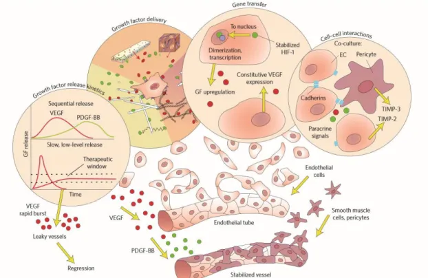
suppressed at the final stage of angiogenesis, which is characterized by the upregulation of endogenous angiogenesis inhibitors (Figure 8).
30
Figure 8. Vascularization of tissue-engineering scaffolds. (Reused from Ref. 19
with permission from Nature Publishing Group).
1.5.2. Biomaterial-Based Therapeutics to Target the Angiogenesis Process
Protein and peptide-based therapies designed to stimulate or inhibit angiogenesis have been clinically tested for a decade; however, only a few therapeutic agents have been approved. Transition of angiogenesis therapy from animals to humans requires comprehensive animal studies with biomaterials-based systems, which are currently limited. However, biomaterials nevertheless show great promise and might emerge as a new platform for the treatment of diseases which are currently known to be unresponsive to clinical intervention.
The discovery of advanced synthesis methods has facilitated the development of a great number of therapeutic reagents and peptide-based drugs. The development of synthetic peptides has progressed considerably with solid-phase peptide synthesis
31
especially in the areas of biotechnology and medicine. Among peptide based biomolecules, peptide amphiphiles are very useful to develop materials with advanced multi-functional properties through simple chemical alterations. Solid-phase peptide synthesis and liquid-phase peptide synthesis are two main methods commonly used in peptide based biomaterials. Solid phase peptide synthesis method depends on the progressive addition of amino acids onto an insoluble resin. After the first amino acid is attached to resin by a linker, following amino acids are added from the carboxy-terminus to the amino-carboxy-terminus. Side chains of each amino acid are bound to protecting groups to prevent any undesirable reaction throughout chain assembly. Protected groups are then removed in a controlled manner in each coupling reaction until the chain is completed. Finally, the synthesized peptide is cleaved from resin and lyophilized. The purity of the peptide molecule can be evaluated by liquid chromatography and mass spectrometry. The peptide can also be purified reverse-phase high-performance liquid chromatography 1.
The optimum length of peptide molecules for scaffold applications is between 10-20 amino acids. Larger peptides typically show solubility and uptake problems, limiting the maximum size that is feasible for use in cellular applications. Chemical structure of certain amino acid sequences complicate synthesis efforts, but in general, hydrophilic sequences are more soluble and known to be easier to synthesize. Peptides can be synthesized either manually or through a peptide synthesis machine. Although the manual methodology requires significant knowledge of peptide chemistry and is labor intensive, it allows much greater flexibility in the design and modification of peptides.
Peptides have favorable properties over antibodies in terms of their solubility, cost, size, and ease of modification and manufacturing 113. Since the angiogenesis process