Giant Post-traumatic Cyst after Motorcycle
Injury: A Case Report with Review of the
Pathogenesis
Yilmaz A.1, Yener O.2
1Department of General Surgery, Istanbul Medipol University, Istanbul, Turkey; 2Department of Surgery, Göztepe Training and Research Hospital, Istanbul, Turkey
Received June 11, 2012; Accepted April 8, 2013.
Key words: Huge cyst – Trauma – Hip
Abstract: Post-traumatic cysts of soft tissue usually occur at the junction of the
subcutaneous fat and deep fascia, most often filled with serosanguinous fluid and lined with fibrous tissue. It appears as complication after severe injuries when crushing and shearing forces cause separation of the skin and subcutaneous fat from the deep fascia and muscle, creating a cavity filled with hematoma and liquefied fat. This rare condition calls Morel-Lavallee lesion, which was first described by this French physician in 1853 (Sterling et al., 1977; Tull and Borrelli, 2003). Magnetic resonance imaging is the modality of choice for detection and revealing the exact size and location of these lesions. The best method of treatment is surgical excision with complete resection.
Mailing Address: Dr. Oktay Yener, Department of Surgery, Göztepe Training
and Research Hospital, Plaj yolu yıldız sok. Kaya apt. No:16 D:10 caddebostan, Istanbul, Turkey; Mobile Phone: 905 337 088 469; e-mail: [email protected]
Introduction
Post-traumatic cysts are well known entity that can arise after blunt, low or high velocity crush trauma in parenchyma organs, brain, bones and soft tissues.
Post-traumatic cysts are those that result from resolution of haematomas, and are commonly located between muscles, fascial or subcutaneous planes. The content is usually straw or brown coloured fluid containing cholesterol crystals. Manifestation of symptoms depends on the size, location and the presence of complications. It commonly appears about the hip region, especially over the great trochanter. In the majority of causes is associated with a pelvic girdle trauma (Hudson et al., 1992). Early surgical approach with percutaneous drainage, washing, irrigation and compression bandaging is a method of choice for these lesions (Kothe et al., 2006).
Case report
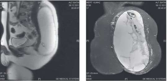
A male patient, 23-year-old, was admitted to the Department of General Surgery. He was complaining on swelling of left hip (Figure 1a and b). Patient’s history commenced 3 months ago, after the polytrauma (motorcycle accident). Two months later, fluctuated lesion was developed around sacrum and hip area with mild progressive enlargement during the time. The tumor-like formation was localized between subcutaneous tissue and deep fascia. The patient did not have a history of anticoagulation or bleeding diathesis. The magnetic resonance (MR) images revealed fusiform formation with well-defined margins. The lesion was 45 cm long (Figure 2a) and its widest diameter was 28 cm (Figure 2b),
appeared to be contained in the deep subcutaneous and perifascial space in the proximal part, generating a palpable bulge or compressive deformity on underlying muscles.
Such findings closely correlate with encapsulated waterlike fluid, in this case seroma. This probably accounts for the long-standing nature of the lesion. Surgical excision with complete resection of cyst had been performed (Figure 3). Cyst was filled with serous fluid and had thin capsule. During the operation, cyst was resected with capsule and sent to histopathology department. The wound was closed with simple multilevel interrupted sutures. Vacuum drainage
was removed twenty days and sutures two weeks after the surgery. During first month after surgery, moderate seroma formation had been evacuated several times. After that no further leakage had been noticed, which was confirmed with ultrasound examination. Control clinical examination, two months after surgery showed normal physical status with full range of motion. Histopathology showed cystic hemoragic cavity situated in the deep subcutaneous fat tissue.
Figure 2a and b – Huge post-traumatic cyst MR findings (sagittal and coronal).
Discussion
There are only a few scientific papers about such kind of lesion in literature. This rare condition was very often neglected in the early stage of expansion when the treatment is much easier to achieve. In the later stage, these lesions can resolve spontaneously, but in some cases can persist and change in pseudocystic formation. Long-standing Morel-Lavallee lesion had usually become a result
of chronic lymphatic extravasations and proliferate phase of healing, with fibroblasts and endothelial cells migrating into the injury site, forming epithelial border
to the healthy tissue (Harma et al., 2004; Tsur et al., 2006).
The subcutaneous fat is prone to trauma or ischemia. Fat necrosis arises due to multiple local or systemic events causing a compromise in the blood supply of the subcutaneous tissue. Its pathogenesis seems to be related to ischemic changes secondary to previous trauma. Encapsulated fat necrosis is a well-defined entity even though several names have been proposed for this condition, including mobile encapsulated lipoma, encapsulated necrosis, or nodular-cystic fat necrosis (Bradshaw et al., 1972). Beside trauma, there are many systemic diseases that can produce this condition such as lipodermatosclerosis, erythema nodosum, necrobiosis lipoidica, sclerosing panniculitis, nodular vasculitis, complication of pancreatic disease etc. (Mukherjee et al., 2007).
The patient was asymptomatic for at least two months when he started noticing a swelling at the site of previous trauma. Clinical assessment revealed a benign looking cyst and operative findings showed a huge cyst situated in the subcutaneous tissue which was highly suggestive of traumatic origin consistent with post-traumatic cyst that may follow resolution of haematoma.
Conclusion
Haematomas occurring in muscle masses may occur anywhere in the body, especially at the loins and anterior lateral aspects of the thigh. Our patient presented because of the massive size of the swelling. Large cysts may compress vital structures, like vessels and nerves causing individual symptoms. Complications such haemorrhage and calcification may follow infection or haemorrhage. Sudden haemorrhage occurring in the cyst leads to painful increase in size.
References
Bradshaw, J. R., Davies, G. T., Edwards, P. W., Richards, H. J. (1972) The radiological demonstration of traumatic cysts due to severe soft tissue trauma. Br. J. Radiol. 45(540), 905–910.
Harma, A., Inan, M., Ertem, K. (2004) The Morel-Lavallée lesion: a conservative approach to closed degloving injuries. Acta Orthop. Traumatol. Turc. 38, 270–273. (in Turkish)
Hudson, D. A., Knottenbelt, J. D., Krige, J. E. (1992) Closed degloving injuries: results following conservative surgery. Plast. Reconstr. Surg. 89, 853–855.
Mukherjee, K., Perrin, S. M., Hughes, P. M. (2007) Morel-Lavallee lesion in an adolescent with ultrasound and MRI correlation. Skeletal Radiol. 36, S43–S45 (Suppl. 1).
Sterling, A., Butterfield, W. C., Bonner, R. Jr., Quigley, W., Marjani, M. (1977) Post-traumatic cysts of soft tissue. J. Trauma 17, 392–396.
Tsur, A., Galin, A., Kogan, L., Loberant, N. (2006) Morel-Lavallee syndrome after crush injury. Harefuah 145, 111–113, 166. (in Hebrew)
Tull, F., Borrelli, J. Jr. (2003) Soft-tissue injury associated with closed fractures: evaluation and management. J. Am. Acad. Orthop. Surg. 11, 431–438.