T.C.
ISTANBUL MEDIPOL UNIVERSITY HEALTH SCIENCES INSTITUTE
MASTER THESIS
EVALUATION OF DIGOXIN AS ANTI-CANCER AGENT IN
COLON CANCER CELL LINE
KALIF MOHAMED MOHAMUD
DEPARTMANT OF BIOCHEMISTRY
SUPERVISOR
Assist. Prof. S. Sibel Erdem ISTANBUL - 2017
III
AKNOWLEDGEMENTS
First and foremost thanks to the Allah who has granted me all these graces to complete this Project and plessed me by his power, mercy, and patience during my life.
It is a pleasure to find the chance to thank all the people that were directy or indirect involved in the success of completion of this master thesis.
First of all I would like to thank my Supervisor Assistant Professor S. Sibel Erdem for her kindly help, her continuous support, and encouragement throughout the completion of this work. Without her guidence, advice, motivation, and her rich source of ideas this thesis would not be what it is today.
I would like to thank Professor Nesrin Emekli and Associate Professor Türkan Yigitbaşı for their enormous amount of support, advice, and encouragement during my master’s study really they are important factors for my successful and jury member Professor Ahmet Ata Alturfan.
I would like to thank Ms. Vildan for her support, and helping during my experiment. I am very grateful to my mother, to my wife and children, to my brothers and sisters for their supporting and encouragement.
I would like to thank all my friends specially Ahmed, Mohamed, and Abdishakur for their support and motivation.
Finally I would like to thank İHH and Zamzam foundation for providing scholarship and financial support.
IV CONTENTS
Page number
THESIS APPROVAL FORM………..…...I DECLARATION………...II ACKNOWLEDGEMENTS………...……...………III CONTENTS………..…...IV-VI ABBREVIATIONS AND SYMBOLS………..…..VII LIST OF TABLE………....VIII LIST OF FIGURES .……..……...………VIII
1. ABSTRACT………...1 2. ÖZET………...2 3. INTRODUCTION………3 3.1. Introduction………...3 3.1.1 Objectives………...5 3.1.2 Problem statement………...5
3.1.3 Purpose of the study………...5
3.1.4 Hypotheses………...6
4. GENERAL INFORMATION………...7
4.1. What is cancer?...………...7
4.2. Morphological and biochemical changes………...9
4.2.1 Morphological changes………...9
4.2.2 Biochemical changes………...9
4.3. Types of cancer………...10
V
4.3.2 Sarcoma………..……….……...10
4.3.3 Lymphoma………..…...10
4.3.4 Leukemia………...11
4.4. Cancer cell metabolism……….11
4.4.1 Metabolic alterations associated with cancer………...12
4.4.2.Regulation of cancer cell metabolism………..12
4.4.3 Hypoxia and cancer cell metabolism………...13
4.4.4 Metabolic signaling in cancer cells………...14
4.4.5 Metabolic behavior of cancer cells………...14
4.5. Colorectal cancer (CRC) ……...15
4.6 Apoptosis………15
4.6.1 Features of apoptosis………...16
4.6.2 Significance of apoptosis………...16
4.6.3 Correlation between apoptosis and proliferation………...16
4.7 Treatment of cancer………17
4.7.1 What is Irinotecan?...17
4.7.1.1 How Irinotecan Works……….19
4.7.1.2 Side effects………...19
4.7.2 Digoxin………...20
VI
4.7.2.2 Cardiac glycosides in cancer treatment………...23
4.7.2.3 Digoxin toxicity………...23
4.7.2.4 Side effects of digoxin………..23
4.7.2.5 Metabolism and excretionof digoxin………...…...24
4.7.3 Combination therapy………...24
5. MATERIALS AND METHOD……….29
5.1 Material………..……...,.29
5.2 Method………29
5.2.1 Cell Culture Experiments………...29
5.2.2 Digoxin Cytotoxicity………...29
5.2.3 Irinotecan Cytotoxicity...,………30
5.2.4 IC50 Determination and Analysis...30
5.2.5 Combination Therapy...31 5.2.6 CompuSyn Analysis………...31 6. RESULTS ………...32 7. DISCUSSİON………...39 8. REFERENCES………...46 9. CURRICULUM VITAE………58
VII ABBREVIATIONS
AFP Alpha fetoprotein ATP Adenosine triphosphate CEA Carcinoembryonic antigen CI Combination index
CRC Colorectal cancer
DNA Deoxyribonucleic acid
DMEM Dulbecco's modified Eagle's medium
DMSO Dimethyl sulfoxide
DRI Dose reduction index Fa Fraction affected
HIF Hypoxia inducible factor HL Hodgkin lymphoma
IC50 Half maximal inhibitory concentration MMS Mitochondrial metabolic signaling
MTT 3-(4,5-dimethylthiazol-2-yl)-25diphenyltetrazolium bromide NADPH Nicotinamide adenine dinucleotide phosphate
NHL Non-Hodgkin lymphoma PBS Phosphate-buffered saline PPP Pentose phosphate pathway UV Ultraviolet
VIII LIST OF TABLES
Table: 4.7.3 Properties of Digitalis Glycosides...21
Table: 6.4 Theoretical and experimental comparison for combinational affect declarement………...36
Table: 6.5 Dose Reduction Index Values of Digoxin and Ironetcan in Combination Therapy...38
LIST OF FIGURES Figure: 4.7.1 Structure of Irinotecan………...18
Figure: 4.7.2 Structure of Digoxin………...20
Figure: 4.7.4 Action mechanism of biochemical ………...22
Figure: 5.2.5 Concentration of digoxin and Irinotecan………...31
Figure: 6 .a.Digoxin treatment with 24 hrs incubation………...32
Figure: 6. b.Irinotecan treatment with 24hrs incubation……….32
Figure:6.1 HCT 116 colon cancer under the light microscope...33
Figure:6.2 Cell viabilty after combination therapy of Digoxin and Irinotecan...33
1 1. ABSTRACT
EVALUATION OF DIGOXIN AS ANTI-CANCERAGENT IN COLON CANCER CELL LINE
Colorectal cancer is a common disease all over the world and the occurrence of colorectal cancer is increasing in developing countries. Generally the occurence and mortality of colorectal cancer are substantially advanced in men than women. Many factors play a significant role in the advancement of cancer. Genetics, diet, alcohol, smoking, and other environmental factors are among the main causes for colorectal cancer growth.
The major goal of this study was to develop an in vitro cell culture based assay to investigate the effect of combination therapy of Digoxin and Irinotecan on chosen Human Colon Cancer Type (HCT 116) colon cancer cell line. Another objective was to examine the anticancer effect of Digoxin on HCT 116 cell line regarding to cell proliferation, apoptosis and inflammation.
Digoxin, which is a commonly used cardiac glycoside, and chemotherapy agent called Irinotecan are chosen for the study. Initially, the colon carcinoma cell line HCT 116 was treated with varying concentrations of Digoxin to study its effect on cell survival and proliferation. Treatment of Digoxin not only inhibited the growth of HCT 116 cell line but also Digoxin induced apoptosis and disrupted cell membrane. IC50 values for Digoxin and Irinotecan were determined as 78 μM and 330 μM, respectively. When Digoxin is used in combination with Irinotecan, the findings clearly showed that two drugs had synergistic effects at all concentrations at certain ratios. The results showed that 55% of the cell death could be achieved if the Irinotecan is used at 94.99 µM in combination with 68.8 µM Digoxin.
Our results showed that the system composed of Digoxin and Irinotecan has psitive outcome on the treatment of colon cancer. The results indicated that these two drugs can be used to generate more effective treatment for colorectal cancer as well as for other cancer types in near future.
2 2. ÖZET
KOLON KANSERİ HÜCRE KÜLTÜRÜNDE DİGOXİN'İN ANTİ KANSER ÖZELLİĞİNİN ARAŞTIRILMASI
Kolorektal kanser dünyada sık karşılaşılan hastalıklardan biri olup, insidansı gelişen ülkelerde artmaktadır. Genel olarak kolorektal kanserden ölüm oranları erkeklerde kadınlardan çok daha sık görülmektedir. Kanserin gelişmesinde etkin pek çok faktör bulunmaktadır. Kolorektal kanser gelişiminde genetik yapı, diyet, alkol, tütün ürünleri ve benzeri çevresel faktörler önem teşkil etmektedir.
Bu çalışmanın ana hedefi Digoxin ve İrinotectan adlı iki ilaçtan oluşan bir kombinasyon terapinin kolorektal kanser tedavisinde etkisini in vitro sistemde test etmektir. Diğer hedefi ise Digoxinin seçili hücre proliferasyon, apoptoz ve enflammasyon ile bağlantılı olan HCT 116 kolon kanser hücre zincirindeki anti-kanser etkisini incelemektir.
Sunulan çalısma için kardiyak glikozit olarak kullanilan Digoxin ile kemoterapi ilacı olan İrinotectan seçilmiştir. Öncelikle, HCT 116 hücreleri değişen Digoxin konsantrasyonları içeren hücre besiyeri ile inkübe edilip hücrelerin proliferasyon ve canlılılığı ölçülmüştür. Digoxin’in konsantrasyona bağlı olarak yalnızca HCT 116 hücre hattının canlılığını engellemediği ayni zamanda apoptozu indüklediği de görülmüştür. Digoxin, İrinotecan ile aynı anda uygulandıgında ise iki ilaç arasinda çalısılan tüm konsantrasyon değerlerinde ve belirli oranlarda sinerjik bir etkinin olduğu saptanmıştır. Digoxin’in IC50 değeri 78 μM ve İrinotecan IC50 değeri 330 μM olarak belirlenmiştir. 94.99 µMİrinotecan,68.8µM Digoxin ile birlikte uygulandıgında ise hücrelerin %55’inin canlılıgını yitirdiği belirlenmiştir.
Elde edilen sonuçlar, Digoxin ve Irinotecan’dan olusan kombinasyon terapinin kolon kanseri hücre hattındaki etkileri olumlu oldugunu göstermektedir. Bu sonuçlar bu iki ilacın kolorektal kanserinde ve diğer kanser çeşitlerinde yakın gelecekte daha etkin tedavi geliştirmek için kullanabileceklerini işaret etmektedir.
3 3. INTRODUCTION AND PURPOSE
3.1. Introduction
Cancer is a fatal disease, which develops as result of rapid and uncontrolled division of cells. When this kind of abnormal development takes place in the colon or rectum it is regarded as colorectal cancer (CRC), which is characterized as the large intestine cancer. Colon (large bowel or large intestine) is a muscular pipe approximately 5 feet lengthypart of the digestive system that absorbs water and salts from the food, whereas rectum reaches 6 inches of the digestive system (1).
CRC is a cancer, which grows in one of two, the colon tissues, which represents the longest branch of the great intestine, and in the rectum tissues, which is the large section of the intestine ahead of the anus. CRC is one of the most important cause of cancer deaths globally. Nearly all CRC beginas of a small development named polyp, such polyps are typically benign, but a few might grow as a cancer after long time, which may be more than 10 years. CRC is the second most common cancer in male and the third most common cancer in female population (2).
The occurrences of CRC differ extensively all over the world and the most frequent occurrence rates were found in developed countries particularly in Australia, New Zealand, Europe and North America, while the least incidence rates were found in Africa and Asia. These geographical discrepanciescan be attributed to variances in dietary and environmental factors. Nevertheless, the occurrence rates of CRC are getting higher in newly developed countries. The increasing rates of cancers are attributed to Westernization of diet, rise in obesity, and inactivity (3).
Many factors such as age, the existence of polyps, inflammatory bowel illness, standard of living, and genetic background have been associated with the risk of CRC. Environmental factors like obesity, physical inactivity, poor diet, and smoking and profound alcohol utilization make up around 80% of all CRC conditions (4).While early
4 CRC might not show any signs, and indications of CRC mainly rely on its position and capability to metastasize (5,6).
The majority of drugs used for the treatment of cancer today are cytotoxic drugs that work by interfering in some way with the operation of the cell’s DNA. Identification of cytotoxic compounds led the development of anti-cancer therapeutic for several decades. Advanced in cancer treatment, however continued to be limited by the identification of unique biochemical aspects of malignances that could be exploited to selectively target tumor cells (7).
Cardiac glycosides, aregroup of natural compounds, and are also important types of molecules for heart failure treatment.They have steroid like structures and inhibit membrane Na+-K+pump leading to activation of Na+/Ca++ exchanger and elevation of intracellular Calcium. Eventually this leads positive intropic effects on cardiac muscle. Among cardiac glycosides, particularly Digoxin has been formerly known and used as an antiarrhythmic agent. In the 1960s, an obvious inhibition of cancer cells by cardiac glycosides was stated (8). In recent years, there are several studies proposed that cardiac glycosides also have anti-cancer effect (9). In the shed of this light, Digoxin’s potential anticancer effects were investigated either by employing Digoxin alone or incombination with other agents(10). Antiproliferative, pro-apoptotic, and chemotherapy sensitization effects of Digoxin have been studied (8). Today, it is known that Digoxin inhibits proliferation of tumor cells when applied at optimal concentrations (11).
Irinotecan is a commonly used chemotherapy drug in clinics. It is generellay employed for the treatment of bowel cancer or CRC. Generally, it is used with other anti-cancer medicines. The mechanism of Irinotecan includes an enzyme known as topoisomerase, which takes role during DNA replication. Inhibition of this enzyme by Irinotecan, DNA replication halts, interfering with the synthesis of proteins required for cancer cell development and replication. Eventually, the cancer cell can not divide and proliferate (12).
5 3.1.1. Objectives
The main objective of this study was to develop an in vitro cell culture combination therapy based assay to study the effect of Digoxin and Irinotecan on colon cancer treatment. Our long-term goal was:
1. To understand the family history characteristics associated with increased CRC risk and with inherited CRC syndromes.
2. To contribute the prolongation of patient’s life, and life quality. 3. To help our understanding and improve knowledge of CRC. 4. To contribute to reduce mortality of cancer.
3.1.2 Problem statement
Treatment choices for CRC include surgery, chemotherapy, radiotherapy or combination of more than one treatment. The drugs that are used in particularly in chemotherapy are toxic and have several drawbacks to healthy cells as well. Thus, there is a need for new treatment regimens that has minimum or no side effect. Using more than one type of drug and trying to catch a synergy in between them to get better treatment efficiency is a way to achieve this goal (13).
3.1.3 Purpose of the study
The main purpose of this study was to plant seeds for a combination therapy for CRC employing two drugs that are already used in clinics. In order to achieve that goal, cell culture based assay used to study the effects of Digoxin and Irinotecan on CRC treatment using Human Colon cancer Type (HCT 116) colon carcinoma cell lines. Both of these drugs are used in clinics for different purposes as elaborated above. Employing varying concentrations of Digoxin and Irinotecan, we aim to obtain preliminary results for a new treatment strategy, which would offer synergism between the two drugs and would have minimum side effect.
6 3.1.4 Hypotheses
1. Digoxin, proportional to its concentration, inhibits the growth of HCT 116 cells. 2. Irinotecan and Digoxin can be used concurrently in HCT 116 cell line to screen the cell viability of the cell.
3. It is possible to determine synergism in between Irinotecan and Digoxin for colon cancer.
7 4. GENERAL INFORMATION
4.1 What is cancer?
Cancer is defined as a fatal disease, which arises when variations in a group of normal cells inside the body inclined to unlimited growth causing formation of a lump called a tumor. This description is applicable to all cancers except Hematological cancers. If the affected cells left untreated, tumors may develop and anchoring to the healthy tissue, or to further spread (metastasis) of the tumor might be possible through bloodstream and lymphatic system.
Health cells in human body are continuously split, differentiate into another cell or recycled based on the variety of signals and pathways. However, cancer cells grow independty from these signaling which yields unlimited development and proliferation. If this proliferation is left untreated and allowed to spread, it may be serious or incurable, as it is known that 90% of cancer associated deaths are due to tumor spread (14).
Cancer is also a disease which may happen again or persist during the lifetime of the cancer patient, some cancer patients are never entirely free of the disease whereas other cancer patients may free of the disease after medication or may be in remission and then the cancer may return. Although cancer encompasses at least varying diseases, all cancer cells have a significant attribute, they are nonstandard cells in which normal cell division disrupted, and also cancer grows from modifications that cause normal cells to gain abnormal functions.
These modifications are often the outcome of inherited mutation or came from environmental causes. Also the majority of cancers are not come from one particular incident or factor, cancer cells have nature of unlimited cell development, and also have capability to attack their close tissues and eventually to create metastasis in remote spaces of human body.
8 Gene mutation or deregulation also helps cell movement that is highly related with tissue attack and the formation of remote metastasis. Cancer from a series of molecular events that basically change the ordinary characteristics of the cell, in cancer cells the normal control systems that stop cell overgrowth and attack of other tissues are put out of action (15).
Despite different types of cancer, which are more than one hundred sorts, a number of similarities that are general to the majority of cancers have been determined. Healthy tissues cautiously control proliferative stimuli, which are conveyed by indicating pathways and rule cell development and division symptoms. These indicating pathways in the cancer cells are often changed, exposing cancer cells to proliferate autonomously. However, unrolled proliferation needs not only the presence of improved proliferative indications but also inactivation of the mechanisms that limit cell proliferation (16).
It is commonly accepted that cancer cells need unlimited replication potential as to produce macroscopic tumors. In healthy tissues, the replication capability is restricted by two major mechanisms. The first one is senescence which preserves cells in a resting but viable condition. The second one is crisis which engages cell loss. The capability of cancer cells to conquer these limitative processes has been demonstrated immortalization, and the enzyme telomerase constitutes a fundamental function in this procedure and is linked with a resistance to initiation of both these mechanisms.
The mortality of cancer depends on the ability of cancer cells for dispersion to other branches of the human body in a multistep process of attacking and metastasis. This process consists of a chain of cell alteration, starting with local invasion, then intravasation into close by blood and lymphatic vessels. Finally the growth of micro metastasis lesions into macroscopic tumors and turn into colonization. Also cancer cells grow alterations in their form as well as in their connection to other cells and to the extracellular matrix. There are a lot of diverse kinds of malignant tumors being
9 determined but more than 50% of recently diagnosed cancers occur in five main organs such as lungs, colon/rectum, breast, prostate, and uterus.
Cancers of the lungs, colon/rectum and prostate are the principal most important causes of deaths in men and in women, breast, colorectal and uterine cancers are the most widespread (17).
4.2 Morphological and biochemical changes
In comparasion to healthy cells, there are several morphological and biochemical changes in cancer cells.
4.2.1 Morphological changes
Have usually rounded shape, larger than health cells.
Cells show nuclear and cellular pleomorphism, hyperchromatism, altered nuclear: cytoplasmic ratio, abundant mitosis, sometimes tumor giant cells.
Transformed cells often grow over one another and form multilayers. Can grow without attachment to the surface in vitro, diminished adhesion. 4.2.2 Biochemical changes
Increased formation of DNA and RNA.
Show increased rate of glycolysis both aerobic and anaerobic. Show alterations of permeability and surface charge.
Changes in composition of glycoproteins and glycosphingolipids on cell surfaces.
Alterations of the oligosaccharide chains.
Increased activity of ribonucleotide reductase and decreased catabolism of pyrimidines.
Secretion of certain proteases and protein kinases.
Alterations of isoenzyme patterns often to a foetal pattern and synthesis of foetal proteins, e.g. carcinoembryonicantigen (CEA), α-fetoprotein (AFP), etc.
10 4.3 Types of cancer
Cancers are categorized into diverse sorts (Carcinomas, Sarcomas, Lymphomas and Leukemia’s), relying on which tissues they originate from by the type of cell that the tumor cells come from (19, 20).
4.3.1 Carcinomas
Carcinomas are cancers that initiate in the epithelial tissues, epithelial cells are the cells that covering the outside of the body as well as the inside parts and cavities, carcinomas might influence the breast, lung, prostate, and colon are also in the midst of the most generalsorts of cancer in adults.
Carcinoma which affect the adrenal glands. Carcinoma which affects the thyroid gland. Carcinoma which affects the nose and pharynx. Malignant melanoma which describes skin cancer. Skin carcinoma other than melanoma.
Other carcinomas including those affecting the salivary gland, colon, appendix, lung and bronchus, cervix, and urinary bladder.
4.3.2 Sarcoma
Cancers of the connective tissue are labeled Sarcomas. This kind of cancer grows in the connective tissue of the body include, fat, blood vessels, nerves, bones, muscles, deep skin tissues, and cartilage (21).
4.3.3 Lymphoma
Lymphoma is a cancer that develops in the white blood cells, which is part of body’s immune system. There are two major types of lymphoma: Hodgkin (HL) and non-Hodgkin (NHL) lymphoma (22).
11 4.3.4 Leukemia
Leukemia is also a cancer of white blood cells, but some leukemia begin in other blood cell sorts, signs of entirekinds of leukemia are associated to reproduction of nonstandard blood cells and substitution of the bone marrow by cancerous cells (23).
4.4 Cancer cell metabolism
Metabolism is the group of life-maintaining chemical revolutions inside the cells of living organisms. These interactions or enzyme-catalyzed reactions permit organisms to develop and reproduce, sustain their composition, and respond to their surroundings. All the cells in our body need specific molecules that can either be burned to create energy (catabolism) or utilized to produce building blocks of the basic structure to maintain proliferation (anabolism).
In relation to prerequisites of nutrients are mainly essential in reproducing cells, because they require to conserve their integrity and execute their physiological tasks and to produce a fresh daughter cell as well (24).
Cancer cells are distinguished by modifications in cell metabolism together withgreater glucose consumption and aerobic glycolysis, normally known as the “Warburg effect” (25). Cancer cells change their metabolism as to hold up their fast reproduction and spreading out through the body. Typical cell replication in the tissues is restricted by the accessibility of growth controlling factors and by the interaction with nearby cells.
The accessibility of nutrients and oxygen, needed for cell replication and metabolism, mainly relies on blood supply, because early tumor development happens in the absence of creation of new blood vessels. Tumor cells have a greatly advanced rate of glucose use through a glycolysis pathway which does not provide pyruvate to the Krebs cycle but that instead changes pyruvate to lactate, many tumors consume this
12 glucose to lactate pathway even in the existence of oxygen, illustrating why the term aerobic glycolysis is frequently employed as an alternative to the Warburg effect (25).
4.4.1 Metabolic alterations associated with cancer
Cancer cells displayed major changes in energy metabolism and mitochondrial respiration weighed against ordinary cells, also cancer cells actively utilized glycolysis for ATP production, even in the existence of an abundant supply of oxygen. Although whether metabolic changes cause tumor genesis or are aconsequence of transformation is still under discussion, but greatreliance on glycolysis is seen in the most of tumors and that glycolysis gives ATP in addition to the metabolic intermediates necessary for cancer cell proliferation and tumor growth.
Metabolic intermediates produced through glycolysis offer significant basics or foundations for the synthesis of DNA, fatty acids and for redox control. For example, glucose-6-phosphate, the product of the first glycolytic reaction, may be channeled to the pentose phosphate pathway (PPP) to produce ribose5phosphate and nicotinamide adenine dinucleotide phosphate (NADPH), which are significant for nucleic acid metabolism and redox homeostasis, respectively.
NADPH and three carbon metabolic intermediates are also essential for lipid biosynthesis.Last but not least, the high levels of lactic acid generated during glycolysis might activate metalloproteinase and matrix remodeling enzymes, thereforeresulting cancer metastasis (26).
4.4.2 Regulation of cancer cell metabolism
The cells of the cancer show metabolic phenotypes, which differentiate them from ordinary tissue cells, in specifically an improved activity of metabolic pathways are essential for cell development. In turn, accumulating evidence presents that major oncogenes, for instance, Ras and Myc, positively control metabolic pathways that are up regulated in cancer cells.
13 Cancer cells obtain alterations to the metabolism of all four main classes of macromolecules carbohydrates, proteins, lipids and nucleic acids. Several comparable alterations are also seen in quickly proliferating normal cells, in which they stand for suitable reactions to physiological development indications as opposed to constitutive cell autonomous adaptations (27).
According to cancer cells, these adaptations should be executed in the stressful and dynamic microenvironment of the solid tumor, where concentrations of critical nutrients like glucose, glutamine and oxygen are temporally heterogeneous. The character and significance of metabolic response in cancer has often been maked owing to the use of tissue culture circumstances in which both oxygen and nutrients are constantly in surplus.
Malignant conversion of cells needs definite adaptations of cellular metabolism to hold up development and survival. In the early twentieth century, Otto Warburg recognized that there is elementary dissimilarity in the central metabolic pathways working in malignant tissue. He displayed that cancer cells use a large amount of glucose, sustain high rate of glycolysis and alter a majority of glucose into lactic acid even under normal oxygen concentrations (28).
4.4.3 Hypoxia and cancer cell metabolism
Hypoxia is a situation where in the human body or an area of the human body is deprived of sufficient oxygen. Hypoxia is frequently a pathological situation. The environmental stress of hypoxia activates molecular alterations that make easy metabolic adaptations. Some of such changes are up-regulated glycolytic enzyme expression along with oxidative phosphorylation inhibition, mitochondrial selective autophagy, and glucose, citrate creation for fatty acid synthesis (29).
14 Hypoxia-inducible factor (HIF) activation is the most recognized and distinguished molecular reaction running many of these changed metabolic pathways of hypoxia. It has been revealed in the recent few years that oncogenes discovered in a extensive diversity of human cancers can directly activate Hypoxia and other elements of glucose metabolism autonomously of hypoxia (30).
4.4.4 Metabolic signaling in cancer cells
Mitochondrial metabolic signaling (MMS) can be defined as the regulation of cellular gene expression by various (energy) metabolites and by-products that accumulate upstream or downstream of the mitochondrion. The feedback of this process takes place under physiological situations and contributes to the control of cell homeostasis, as well as the diverse pathologies MMS is altered by the abnormal accumulation of indicating metabolites, which interferes with physiological processes and contributes to the progression of the illness and also phenotypic variability.
As mitochondria hosts an excess of cellular metabolic reactions, this organelle is essential for metabolic signaling, like vascular endothelial growth factor, and glucose transporter, which are engaged in the support of tumor proliferation through a redefinition of the metabolic profile (26).
4.4.5 Metabolic behavior of cancer cells
Cancer cells attain an abnormal glycolytic behavior relative to a large extent, to their intracellular alkaline pH. This effect is part of the metabolic changes obtained in most, if not all, cancer cells to deal with adverse situations, primarily hypoxia and low nutrient supply, in order to conserve its evolutionary trajectory with the production of lactate after ten steps of glycolysis. Therefore, cancer cells reprogram their cellular metabolism in a way that provides them their evolutionary and thermodynamic benefit. Tumors have highly heterogeneous microenvironment and cancer cells survive within any of the various habitats that lie within tumors (31).
15 4.5 CRC
Among various cancer types, CRC is a cancer that develops either in the tissues of the colon, the longest part of the large intestine, or in the tissues of the rectum, the least part of the large intestine before the anus (32). Genetics, hormonal changes, environmental factors, alcohol, weight, age, physical activity and diet has role in the development of CRC (33, 34, 35, 36, 37, 38, 39, 40, 41, 42).
4.6 Apoptosis
Apoptosis is a Greek origin, which means "falling off or dropping off" (43). Apoptosis is described as a morphologic alteration resulting in controlled cellular self-destruction by a process obviously different from necrosis. Since the 19th century, several studies have displayed that cell loss contributes to throughout physiological prograssion of multicellular organisms, especially through embryogenesis and metamorphosis (44).
The apoptotic form of cell loss is an active and described process, which plays a significant role in the development of multicellular organisms and in the control and protection of the cell populations in tissues upon physiological and pathological situations. Cell loss is a necessary part of the usual growth and persists into adulthood. Apoptosis contributes to a crucial part in limiting a lot of cells in development and during the life of an organism by the elimination of cells at the suitable moment.
Changes of this regular process may lead to the disturbance of the slight balance between cell replication and cell loss as well as could result in a diversity of diseases. For instance, in several types of cancer, most important propoptotic proteins are mutated or antiapoptotic proteins are up controlled, causing the accumulation of cells and the incapability to react to damaging mutations, DNA damage, or chemotherapeutic agents (45).
16 4.6.1 Features of apoptosis
Cell loss might take place through at least two generally described mechanisms: necrosis or apoptosis. Necrosis is a passive, catabolic, pathological cell death process which commonly happens in reaction to external toxic factors like inflammation, ischaemic or toxic injury.It is not thought to ever happen under physiological situations. On the other hand, apoptosis is an active, metabolic, genetically encoded and evolutionarily chosen death pathway. It takes place under either physiological or pathological situations (46).
4.6.2 Significance of apoptosis
The growth and preservation of multicellular biological systems relay on complicated interaction among the cells composing the organism. It occasionally tends to engage an altruistic performance of individual cells in favor of the organism. .As a whole, apoptotic processes are common biological importance, being engaged in e.g. growth, differentiation, replication/homoeostasis, control and function of the immune system. Therefore, dysfunction or dysregulation of the apoptotic program is implicated in a variety of pathological situations (47).
4.6.3 Correlation between apoptosis and proliferation
The balance between cell proliferation and cell death throughout apoptosis identifies how fast a tumor develops. Most colorectal adenomas are steady for a longtime before, if ever, altering to malignancies. Traditionally, an increase in cell replication, rather than a change in apoptosis, has been used to forecast later tumor growth and prognosis. Nevertheless, in an animal model, the best estimator of tumour development was the level of apoptosis, whereas there is common conformity that deregulation of apoptosis plays a significant role in malignant transformation. The potential estimated or prognostic value of the level of apoptosis in CRC is in question (48).
17 4.7 Treatment of cancer
Treatment is the series of contribution, including psychosocial help, surgery, radiotherapy, and chemotherapy that is targeted curing the disease or significantly prolonging life and life quality of patient. Cancer diagnosis is the first step to cancer management. It includes combination of cautious clinical evaluation and diagnostic examinations together with endoscopy, imaging, histopathology, cytology and laboratory researches (13).
Treatment of cancer, especially CRC, might require surgery, chemotherapy (use of anti-cancer drugs), or radiotherapy (use of x-ray to target and kill cancer cells), hormonal therapy alone or in combination (49). CRC treatment may be managed towards three different phases of the illness. The first is the tumor development, the second is the original cancer tissue and the third is the systemic disease. In the near future, the greatest advantage in CRC treatment might not be at the degree of the primary tumor, but in identifying initial steps in disease progress with respect to prevention, initial discovery and treatment (50).One of the most frequent and advantageous chemotherapeutic drugs used for colon cancer is Irinotecan and the treatment efficiency is dose dependent (51).
4.7.1 What is Irinotecan?
Irinotecan is a chemotherapy agent for bowel cancer and CRC. It is typically used in combination with other anti-cancer medicines.Irinotecan works by inhibiting an enzyme known as topoisomerase. It is an essential enzyme for cells since they require this enzyme to proliferate and develop as of two new cells. The rule applies to helthy cells as well as cancer cells. During cancer treatment, Irinotecan inhibits this enzyme, then the DNA of cellsbecomes tangled up and the cancer cells cannot divide (52).
18 Figure 4.7.1Structure of Irinotecan
For more than three decades, the therapeutic choices for patients with complex CRC have almost completely been rooted in fluoropyrimidines. With the determination that topoisomerase-I (TOP-I) is a significant therapeutic target in cancer therapy, Irinotecan, a semisynthetic TOP-I–interactive camptothecin derivative, has been clinically established in the treatment of CRC. (53).
Irinotecan is an anti-cancer chemotherapy agent. This drug is known as a "plant alkaloid" and/or "topoisomerase I inhibitor." It is a topoisomerase I inhibitor used to treat variety of solid tumors and it is being usually used in combination with other chemotherapeutic agents for the treatment of CRC. (54). The dose of irinotecan that patient recieves rely on many factors such as height and weight, general health and/or other health problems as well as the stage of the cancer being treated. (55).
Chemotherapy treatments are the most effective when provided in other drugs in combination chemotherapy. As it is concluded from the results that were obtained in this thesis as well, the foundation for combination chemotherapy is to utilize medications that function by diversified mechanisms, consequently diminishing the chance that
19 resistant cancer cells will grow. When treatments with diverse impacts are joined, each drug can be used at its optimal dose, without intolerable side effects. (56).
4.7.1.1How Irinotecan Works:
Cancerous tumors are attributed to cell division and grow, which is no longer limited as it is in normal tissue. "Normal" cells control/prevent proliferation when they come into contact with such cells, a mechanism labeled as contact inhibition. Cancerous cells lose this capacity. Cancer cells no longer have the normal check points for cell cycle and balances in place that control and limit cell division. The progress of cell splitting up, whether regular or cancerous cells, is throughout the cell cycle. The cell cycle leaves from the inactive phase, through active developing phases, and then to mitosis (division). The capacity of chemotherapy to eliminate cancer cells relays on its capability to stop cell division. Generally, the treatments work by damaging the RNA or DNA that alerts the cell how to copy itself in division. If the cells could not split, they would die. The quicker the cells are splitting up, the more possibly it is that chemotherapy will eliminate the cells, resulting the tumor to contract. They also induce cell suicide (self-death or apoptosis) (55). Irinotecan follows the similar mechanism, inhibition of TOP-I, to stop cell proliferation
4.7.1.2 Side effects of Irinotecan
Like other chemotherapy agents, Irinotecan has several side effects which lowers life quality of cancer patient (57). These side effects includes but not limited to
Increased sweating Diarrhea
Increased production of saliva Watery eyes
20 4.7.2 Digoxin
Digoxin is one of the cardiac glycosides that directly associated with a collection of medicines having in common definite impacts on the myocardium (muscular wall of the heart). Digoxin contains steroidal nucleus and sugar at position 17 (Figure: 4.7.2). The attached sugars such as glucose, galactose, mannose, and digitalose identify the pharmacodynamic and pharmacokinetic actions of each cardiac glycosides. Digoxin is used in the treatment of cardiac pathology like atrial fibrillation and heart failure (58).
Figure: 4.7.2 Structure of Digoxin
Cardiac glycosides are a group of naturally happening compounds with a steroid like structure can inhibit Na+-K+ ATPase, which can lead to activation of Na+/Ca++ exchanger and elevation of intracellular Calcium, and eventually lead a positive intropic effects on cardiac muscle. Digitoxin and Digoxin are the two commonly prescribed cardiac glycosides. Chemical structures of these two glycosides have three essential structural elements: an unsaturated lactone ring, a steroid nucleus, and many sugar moieties (which mainly effect the pharmacokinetic properties). Digoxin has an additional hydroxyl group compared to Digitoxin (59). Digitalis Glycosides have different properties as shown in Table 4.7.3.
21 Table: 4.7.3 Properties of Digitalis Glycosides
Property Digoxin Digitoxin
Lipid Solubility Medium High
Oral Absorption 75% >90%
Plasma Protein Binding 20-40% >90%
Plasma Half-Life 1.6 days 7 days
Route of Elimination Kidney Liver
Despite their different structure, both of them are inhibitors of the plasma membrane Na+-K+-ATPase. As they inhibit Na+-K+-ATPase, intracellular Na+ concentration increases while intracellular K+concentration decreases, resulting in an increase in cardiac contraction. This action acts as the synthesizing of signaling molecules like Caveolin, SRC kinase, epidermal growth factor receptor (EGFR) and the inositol triphosphate receptor. The inhibitory effects of cardiac glycosides on Na+-K+ -ATPase could account the biological properties of these compounds including their anticancer activities (60).
The mechanism whereby cardiac glycosides lead to a positive inotropic impact and electrophysiological alterations is still not entirely clear. Many mechanisms have been projected, but the main generally agreed engages the capability of cardiac glycosides to hold back the membrane bound Na+-K+-ATPase pump in charge of Na+-K+ exchange (61). The process of muscle contraction may be depicted as shown in Figure: 4.7.4.
22 Figure: 4.7.4Biochemical mechanism of action
4.7.2.1 Anti-cancer and Anti-proliferative effects properties of digoxin
Several studies proposed that cardiac glycosides can have anticancer characteristics. Also Digoxin inhibit proliferation of human tumor cells when applied at optimal concentrations (11).
Increased rate of cell proliferation is hallmark of cancer development. Cardiac glycosides have anti-proliferative activities through their control of the cell cycle. Anti-proliferative impacts of cardiac glycosides against human breast and prostate cancer cells have also been stated (62). The growing of intracellular calcium concentration followed treatment with digoxin is related to the anti-proliferative impacts of this composite in androgen-reliant and androgen-autonomous prostate cancer cell lines (63).
23 4.7.2.2 Cardiac glycosides in cancer treatment
Preclinical and clinical data recommend that Digoxin may be used as anti-cancer agent in cancer therapy.In addition, recent reports have shown that Digoxin can prevent development of cells and induce apoptosis in cancer cells at certain concentrations recommended for cardiac patients. Cardiac glycosides used for the medication of cardiac problems might also be beneficial as anticancer agents (64).
Several epidemiological researches concluded that Digoxin is effective in a lot of cancers including lung, colon, prostate, and ovary. However, among middle aged women using Digoxin, breast cancer and uterus cancers rates have increased since both of these cancers are often estrogen-sensitive (60, 65).
4.7.2.3 Digoxin toxicity
Digoxin toxicity takes place when Digoxin concentration exceeds tolerable limits in a human body. Digoxin toxicity causes hyperkalemia, which is known as high potassium concentration in blood. The sodium/potassium ATPase pump push sodium out of the cell and uptake potassium from the cellular fluid into the cell. Blockage of this mechanism results in high potassium levels in blood and eventually to hyperkalemia. Digoxin toxicity is declined in condition called hypokalemia (low potassium level in blood) since Digoxin competes with potassium to bind to the ATPase pump on the same location. When potassium levels are low, digoxin can more simply attach to the ATPase pump and its inhibitor function gains great impacts. There are some factors such as age, kidney diseases, hypothyroidism, heart disease and use of medicines that interact with digoxin that increasethe risk for digoxin toxicity (66).
4.7.2.4 Side effects of digoxin
The major reactions of digoxin arise when there is an excess of it in the blood stream. These impacts may range from gastrointestinal impacts like appetite, abdominal pain, intestinal ischemia, hemorrhagic necrosis of the intestines, vomiting, and diarrhea
24 or behavior changes to severe heart abnormalities. Furthermore, digoxin might bring about visual disturbances, headache, weakness, dizziness, apathy, confusion, mental disturbances, and skin reaction (67).
4.7.2.5 Metabolism and excretion of digoxin
Following intravenous administration of Digoxin to healthy volunteers, 50% to 70% of the drug dose is excreted unaltered way in the urine. In healthy volunteers with healthy renal function, digoxin has a half-life of 1,6 to 2 days. The urinary metabolites, which encompasses, digoxigenin bisdigitoxoside, dihydrodigoxin and their glucuronide as well as sulfate conjugates, are polar in nature and are believed to formas a result of hydrolysis, oxidation, and conjugation (68).
4.7.3 Combination therapy
The overall goal of cancer therapy is to destroy all the cancer cells at the same time with minimal harm to the healthy tissue. This may be reached in several ways, either directly or indirectly by depriving the cancer cells of signals required for cellular replication or by stimulating an immune reaction. Combination therapy is one way to improve effectivenss of the cancer treatment (69).
Combination therapy is a wide expression for the use of various treatments or therapies as to fight the one single disease. Although it normally represents the use of two or more medicines, it may also include immunotherap, radiotherapy, chemotherapy, surgery, non-medical therapies along withpsychological support.
The practice of combination therapy especially for cancer patientsis not new.Combination therapy may provide an obvious benefit to the patient. For instance, while one medicine might be used to reduce inflammation or slow down the development of disease, another may directly devastate the foreign cells. Therefore, in order to get best outcome, it is beneficial to apply more than one therapy to treat cancer
25 like diseases (70).Over the past decade, since many doctors, oncologists, and other medical professionals suggest combination therapies for a variety of diseases and conditions, the number of accepted and examined combination therapies significantly raises. The use of more than one kind of therapy is a hallmark of cancer medication. The complexity of the disease, rate of metastasis, fast advancement in stages, as well as its genetic variety highlights the need for a different of methods to invade it. For well over half a century, physicians have had three major sorts of cancer medications: surgery, radiotherapy, and chemotherapy. The use of a definite treatment or combination of treatments relays on an array of aspects, including the type of cancer, its aggressiveness, and it’s potential to metastasize, or spread to other parts of the body. With today’s technology and knowledge, it is essential to identify how this method can assist in treatment of various cancer typeswith minimal side effects to the patient (71).
Combination therapy, a type of treatment that unites two or more therapeutic agents, is a foundation of cancer therapy (72). The mixture of anti-cancer treatments improves efficiency weighed against the mono-therapy method since it aims at significant and/or different pathways in a characteristically synergistic or an additive way. This way mainly diminishes drug resistance, whereas concurrently offering therapeutic anti-cancer advantages, such as degreasing tumor development and metastatic potential, arresting mitotically active cells, degreasing cancer stem cell populations, and inducing apoptosis (73).
The complication of the disease, its inclination to extend outside of its original place, and development of resistance to definite treatments as well as its genetic variety highlights the necessity for a diversity of methods to attack it. In several situations, combination therapy not only raises the probability of a treatment or long-term remission, but also diminishes harm to essential organs and tissues more than a single method (74).
26 For almost more than half a century, physicians have had three major sorts of cancer medications in their arsenal: surgery, radiation therapy, and chemo-, or drug, therapy. The use of a specific medication or mixture of medications relies on a range of aspects, together with the kind of cancer, its aggressiveness, and its potential to metastasize, or extend to other branches of the body. As a common law, solid tumors (which shape masses within individual organs) are typically cured by surgery, if limited to the place they began in, and/or radiation therapy to take away or decrease the tumor, followed by chemotherapy to remove any residual tumor cells. Blood-associated cancers suchas leukemia, lymphoma, and multiple myeloma are commonly cured by chemotherapy agents and/or radiation therapy since the disease is dispersed over wide parts of the body (73).
One sort of treatment may occasionally formulate a tumor more susceptible to a second sort. Specific chemotherapy treatments, for instance, may raise tumors’ susceptibility to radiation therapy. In other conditions, treatments work together, each improving the others’ strength, so their joint effectiveness is superior than would be forecasted by their individual effect. Combinations of diverse chemotherapy agents are often employed to obstruct cancer’s “escape routes.” A tumor that develops into resistant to one chemotherapy agent might surrender to treatment with a second or third. (75).
In the latest years, a diversity of latest treatments has been commenced that supplement the traditional triad of surgery, radiation therapy, and chemotherapy. These comprise “targeted” drugs that zero in on specific irregular genes or proteins in tumor cells, anti-angiogenic drugs that stop tumors from siphoning off nutrients from the bloodstream to maintain their unrestricted development, that impede cancer cells’ capability to refurbish smashed DNA, and immunotherapy agents that promote an immune system attack on tumors. (76).
Over a half-century has gone since cancer chemotherapy started. The chemotherapy agents now in use initiated as biologically targeted therapy. Hitchings and
27 Elion built up certain inhibitors of purine synthesis, such as 6-MP; Heidelberger targeted RNA synthesis with 5-FU; and Farber targeted the reduced folate pathway with aminopterin (77). These improvements not only offered the basis for cancer medication but also turned into tools for finding of the basic workings of transformed cancer cells. Natural products like the Vinca alkaloids, anthracylines, and taxanes were chosen specially for activity against cancer propagation. These agents were joined with other classes of agents, such as hormones, alkylating agents, and irradiation that are active against reproducing cells. (78).
For several cancers, the most excellent methodology is a combination of surgery, radiation therapy, and/or chemotherapy. Surgery or radiation therapy cures cancer that is limited to its site, whereas chemotherapy also eliminates the cancer cells that have extended to remote places. Sometimes radiation therapy or chemotherapy is provided before surgery to contract a tumor, as a result leading the opportunity for complete surgical elimination. Radiation therapy and low-dose chemotherapy after surgery assist to devastate any residual cancer cells. The stage and type of the cancer frequently reveals whether single therapy or amalgamation therapy is required. For instance, early-stage breast cancer might be cured with surgery alone or surgery joined with radiation therapy, chemotherapy, or with all three medications, relying on the size of the tumor and the exposure of reappearance. For instance, locally advanced breast cancer is commonly cured with chemotherapy, radiation therapy, and surgery (79).
Sometimes combination chemotherapy is employed not to treat but to diminish signs and prolong life. Combination chemotherapy can be helpful for people with complex cancers that are not appropriate for radiation therapy or surgical treatment (for example, people with non–small cell lung cancer, esophageal cancer, or bladder cancer that cannot be entirely eliminated by surgery) (80).
Combinations of diverse chemotherapy agents are regularly utilized to block cancer’s “escape routes.” A tumor that turns into resistant to one chemotherapy agent may succumb to treatment with a second or third. In many conditions, combination
28 therapy not only increases the chances of a cure or long-term remission, but also reduces damage to vital organs and tissues more than a single approach. The stage and type of the cancer often identifies whether single therapy or combination therapy is necessary. For instance, early-stage breast cancer might be healed with surgery alone or surgery combined with radiation therapy, chemotherapy, or with all three medications, relaying on the size of the tumor and the danger of repetition. (81)
Chemotherapy treatments that influence cells only when they are splitting up are called cell-cycle specific. Chemotherapy treatments that influence cells when they are at rest are called cell-cycle non-specific. The scheduling of chemotherapy is set according to the sort of cells, rate at which they split up, and the time at which a given drug is expected to be effective. This is the reason why chemotherapy is usually given in cycles. Chemotherapy is mainly efficeint at eliminating cells that are quickly splitting up. Unfortunately, chemotherapy does not determine the difference between the cancerous cells and the regular cells. The "normal" cells will develop back and be healthy but in the meantime, side effects happen. The "normal" cells most generally affected by chemotherapy are the blood cells, the cells in the mouth, stomach and bowel, and the hair follicles; resulting in low blood counts, mouth sores, nausea, diarrhea, and/or hair loss. Different drugs may affect different parts of the body (55).
Occasionally combination chemotherapy is used not to cure but to diminish signs and prolong life. Combination chemotherapy can be helpful for people with complex cancers that are not suitable for radiation therapy or surgical treatment (for example, people with non–small cell lung cancer, esophageal cancer, or bladder cancer that cannot be completely removed by surgery) (81).
29
5. MATERIAL AND METHOD 5.1 Material
This studyof HCT116 involving routine cell culture was analysed in the Medipol University Laboratory by using the following Solutions and chemicals;
HCT 116, Human Colon Cancer Cell Line (ATCC) DMEM with L-Glutamine (CAPRICORN)
Fetal Bovine Serum (FBS) (BIOWEST)
Phosphate-Buffered Saline (PBS) (CAPRICORN) Antibiotic (penicillin-streptomycin)
Trypsin- EDTA (0.05%) in DPBS (CAPRICORN) Dimethyl sulphoxide (DMSO) (MERCK)
MTT (3-(4,5-Dimethylthiazol-2-yl)-2,5-Diphenyltetrazolium Bromide) SpectraMax Plate Reader
Digoxin (C41H64O14) Irinotecan (C33H38N4O6)
5.2 METHOD
5.2.1 Cell Culture Experiments
HCT-116 human colon cancer cell line is cultured by the DMEM medium with 10% FBS (fetal bovines serum) and 100U/ml Penicillin/Streptomycin under the cell culture conditions. Cells are passaged in every three days.
5.2.2 Digoxin Cytotoxicity
HCT-116 human colon cancer cells seeded into 96 well plates by 10 000 cells/well and incubated 24 hours for complete adhesion. 7 different Digoxin solutions prepared between 0.001 µM to 20 µM concentration and in every well 200 µl related concentration was applied, incubated for 24 hours. One well was marked as control
30 group, treated with only complete medium and another one is as positive control and treated with %0.25 Triton-X. All experiments repeated in triplacate. After incubation, medium with Digoxin is removed and changed with 200 µL fresh medium with 5 ng MTT (3-(4,5-Dimethylthiazol-2-yl)-2,5-Diphenyltetrazolium Bromide). Cells were incubated with MTT solution at 37 C for 3 hours. Then MTT solution removed and 200 µL DMSO was added to all wells.After shaking at 300 rpm for 20 minutes to provide full dissolution of formazan and absorbance is measured at 570 nm by plate reader. 5.2.3 Irinotecan Cytotoxicity
HCT-116 human colon cancer cells seeded into 96 well plates by 10 000 cells/well and incubated 24 hours for complete adhesion. 7 different Irinotecan solutions prepared between 0.1 µM and 500 µM and in every well 200 µl related concentration was applied, incubated for 24 hours. 1 well was marked as control group, treated with only complete medium and another one is as positive control and treated with %0.25 Triton-X. All experiments were done in triplicate. After treatment, medium with digoxin is removed from cells and changed with 200 µL medium with 5 ng MTT (3-(4,5-Dimethylthiazol-2-yl)-2,5-Diphenyltetrazolium Bromide). Cells was incubated with MTT solution at 37 C for 3 hours. Then MTT solution removed and 200 µL DMSO was added to all well shaked at 300 rpm 20 minutes to provide full dissolution of formazan and absorbance is measured at 570 nm by plate reader.
5.2.4 IC50 Determination and Analysis
The drug concentrations used in most of the combination studies were based on the IC50 values, which are characterized as the drug concentration responsible for 50% growth inhibition. In this study, GraphPad Prism 6 was used to determine IC50 values and to produce following graphs. For determination of these data, untreated cell control group is accepted as 100 and Triton-X treaated control group was accepted as 0. By this process other absorbance values were normalised and became comparitable results. According to the IC50 values of each drug combination therapy experiment was set up.
31 5.2.5 Combination Therapy
HCT-116 human colon cancer cells seeded into 96 well plates by 10 000 cells/well and incubated 24 hours for complete adhesion. 10 different concentration points has been applied between 0.187X and 6X where X is IC50 values for each drug (Figure 5.2.5). Drugs were applied to the cells both at the same time, this condition requires 2 times more concentrated solutions for each drug since same time application causing volume decreasing to the half of the usual volume. After the treatment, medium with drug combination is removed from cells and changed with 200 µL medium with 5 ng MTT (3-(4,5-Dimethylthiazol-2-yl)-2,5-Diphenyltetrazolium Bromide). Cells were incubated with MTT solution at 37 C for 3 hours. Then MTT solution removed and 200 µL DMSO was added to all well shaked at 300 rpm 20 minutes to provide full dissolution of formazan and absorbance is measured at 570 nm by plate reader. Results were evaluated in CompuSyn program.
Figure: 5.2.5This is showing concentration of Digoxin, Irinotecan, and combination of both drugs, also IC50 values, concentration (or dose) required to inhibit (or to affect) a system by 50%.
5.2.6 CompuSyn Analysis
The analysis of effect of obtained results between separately applied treatments for each drug and combinational therapy was determined by Prof. Chou’s directions (Chou and Martin, 2005) Compusyn program.
32 6. RESULTS
After the application of 7 different concentrations (0.001 µM to 20 µM) of Digoxin and (0.1 µM -500 µM) Irinotecan, results was normalized and graphs were drawn (Figure 6a and Figure 6b) on HCT-116 we obtained from the results, the effect between drug molecules separately and the combinative application (Figure 6.2). The IC50 values (concentration required to reduce the initial cell number by 50%) of Digoxin and Irinotecan were calculated 78 μM and 330 μM respectively. To apply combination therapy we need to apply drugs at these IC50 concentrations and by the help of CompuSyn program, the effect of thee combination therapy (synergism, antagonism or addititive) and clinical beneficially became clear. Combination therapy was used in a constant ratio as it was mentioned above and according to the IC50 values ratio was calculated as 1:4.25 (78 μM: 330 μM).
Figure 6: HCT 116 human colon cancer cell viability after a) digoxin treatment with 24 hour incubation b) Irinotecan treatment with 24 hour incubation
a)
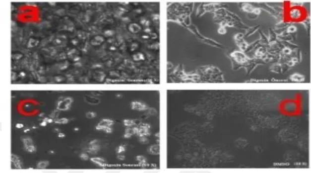
33 Figure:6.1: HCT 116 colon cancer under the light microscope images of cells a) After 24 hours after Digoxin incubation (20x), (b)Before incubation with Digoxin (40x), (c) 24 hours after Digoxin incubation (10x) (d)24 hours after DMSO application (10x)
Figure 6.2:HCT 116 human colon cancer cell viability after combination therapy of Digoxin and Irinotecan at constant ratio of 1:4.25
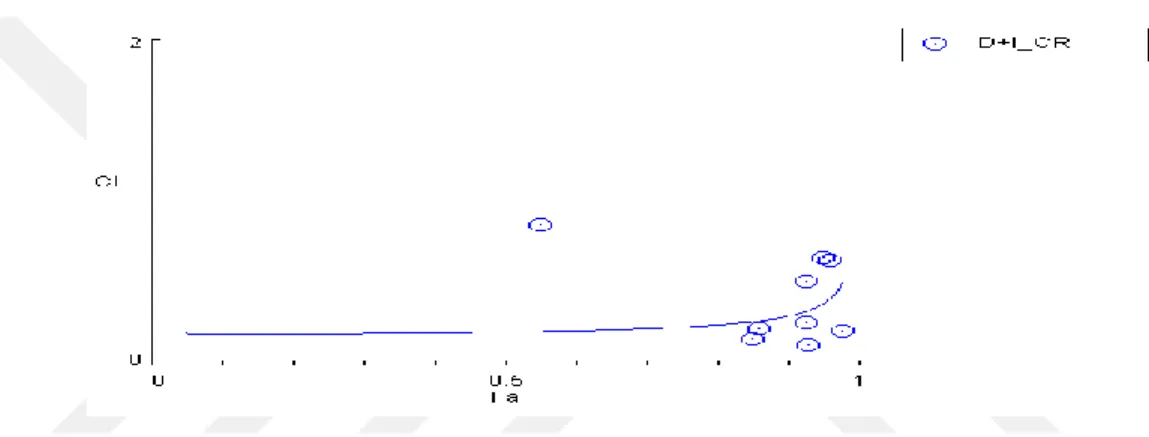
34 According to the CompuSyn program database, combinational therapy results at all the applied points show the synergistic feature (Figure 6.3) which is also consistent with the theoretical data produced by the software (Table 6.4). Synergisim is determined according to the value called combinational index (CI). If CI value is smaller than 1, results are accepted to show synergistic effect, if it is equale to 1 showing additive affect and if it is bigger than one showing antagonistic effect.
Figure 6.3: Combinational Index Graph for Digoxin and Irinotecan Combination Therapy.
Another factor that determines the effectiveness and usefulness of combination therapy is called dose reduction index (DRI). The dose reduction index (DRI) is calculated using the Calcusyn software as well. DRI is used to estimate the IC50 concentration of each drug against the indicated cell lines. DRI provides valuable information to translate basic research to clinics. It (DRI) basically estimates the extent to which the dose of one or more drugs in the combination can be reduced to achieve the similar affects, which would be in comparasion to those achieved with single agents.
35 The software has estimated DRI values for Irinotecan and Digoxin. The results are presented in Table 6.6. The results showed that 55% of the cell death could be achieved if the Irinotecan is used at 94.99 µM in combination with 68.8 µM Digoxin. At these combination ratios, it is possible to reduce Irinotecan’s concentration 1.5 times as compared to Irinotecan alone treatment (Table 6.5). This is very promising outcome of this study since the dose reduction of highly toxic drug for cancer therapy would be great advancement in the area. However, if one desires to achieve higher cell death (>55%), extremely high concentrations of Irinotecan has to be used, which can not be translated to clinics (Table 6.5).
36 Table 6.4 Theoretical and experimental comparison for combinational affect of two drugs when used in 1:4.25 ratio
THEORETICAL EXPERIMENTAL
Doses CI Values Fa Affect Doses CI Values Affect
0.20598 0.19570 0.05 SYNERGISM 76.6572 0.86559 SYNERGISM 0.60034 0.19293 0.1 SYNERGISM 102.0 0.16498 SYNERGISM 1.16419 0.19251 0.15 SYNERGISM 153.005 0.22477 SYNERGISM 1.91680 0.19304 0.2 SYNERGISM 204.007 0.12335 SYNERGISM 2.89361 0.19414 0.25 SYNERGISM 408.014 0.26808 SYNERGISM 4.14658 0.19568 0.3 SYNERGISM 816.0 0.51891 SYNERGISM 5.74919 0.19761 0.35 SYNERGISM 1224.04 0.21545 SYNERGISM 7.80536 0.19994 0.4 SYNERGISM 1632.0 0.66654 SYNERGISM 10.4646 0.20271 0.45 SYNERGISM 2040.0 0.64890 SYNERGISM
37 13.9471 0.20599 0.5 SYNERGISM 18.5887 0.20990 0.55 SYNERGISM 24.9216 0.21461 0.6 SYNERGISM 33.8347 0.22040 0.65 SYNERGISM 46.9115 0.22771 0.7 SYNERGISM 67.2249 0.23726 0.75 SYNERGISM 101.483 0.25046 0.8 SYNERGISM 167.088 0.27025 0.85 SYNERGISM 324.022 0.30449 0.9 SYNERGISM 944.374 0.38570 0.95 SYNERGISM 2021.60 0.47863 0.97 SYNERGISM Fa: Fraction Affected, CI: Combination Index
38 Table 6.5 Dose Reduction Index Values of Digoxin and Ironetcan in Combination Therapy.