91
Review
Bleaching of Nonvital Teeth: A Review Soner Şişmanoğlu1 ORCID: 0000-0002-1272-5581
1Department of Restorative Dentistry, School of Dentistry, Altınbaş University, Istanbul, Turkey
Submitted: January 21, 2020; Accepted: March 12, 2020
Abstract: The teeth frequently become discolored in time due to the endodontic treatment residues in the pulp chamber or hemolytic products accumulated in the dentine tubules after trauma. This condition may cause psychosocial problems for patients. Nonvital bleaching has gained popularity due to its conservative nature and low cost to overcome this unpleasant condition. This article will give an overview of nonvital bleaching techniques, materials and regimens used, bleaching procedure and side-effects.
Keywords: Discoloration, trauma, bleaching, whitening, nonvital bleaching
Address of Corresponding: Soner Şişmanoğ[email protected] Tel.: +90(212)7094528; Fax:
+90(212)5250075. Department of Restorative Dentistry, Faculty of Dentistry, Altınbaş University, Zuhuratbaba, İncirli Caddesi No: 11-A, 34147 Bakırköy, Istanbul, Turkey
1. Introduction
Nowadays, people often refer to dentists to have neatly arranged, light-colored, natural-looking teeth. It is even possible to say that aesthetical expectations almost surpass functional needs. However, the dental profession should maintain high ethical standards and do not recommend cosmetic adjustments to suit the patient’s demands.
Central and lateral incisors are the most affected teeth as a result of trauma, 69% and 20% respectively (Abbott, 1997). After dental trauma, the teeth may become discolored in time due to the endodontic treatment residues in the pulp chamber or hemolytic products accumulated in the dentine tubules (Abbott and Heah, 2009). The discoloration in the anterior region is a cosmetic problem that needs to be treated since the discolorations in the nonvital teeth are easily distinguishable. Discoloration in the nonvital teeth is usually asymmetrical and concerns a single tooth; however, it can rarely involve several teeth due to trauma. Endodontic materials in the pulp chamber (especially silver-containing pastes) and necrotic byproducts are the main causes of the discoloration. Inadequate irrigation after pulp extirpation is the most frequent iatrogenic cause. Although the exact mechanism of the discolorations caused by pulp
92
degeneration is not very clear, it is thought to be caused by hemolytic products penetrating to dentin. These products are hemosiderin, hemin, and hematoporphyrin which release iron as the colorant. These substances extending to the dentin tubules can combine with bacterial byproducts and lead to yellow-brown discolorations (Eisenberg, 1975).
Although the discoloration can be treated with restorative treatments, it can also be successfully treated with bleaching. Bleaching is a more conservative option than other restorative options and can be applied easily and safely (Kihn, 2007). In addition, bleaching is cheaper than complicated restorative treatments. Although the mechanism of bleaching is not fully understood (Sulieman, 2004), it is widely accepted that the bleaching agent penetrates hard tissue to oxidize chromophores and reduce discoloration (Joiner, 2004). The success of the nonvital teeth bleaching mainly depends on the etiology of the discoloration, the correct diagnosis of the problem, and the selection of proper bleaching techniques.
Bleaching of discolored, pulpless (nonvital) teeth were first proposed by Truman in 1864 (Truman, 1864). Various agents, such as chloride, sodium hypochlorite, sodium perborate, and hydrogen peroxide have been applied alone or in combination (Howell, 1980). Heat is also often used for the activation of the bleaching agent (Dahl and Pallesen, 2003). In 1961, walking bleach technique was introduced based on keeping sodium perborate and water mixture in the pulp chamber between patient’s appointments (Spasser, 1961). This technique was modified by mixing sodium perborate with 30-35% liquid hydrogen peroxide instead of water to increase the bleaching efficiency (Nutting and Poe, 1963). In the 1960s, 10% carbamide peroxide, which its bleaching effect was noticed coincidentally, gained popularity with the nightguard vital bleaching technique in 1989 and started to be used in nonvital bleaching techniques (Haywood and Heymann, 1989).
Today’s bleaching systems mainly based on the use of hydrogen peroxide, sodium perborate, or carbamide peroxide with an activation such as heat or light. They can be applied externally or internally to the discolored nonvital tooth to oxidize chromophores in the dentin (Sulieman, 2008).
2. Discolorations
The appearance of the teeth differs according to the light reflecting and absorbing properties of dental tissues consisting of enamel, dentin, and pulp. Although the natural tooth shade is similar to dentin, the transparency and thickness of the enamel are also effective in the appearance (Greenwall, 2001). Dental discolorations are classified as extrinsic, intrinsic and internalized staining (stain internalization). Extrinsic discolorations occur when external chromogens settle on the tooth surface or in the pellicle. Intrinsic discolorations occur either locally due to the chromogens presenting inside the dentin or systematically. It is stated that internalized discolorations begin externally and they spread internally through the defects found in enamel (Sulieman, 2008). The discoloration of nonvital teeth often involves dentin and is of intrinsic origin.
93
3. The Etiology of Intrinsic Discolorations
The causes of internal discoloration are genetic disorders, drug administration (especially tetracycline), fluorosis, childhood diseases with high fever, dental traumas, iatrogenesis and intracanal medicaments used for endodontic treatment (Plotino et al., 2008) (Table 1). After root canal treatment, the teeth may become discolored due to endodontic materials, pulp remnants, hemorrhage during root canal treatment (Watts and Addy, 2001). The treatment of intrinsic discolorations is more complex than that of extrinsic discolorations. Although the discolored tooth regains its natural color with intracoronal bleaching, the prognosis of the bleaching varies depending on the endodontic sealer type and duration after the endodontic treatment (van der Burgt and Plasschaert, 1986). For instance, stains made by metallic ions are difficult to bleach. Therefore, all residues inside the pulp chamber should be removed with burs, ultrasonic scalers, or air-abrasion before starting intracoronal bleaching procedure. The reasons for intrinsic discoloration with the indication for nonvital bleaching are as follows:
3.1. Pulp Necrosis
Intrinsic discolorations due to pulp necrosis are caused by the accumulation of hemorrhagic products in dentine tubules (Grossman et al., 1995; Ho and Goerig, 1989). Pulp inflammations due to bacterial, mechanical or chemical irritations can result in necrosis. Necrotic products penetrating from the pulp complex into the dentin tubules as a result of necrotic processes cause discoloration in the dentin (Attin et al., 2003). The nature of the discoloration depends on the time elapsed after pulp necrosis; i.e. the longer the chromogenic components remain in the pulp chamber, the greater the intensity of discoloration. Such discolorations are generally bleached intracoronally (Rostein, 2002).
Table 1. The etiology of the intrinsic discolorations (Plotino et al., 2008)
Pre-eruptive factors Post-eruptive factors
• Dental trauma • Aging
• Genetics (hyperbilirubinemia, amelogenesis imperfecta, cystic fibrosis of the pancreas)
• Endodontic materials, medications, sealers • Intrapulpal hemorrhage
• Pulpal necrosis
• Pulp tissue remnants after endodontic treatment • Restorative materials
• Root resorptions • Medications (Tetracycline)
94
3.2. Intrapulpal Hemorrhage
Pulp extirpation or severe dental trauma can induce hemorrhage by damaging the blood vessels in the pulp tissue. Chromogenic components in the blood also penetrate the dentine tubules and cause discoloration (Arens, 1989; Goldstein and Garber, 1995). Initially observed pink discoloration becomes darker and affects the whole tooth with the hemolysis of the blood cells (Guldener and Langeland, 1993; Watts and Addy, 2001). If pulp necrosis does not occur after trauma, it is reported that this pink discoloration may disappear within a few months due to the revascularization (Andreasen, 1986; Watts and Addy, 2001).
3.3. Pulp Tissue Remnants
Discoloration that occurs after endodontic treatment can be seen as a result of excessive bleeding during pulp extirpation or incomplete removal of the pulp tissue. Pulp tissue remnants may remain in the pulp chamber when the endodontic access cavity is insufficiently prepared (Brown, 1965; Faunce, 1983). The discoloration mechanism of these remnants is similar to pulp hemorrhages. With intracoronal whitening, the tooth can be successfully restored to its original shade, but it would be more accurate to perform more careful endodontic treatment at the beginning and to leave no pulp remnants.
3.4. Endodontic Materials
Dental discoloration caused by endodontic materials is a common problem for both clinicians and patients and may impair the aesthetic appearance of endodontically treated teeth (van der Burgt et al., 1986a; van der Burgt et al. 1986b,). Endodontic filler, sealer or medication residues left in the pulp chamber cause discoloration (van der Burgt and Plasschaert, 1985; van der Burgt and Plasschaert, 1986; Kim et al., 2000). The discoloration caused by these material residues depends on contact time with the dentin. Therefore, although there seems to be no problem at the beginning, the shade of the tooth darkens over time (Vogel, 1975). To avoid this problem, root canal filling should be completed at the proximal bone level.
Endodontic drugs containing barium, iodine or silver, gutta-percha, and root canal sealers may also cause intrinsic discoloration (Bizhang et al., 2003; Grossman et al., 1995). Discolorations caused by endodontic drugs or root canal pastes may be seen in orange-red, dark red, gray or black colored (Bizhang et al., 2003). Solutions containing phenol, cresatin, and penicillin, streptomycin or chloramphenicol cause slight discoloration in the dentin. The most severe discoloration is caused by N2 pastes and polyantibiotic pastes containing Terramycin® and tetracycline (particularly Declomycin®) (Gutiérrez and Guzmán, 1968).
The first developed mineral trioxide aggregate (MTA) was gray colored. It is well-known that the gray MTA causes tooth discoloration. The discoloration is seen in 60% of pulpotomy treatments using gray MTA (Hegde and Naik, 2005; Maroto et al., 2006). Therefore, white MTA was developed and white MTA did not show a significant difference in pulp response compared to gray MTA (Holland et al., 2001). The
95 major difference in chemical composition between white MTA and gray MTA is the concentration of metal
oxides such as aluminium-oxide, magnesium-oxide, and iron-oxide, which are considered to be the main causes of discoloration. Nevertheless, tooth discoloration has been reported after the use of white MTA for the treatment of the vital pulp (Belobrov and Parashos, 2011; Boutsioukis et al., 2008). However, this discoloration occurs in the material itself, not in dentin. Therefore, a significant improvement was achieved in dentin shade after the removal of MTA (Belobrov and Parashos, 2011).
4. Bleaching Agents
In dentistry, hydrogen peroxide and its derivatives are preferred as bleaching agents (Goldstein and Garber, 1995). Hydrogen peroxide can be used directly for intracoronal bleaching, as well as materials such as carbamide peroxide, sodium perborate, which disintegrates into hydrogen peroxide in different ratios as a result of chemical degradation. These bleaching agents can be used separately or in combination (Attin et al., 2003). For instance, sodium perborate can be used by mixing with distilled water or by mixing with liquid hydrogen peroxide to increase the bleaching outcome.
4.1. Hydrogen Peroxide
Most of the bleaching agents contain hydrogen peroxide as an active ingredient. Hydrogen peroxide plays a role as a strong oxidizing agent through the formation of free radicals, reactive oxygen molecules, and hydrogen peroxide anions. These reactive molecules react with long-chained dark chromophores to separate them into smaller, less-colored molecules (Dahl and Pallesen, 2003). Hydrogen peroxide may be administered directly or produced by a chemical reaction from sodium perborate or carbamide peroxide.
4.2. Sodium Perborate
Another commonly used bleaching agent is sodium perborate. Sodium perborate is stable when it is dry. However, in the presence of an acid, hot air, or moisture the sodium perborate disintegrates into hydrogen peroxide and free oxygen (Rotstein and Friedman, 1991). Sodium perborate is produced by the reaction of disodium tetraborate pentahydrate, hydrogen peroxide, and sodium hydroxide. Sodium perborate monohydrate, trihydrate, and tetrahydrate forms are present, and the amount of oxygen released depends on its form (Ari and Üngör, 2002). The monohydrate form breaks down better than tetrahydrate and has higher temperature stability (Schubert and Brotherton, 2011).
4.3. Carbamide Peroxide
Carbamide peroxide produces hydrogen peroxide and urea which decomposes into carbon dioxide and ammonia (Dahl and Pallesen, 2003). The activity of carbamide peroxide in vital and nonvital teeth varies according to its concentration (Lim, 2004). Carbamide peroxide gel at a concentration of 10% is often used in home bleaching for 4 to 8 hours a day for 2 weeks or more (Sulieman, 2008).
96
5. Mechanism of Bleaching
The mechanism of tooth bleaching is not fully understood (Sulieman, 2004). It is widely accepted that peroxide penetrates hard tissue and free radicals oxidize to organic chromophores and reduce discoloration (Joiner, 2004).
Bleaching is also known as an oxidation-reduction reaction. According to the chemical theory explaining the bleaching reaction of hydrogen peroxide, peroxides are converted to unstable free radicals. The free radicals formed as a result of the decomposition of hydrogen peroxide (Fasanaro, 1992; Plotino et al., 2008) diffuse into the interprismatic region of the enamel and carry the small molecules that they break down from large organic molecules (chromophores) out to the surface with its foaming properties. These free radicals react with chromophores which cause discoloration in enamel and resulted in simple molecules that reflect less light (Chng et al., 2005; Joiner, 2004; McEvoy, 1989). As the bleaching process is continued, only the hydrophilic colorless structures remain, which is called the saturation point. Bleaching slows down at this point. If the bleaching is continued, the carbon-containing materials and the carbon bonds of the proteins are destroyed. Hydroxyl groups begin to divide and the substrate is divided into much smaller pieces. The remaining substrate rapidly converts into carbon dioxide and water, accelerating the enamel loss (Vilhena et al., 2019).
Benetti et al., (2004) reported that the concentration of peroxide increased during the bleaching time. Factors such as light and heat facilitate this reaction and accelerate bleaching (Dostalova et al., 2004; Ziemba et al., 2005).
Carbamide peroxide can be used in different concentrations. Tooth whitening mechanism with carbamide peroxide differs from hydrogen peroxide (Bulut et al., 2006). First, the carbamide peroxide disintegrates into hydrogen peroxide and urea. 10% carbamide peroxide disintegrates into 6.6% urea and 3.4% hydrogen peroxide. Then urea is broken down into carbon dioxide and ammonia (Christensen, 2003).
6. Nonvital Tooth Bleaching Techniques
Dental radiographs of the tooth should be recorded to assess the quality of root canal obturation and apical tissues before any bleaching procedure. If there is a failure or problem in the canal treatment, retreatment should be done before bleaching. Nonvital bleaching techniques include walking bleach, modified walking bleach, nonvital power bleaching (also known as heat- or light-activated bleaching), and inside/outside bleaching.
6.1. Walking Bleach
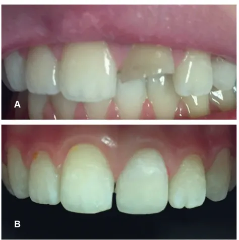
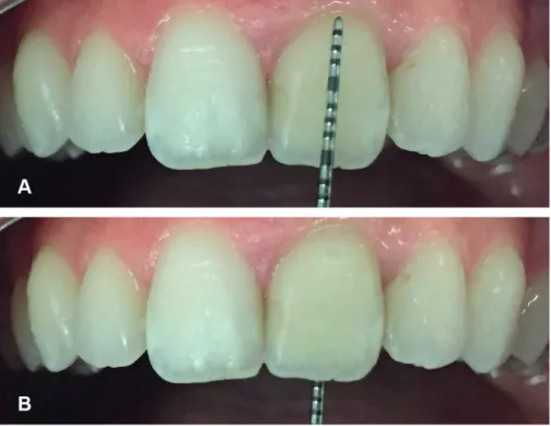
This technique was first described by Spasser and Herbert (1961) and is described as placing the mixture of sodium perborate and water in the pulp chamber. Next appointment, the procedure would be repeated until the desired shade reached. The application protocol of this technique is given in Table 2 and a case representing the walking bleach technique is presented in Figure 1.
97 Table 2. The application of the walking bleach technique
1. After the shade determination and initial photography taken, discolored tooth was isolated by rubber-dam or gingival barrier.
2. The existing restoration is removed and the endodontic access cavity is modified to ensure that no pulp horns, pulp remnants, or endodontic material residues remained. Because, these remnants will cause recurrence.
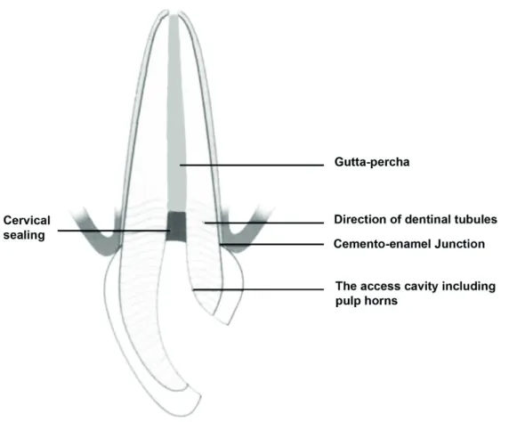
3. It should be ensured that all the restorative material is removed until the dentin surface is reached. Also, if there are superficial stains, they should also be removed using non-aggressive methods such as air-abrasion.
4. To prevent cervical root resorption, the root canal filling should be removed up to the proximal bone level with Gates-Glidden bur. This level is approximately 2-3 mm apical of cemento-enamel junction (CEJ) and can be determined with the help of periodontal probe. Then, the root canal filling should be sealed coronally with calcium hydroxide layer and glass ionomer cement.
5. After the root canal system sealed, the bleaching agent is applied to the pulp chamber. For the classical walking bleach technique, sodium perborate is mixed with distilled water to produce a semi-thick, viscous paste as a whitening agent. In combination technique (also called as modified walking bleach) 30% hydrogen peroxide is used instead of distilled water. After the paste inserted to pulp chamber the excess water is removed by a cotton pellet. Nowadays, 10% carbamide peroxide gel is preferred as a bleaching agent in general. The pulp chamber is sealed with a temporary restoration such as glass ionomer cement.
6. The patient was recalled depending on the bleaching material used and the treatment repeated if the desired shade was not reached.
The walking bleach technique was modified by placing a mixture of 30% hydrogen peroxide and sodium perborate in the pulp chamber. This technique is called modified or a combination walking bleach technique (Nutting and Poe, 1963). Hydrogen peroxide mixed with sodium perborate increases its effect and provides a better outcome. This process is faster and therefore results can be obtained after 1 week (Rotstein, 2001). In today’s walking bleach technique, 10% carbamide peroxide is inserted into the pulp chamber with a syringe instead of sodium perborate mixture and the patient is examined every 3 to 5 days (Sulieman, 2008).
98
Figure 1. A case representing the walking bleach technique (A) Initial photography of trauma induced discoloration and fracture (B) 6-month follow-up photography of the case postbleaching with 10% carbamide peroxide
6.2. Nonvital Power Bleaching
This technique is the least preferred technique due to the use of high temperature causing an increased risk of cervical root resorption. The hydrogen peroxide gel is applied to the pulp chamber at a concentration of 30-35% and activated by light or heat. The temperature is usually around 50-60°C and the heat activation should be interrupted after 5 minutes for 5 minutes to cool down (Rotstein, 2001). The tooth is reevaluated after 2 weeks to determine whether additional treatment is required and, if necessary, the walking bleach technique is applied (Sulieman, 2008) (Table 3). A case representing nonvital power bleaching is presented in Figure 2.
99 Table 3. The application of the nonvital power bleaching technique
1. The tooth is prepared as in the walking bleach technique.
2. The hydrogen peroxide gel (30-35%) is placed in the pulp chamber as a bleaching agent and activated by heat or light. The tooth is exposed to the activated bleaching gel for 5 minutes with the temperature usually between 50-60°C. Then, the tooth is allowed to cool down for 5 minutes and the bleaching agent is washed away for 1 minute. The tooth is dried and the walking bleach technique is applied.
3. Another variation of this technique is applied using 35% hydrogen peroxide without a heat activation. In this technique, the whitening agent is applied both to the pulp chamber and to the facial surface of the tooth. As with vital tooth whitening treatment, activation is provided with the light source. After applying 3 sessions of 5 minutes, the tooth is washed with water and the pulp chamber is closed with temporary restoration. The patient is recalled after 2 weeks to assess if further treatment is necessary or is ready for the definitive restoration.
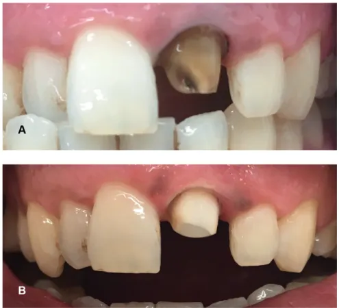
Figure 2. A preprosthetic case representing the nonvital power bleaching using 38% hydrogen peroxide with light activation (modified nonvital power bleaching) (A) Initial photography of endodontically treated tooth (B) Postbleaching photography after 2 weeks
100
6.3. Inside/Outside Bleaching
Settembrini et al., (1997) proposed a bleaching technique called inside/outside bleaching technique using a combination of intracoronal and extracoronal bleaching techniques with carbamide peroxide. In this technique, patients are responsible for the daily at-home use of the bleaching agent and therefore the bleaching effect is directly dependent on their compatibility. The inside/outside bleaching technique uses carbamide peroxide at varying concentrations of 5%, 16%, 22% or 35%. The application protocol of this technique is given in Table 4 and a case representing inside/outside bleach technique is presented in Figure 3.
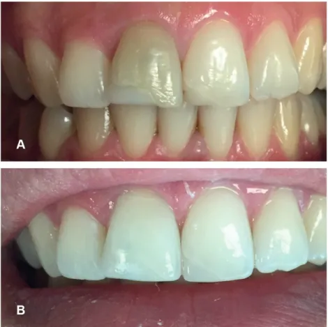
Figure 3. Inside/outside bleaching case with 10% carbamide peroxide (A) Initial photography of the patient with a trauma induced discoloration (B) 7-month follow-up photography of the case
101 Table 4. The application of the inside/outside bleaching technique
1. A custom fitting tray is prepared for the patient and the pulp chamber is covered only with a cotton pellet. The patient removes the cotton pellet with a toothpick and inject the bleaching gel into the cavity to start bleaching session. The patient also applies bleaching gel to the corresponding portion of the bleaching tray and inserts the bleaching tray to the mouth. After the insertion, the patient removes the excess gel with a cotton bud or a toothbrush.
2. After 2-hours of bleaching session, the patient cleans the cavity with a provided syringe and inserts a clean cotton pellet. Also, after every meal, the cotton pellet is renewed in the same way.
3. The patient must protect the cavity from food impaction by placing a cotton pellet to the cavity, and should not eat anything during the bleaching session.
4. In general, it is reported that results are obtained after 5-8 applications and depending on the frequency of the application. Therefore, the patient is recalled for assessment after 3-7 days. 5. When the desired shade is reached, the pulp chamber is closed with temporary restoration, and the
definitive restoration is placed after 2-week of delay.
This technique is simply a combination of at-home bleaching technique and intracoronal bleaching technique. The advantage of this technique in bleaching nonvital teeth is that the bleaching agent is applied both intracoronally and extracoronally. In addition, vital tooth bleaching (at-home bleaching) and nonvital bleaching can be combined simultaneously with the inside/outside bleaching technique (Carrillo et al., 1998). A lower concentration, generally the use of 10%, carbamide peroxide is thought to reduce the risk of cervical root resorption. The main disadvantage of this technique is that patient compliance is necessary and that hand ability is required to place the bleaching agent into the pulp chamber (Sulieman, 2008).
7. Neutralization
Bleaching of endodontically treated teeth with carbamide peroxide, sodium perborate or hydrogen peroxide is a commonly used method for regaining natural color. However, it has been reported that this procedure is followed by cervical root resorption (Friedman et al., 1988; Harrington and Natkin, 1979). Some researchers have claimed that bleaching materials penetrate periodontium and initiate an inflammatory process following cervical root resorption (Fuss et al., 1989). Therefore, the neutralization effects of the following agents were investigated.
7.1. Calcium Hydroxide
Today, intracoronal bleaching is routinely performed as a low-risk treatment method to correct the aesthetics of nonvital teeth. The major side effect of nonvital bleaching is cervical root resorption, which
102
can occur due to inflammation in the periodontium. The risk of cervical root resorption can be reduced by adequate cervical sealing and by avoiding high doses of bleaching agents (Zimmerli et al., 2010). Harrington and Natkin (1979) reported that the leakage of bleaching agents through dentin tubules can directly damage periodontal tissues and initiate an inflammatory response in the cervical region. Similarly, Lado et al., (1983) reported that bleaching agents could penetrate the dentin tubules and denature dentine in the cervical region.
Calcium hydroxide has been used in the treatment of cervical root resorption (Fuss et al., 1989). The mechanism that makes calcium hydroxide effective is not yet known. It has been suggested that the diffusion of ions from the root canal increases the pH of dental tissues, thereby inhibiting osteoclastic activity and activates alkaline phosphatases (Tronstad et al., 1981). On the other hand, the study by Lambrianidis et al., (2002) suggested that the use of calcium hydroxide as a barrier during intracoronal bleaching did not have a significant effect on preventing acidic pH on the external root surface. However, further studies are needed for the clarification of the exact mechanism.
7.2. Sodium Ascorbate
Many methods have been proposed to improve the bond strength of restorative materials to dental tissues after bleaching treatment. The first method that comes to mind is “delayed bonding”. Although it varies according to the bleaching agent used and its concentration, it is generally recommended that delaying the bonding for 1 to 2 weeks to recover bond strength (Lago and Garone-Netto, 2013; Miranda et al., 2013). Benni et al., (2014) state that the use of ethanol or acetone-based bonding agent can also improve bond strength to bleached enamel.
Another frequently used method is the application of antioxidant agents to the enamel surface after bleaching. Sodium ascorbate, α-tocopherol (vitamin E), grape seed extract (proanthocyanidins), lycopene, epigallocatechin gallate (green tea) are among the known antioxidants (Abraham et al., 2013; Guler et al., 2013; Khamverdi et al.,2013). In recent studies, sodium ascorbate has been used as an antioxidant to remove oxidative compounds, especially free radicals (Gutteridge, 1994; Rose and Bode, 1993; Soeno et al., 2008). Soeno et al., (2008) reported that ascorbic acid acts as an antioxidant agent, and ascorbic acid and ferric chlorite increase the bond strength to dentine. Turkun et al., (2009) reported that adhesion to dentin increased by 35% after applying sodium ascorbate as an antioxidant to bleached dentin.
8. Recurrence and Efficiency
Increasing rates of recurrence have been reported ranging from 10% to 49% in the literature with the time elapsed after bleaching (Friedman, 1997; Friedman et al., 1988; Glockner et al., 1999; Holmstrup et al., 1988). Lise et al., compared two different intracoronal bleaching techniques (walking bleach and inside/ outside bleaching) and reported that both techniques were effective after 1-year clinical follow-up with no recurrence (Lise et al., 2018). Deliperi detected recurrence in 15 of 25 teeth with intracoronal bleaching as a result of a 5-year clinical follow-up, however, he added that the recurrence had a maximum of 6 shades on the VITA color scale (Deliperi, 2008) (Table 5). In another study conducted by Deliperi and Bardwell (2005),
103 the researchers reported a similar recurrence rate of approximately half of the 26 bleached teeth with up
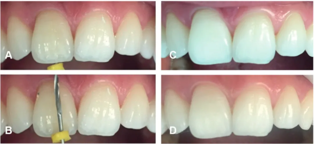
to 4 shades. Glockner et al., reported 79% of clinical success as a result of a 5-year clinical follow-up. In addition, they reported that if only the endodontic access cavity was prepared and the remaining tooth tissues were intact, the success rate increased to 91% (Glockner et al., 1999). According to Amato et al., (2006), the success rate of intracoronal bleaching after 16 years was 62.9%. In the same study, no cervical root resorption was observed in radiological examinations (Amato et al., 2006). Recurrence is relatively common in intracoronal bleaching. For this reason, some researchers recommend overbleaching the teeth to compensate recurrence (Bersezio et al., 2017) (Figure 4).
Figure 4. Endodontic treatment related discoloration (A) and (B) removal of endodontic material to the proximal bone level (C) overbleached tooth (D) 11-month follow-up photography
Table 5. Reordering of the Vita Shade Guide by value (light to dark by manufacturer)
Tab B1 A1 B2 D2 A2 C1 C2 D4 A3 D3 B3 A3.5 B4 C3 A4 C4
Rank 1 2 3 4 5 6 7 8 9 10 11 12 13 14 15 16
Bersezio et al., evaluated the effectiveness of walking bleaching with different bleaching agents (35% hydrogen peroxide and 37% carbamide peroxide). They also investigated the effect of intracoronal bleaching on patients’ psychosocial and aesthetic self-perceptions. As a result of their study, it was reported that effective and satisfactory results were obtained for patients after intracoronal bleaching (Bersezio et al., 2017). Gupta et al., reported 95% of patient satisfaction as a result of intracoronal bleaching performed on 41 patients with discoloration due to the trauma (Gupta and Saxena, 2014).
Color parameters L*, a*, b* established by the Commission Internationale de L’Eclariage (CIE) in 1978 (CIELAB). This system includes a lightness variable (L*) and chromatic coordinates (red/green, a* and yellow/ blue, b*) by following human color perception. With this system, the difference between two different color measurements can be determined quantitatively (ΔE). Studies reported that 5 units ΔE (according to CIELAB color space) change is required for successful results of bleaching treatment (Bersezio et al.,
104
2017). Hence, it can be interpreted that intracoronal bleaching treatment provides effective bleaching (Bersezio et al., 2017; Deliperi, 2008; Poyser et al., 2004). On the other hand, it was reported in a study that dentists were more critical in evaluating the bleaching outcome compared to patients. As a result of a 5-year clinical follow-up of intracoronal bleached teeth, the success rate was rated as 75% and 98% according to the dentists and patients, respectively (Glockner et al., 1999).
9. Cervical Root Resorption
Although cervical root resorption is the most serious adverse effect of intracoronal bleaching (Feiglin, 1987; MacIsaac and Hoen, 1994), the underlying mechanism is not yet fully understood. The first cases of cervical root resorption were reported by Harrington and Natkin (1979). According to Heithersay (1999a), cervical root resorption was seen only 3.9% of the intracoronal bleaching cases. He also reported that cervical root resorption was due to orthodontic treatment (24.1%), dental trauma (15.1%) and surgical procedures such as periodontal or transplantation (5.1%), respectively. However, in combination with intracoronal bleaching and dental trauma, the cervical root resorption rate increases even more (Heithersay, 1999b). Several long-term follow-up studies have reported that cervical root resorption can be seen even years after intracoronal bleaching (Abou-Rass, 1998; Aldecoa and Mayordomo, 1992; Anitua et al., 1990; Harrington and Natkin, 1979; Holmstrup et al., 1988; Lado et al., 1983).
It is thought that the cervical root resorption due to intracoronal bleaching is caused by the penetration of the bleaching agent to the periodontium. The presence of several predisposing factors that increase the penetration of the bleaching agent is mentioned (Baratieri et al., 1995; Friedman et al., 1988; Niederman et al., 1998). Dietschi (2006) recommends the use of low-concentration bleaching agents or sodium perborate mixed with distilled water with thin dentin walls. In this way, the penetration possibility of the bleaching agent into the periodontium would be reduced. In addition, an increased incidence of cervical root resorption has been reported in patients undergoing intracoronal bleaching at a young age (Abou-Rass, 1998; Aldecoa and Mayordomo, 1992; Anitua et al., 1990; Friedman et al., 1988; Harrington and Natkin, 1979; Holmstrup et al., 1988; Lado et al., 1983), due to having relatively larger dentine tubules. Heat activation is known to increase the efficacy of bleaching agents. It is reported that hydrogen peroxide applied to the pulp chamber by thermocatalytic technique can penetrate the outer surface of the tooth (Al-Nazhan, 1991; Dahlstrom et al., 1997; Farmer et al., 2006; Friedman et al.,1988; Friedman, 1989; Gimlin and Schindler, 1990; Goon et al., 1986; Lado et al., 1983; Latcham, 1986; Latcham, 1991; Madison and Walton, 1990; Montgomery, 1984; Szajkis et al., 1986). Therefore, the thermocatalytic technique is not preferred today due to the high risk of cervical root resorption (Attin et al., 2003; Friedman, 1997; Madison and Walton, 1990). On the contrary, cervical root resorption was not observed for sodium perborate – hydrogen peroxide solution with walking bleaching technique (Madison and Walton, 1990). Nowadays, carbamide peroxide is frequently preferred as an intracoronal bleaching agent (Bersezio et al., 2017; Ganesh et al., 2013; Shaheen et al., 2017; Valera et al., 2009). According to Lee et al. (2004), 35% carbamide peroxide showed the least extraradicular diffusion, followed by sodium perborate and 35% hydrogen peroxide, respectively. In addition, carbamide peroxide was found biocompatible than the hydrogen peroxide (Llena et al., 2019).
105 Another important predisposing factor of cervical root resorption observed after intracoronal bleaching is
cervical sealing (Plotino et al., 2008). However, cervical root resorption rate can be reduced to 1.9% at 16-19 years follow-up (Amato et al., 2006; Heithersay et al., 16-1994), with a proper cervical sealing (Heller et al., 1992). Dietschi (2006) reports that cervical root resorption is not observed after 20 years of intracoronal bleaching with 30% hydrogen peroxide due to correct cervical sealing. To ensure proper cervical sealing, it is necessary to reduce the root canal filling to 3 mm apical of the cemento-enamel junction (CEJ) and then a layer of calcium hydroxide covered with a cement material such as zinc phosphate or glass ionomer applied prior to intracoronal bleaching (Rotstein et al., 1992). Ideally, cervical sealing should be determined according to the visible crown length of the discolored tooth. For this, it would be appropriate to remove the existing root canal filling up to the proximal bone level to seal interproximal dentin tubules (Carrillo et al., 1998; Steiner and West, 1994) (Figures 5 and 6).
106
Figure 6. Determination of the cervical sealing level (A) Measuring the clinical crown length with the periodontal probe (B) Checking that the endodontic material is removed to the specified level
Cervical root resorption is generally clinically asymptomatic (Plotino et al., 2008). Therefore, the bleached teeth should be examined radiographically in the first five years following the intracoronal bleaching. Early diagnosis of cervical root resorption can be treated, whereas extraction is the only option, in severe root resorption (Goon et al.,1986; Latcham, 1986).
Conclusion
The discolorations in nonvital teeth, particularly in the anterior region cause aesthetic problems for patients and can affect everyday life. Although 100% success is not achieved in all intracoronal bleaching and certain rates of recurrence are encountered, it is a conservative treatment that can be preferred at least for delaying invasive restorative treatments.
Cervical root resorption is observed in nonvital bleached teeth, especially in the presence of dental trauma history. Therefore, it should be ensured that adequate cervical sealing is performed firstly. On the other hand, the thermocatalytic technique and high-dose hydrogen peroxide applications should be avoided. Recurrence appears to be inevitable after Intracoronal bleaching, hence more clinical trials are needed on color stability for better understanding.
107
Acknowledgement
The author presents his gratitude to Dr. Rana Turunc-Oguzman for her contribution on the nonvital power bleaching case.
Conflict of Interests
Author declares no conflict of interests.
References
Abbott, P., Heah, S. Y. S. (2009). Internal bleaching of teeth: an analysis of 255 teeth. Australian Dental Journal, 54(4), 326–333.
Abbott, P. V. (1997). Aesthetic considerations in endodontics: Internal bleaching. Practical Periodontics and Aesthetic Dentistry, 9(7), 833–842.
Abou-Rass, M. (1998). Long-term prognosis of intentional endodontics and internal bleaching of tetracycline-stained teeth. Compendium of Continuing Education in Dentistry, 19(10), 1034–1038, 1040–1042, 1044 passim.
Abraham, S., Ghonmode, W. N., Saujanya, K. P., Jaju, N., Tambe, V. H., Yawalikar, P. P. (2013). Effect of grape seed extracts on bond strength of bleached enamel using fifth and seventh generation bonding agents. Journal of International Oral Health, 5(6), 101–107.
Al-Nazhan, S. (1991). External root resorption after bleaching: a case report. Oral Surgery, Oral Medicine, Oral Pathology, 72(5), 607–609.
Aldecoa, E. A., Mayordomo, F. G. (1992). Modified internal bleaching of severe tetracycline discoloration: a 6-year clinical evaluation. Quintessence International, 23(2), 83–89.
Amato, M., Scaravilli, M. S., Farella, M., Riccitiello, F. (2006). Bleaching teeth treated endodontically: long-term evaluation of a case series. Journal of Endodontics, 32(4), 376–378.
Andreasen, F. M. (1986). Transient apical breakdown and its relation to color and sensibility changes after luxation injuries to teeth. Dental Traumatology, 2(1), 9–19.
Anitua, E., Zabalegui, B., Gil, J., Gascon, F. (1990). Internal bleaching of severe tetracycline discolorations: four-year clinical evaluation. Quintessence International, 21(10), 783–788.
Arens, D. (1989). The role of bleaching in esthetics. Dental Clinics of North America, 33(2), 319–336. Ari, H., Ungor, M. (2002). In vitro comparison of different types of sodium perborate used for intracoronal bleaching of discoloured teeth. International Endodontic Journal, 35(5), 433–436.
108
Attin, T., F. Ajam, P. F., Lennon, A. M. (2003). Review of the current status of tooth whitening with the walking bleach technique. International Endodontic Journal, 36(5), 313–329.
Baratieri, L. N., Ritter, A. V., Monteiro, S. Jr., Caldeira de Andrada, M. A., Cardoso Vieira, L. C. (1995). Nonvital tooth bleaching: guidelines for the clinician. Quintessence International, 26(9), 597–608.
Belobrov, I., Parashos, P. (2011). Treatment of tooth discoloration after the use of white mineral trioxide aggregate. Journal of Endodontics, 37(7), 1017–1020.
Benetti, A. R., Valera, M. C., Mancini, M. N., Miranda, C. B., Balducci, I. (2004). In vitro penetration of bleaching agents into the pulp chamber. International Endodontic Journal, 37(2), 120–124.
Benni, D. B., Naik, S. N., Subbareddy, V. V. (2014). An in vitro study to evaluate the effect of two ethanol-based and two acetone-ethanol-based dental bonding agents on the bond strength of composite to enamel treated with 10% carbamide peroxide. Journal of the Indian Society of Pedodontics and Preventive Dentistry, 32(3), 207–211.
Bersezio, C., Martin, J., Peña, F., Rubio, M., Estay, J., Vernal, R., Junior, O. O., Fernández E. (2017). Effectiveness and impact of the walking bleach technique on esthetic self-perception and psychosocial factors: a randomized double-blind clinical trial. Operative Dentistry, 42(6), 596–605.
Bizhang, M., Heiden, A., Blunck, U., Zimmer, S., Seemann, R., Roulet, J. F. (2003). Intracoronal bleaching of discolored non-vital teeth. Operative Dentistry, 28(4), 334–340.
Boutsioukis, C., Noula, G., Lambrianidis, T. (2008). Ex vivo study of the efficiency of two techniques for the removal of mineral trioxide aggregate used as a root canal filling material. Journal of Endodontics, 34(10), 1239–1242.
Brown, G. (1965). Factors influencing successful bleaching of the discolored root-filled tooth. Oral Surgery, Oral Medicine, Oral Pathology, 20(2), 238–244.
Bulut, H., Turkun, M., Demirbas, Kaya, A. (2006). Effect of an antioxidizing agent on the shear bond strength of brackets bonded to bleached human enamel. American Journal of Orthodontics and Dentofacial Orthopedics, 129(2), 266–272.
van der Burgt, T. P., Mullaney, T. P., Plasschaert, A. J. (1986a). Tooth discoloration induced by endodontic sealers. Oral Surgery, Oral Medicine, Oral Pathology, 61(1), 84–89.
van der Burgt, T. P., Plasschaert, A. J. (1985). Tooth discoloration induced by dental materials. Oral Surgery, Oral Medicine, Oral Pathology, 60(6), 666–669.
van der Burgt, T. P., Plasschaert, A. J. (1986). Bleaching of tooth discoloration caused by endodontic sealers. Journal of Endodontics, 12(6), 231–234.
van der Burgt, T. P., Eronat, C., Plasschaert, A. J. (1986b). Staining patterns in teeth discolored by endodontic sealers. Journal of Endodontics, 12(5), 187–191.
109 Carrillo, A., Arredondo Trevino, M. V., Haywood, V. B. (1998). Simultaneous bleaching of vital teeth and an
open-chamber nonvital tooth with 10% carbamide peroxide. Quintessence International, 29(10), 643–648. Christensen, G. (2003) New generation in-office vital tooth bleaching, part 2. Clinical Research Associates (CRA) Newsletter, 3(27), 1–3.
Chng, H. K., Ramli, H. N., Yap, A. U. J., Lim, C. T. (2005). Effect of hydrogen peroxide on intertubular dentine. Journal of Dentistry, 33(5), 363–369.
Dahl, J. E., Pallesen, U. (2003). Tooth bleaching-a critical review of the biological aspects. Critical Reviews in Oral Biology and Medicine, 14(4), 292–304.
Dahlstrom, S. W., Heithersay, G. S., Bridges, T. E. (1997). Hydroxyl radical activity in thermocatalytically bleached root-filled teeth. Endodontics and Dental Traumatology, 13(3), 119–125.
Deliperi, S. (2008). Clinical evaluation of nonvital tooth whitening and composite resin restorations: five-year results. The European Journal of Esthetic Dentistry, 3(2), 148–159.
Deliperi, S. and Bardwell, D. N. (2005). Two-year clinical evaluation of nonvital tooth whitening and resin composite restorations. Journal of Esthetic and Restorative Dentistry, 17(6), 369–378.
Dietschi, D. (2006). Nonvital bleaching: general considerations and report of two failure cases. The European Journal of Esthetic Dentistry, 1(1), 52–61.
Dostalova, T., Jelinkova, H., Housova, D., Sulc, J., Nemec, M., Miyagi, M., Brugnera, Jr. A., Zanin, F. (2004). Diode laser-activated bleaching. Brazilian Dental Journal, 15 Spec No: SI3-8.
Eisenberg, E. (1975). Anomalies of the teeth with stains and discolorations. The Journal of Preventive Dentistry, 2(1): 7–14, 16–20.
Farmer, D. S., Burcham, P., Marin, P. D. (2006). The ability of thiourea to scavenge hydrogen peroxide and hydroxyl radicals during the intra-coronal bleaching of bloodstained root-filled teeth. Australian Dental Journal, 51(2), 146–152.
Fasanaro, T. S. (1992). Bleaching teeth: history, chemicals, and methods used for common tooth discolorations. Journal of Esthetic and Restorative Dentistry, 4(3), 71–78.
Faunce, F. (1983). Management of discolored teeth. Dental Clinics of North America, 27(4), 657–670. Feiglin, B. (1987). A 6-year recall study of clinically chemically bleached teeth. Oral Surgery, Oral Medicine, Oral Pathology, 63(5), 610–613.
Friedman, S., Rotstein, I., Libfeld, H., Stabholz, A., Heling, I. (1988). Incidence of external root resorption and esthetic results in 58 bleached pulpless teeth. Dental Traumatology, 4(1), 23–26.
Fuss, Z., Szajkis, S., Tagger, M. (1989). Tubular permeability to calcium hydroxide and to bleaching agents. Journal of Endodontics, 15(8), 362–364.
110
Ganesh, R., Aruna, S., Joyson, M., Manikandan, Deepa. (2013). Comparison of the bleaching efficacy of three different agents used for intracoronal bleaching of discolored primary teeth: an in vitro study. Journal of Indian Society of Pedodontics and Preventive Dentistry, 31(1), 17–21.
Gimlin, D. R. and Schindler, W. G. (1990). The management of postbleaching cervical resorption. Journal of Endodontics, 16(6), 292–297.
Glockner, K., Hulla, H., Ebeleseder, K., Städtler, P. (1999). Five-year follow-up of internal bleaching. Brazilian Dental Journal, 10(2), 105–110.
Goldstein, R. E. and Garber, D. A. (1995). Complete dental bleaching. Quintessence Pub. Co.
Goon, W. W. Y., Cohen, S., Borer, R. F. (1986). External cervical root resorption following bleaching. Journal of Endodontics, 12(9), 414–118.
Greenwall, L. (2001). Bleaching techniques in restorative dentistry: an illustrated guide. CRC Press 1th edition. Grossman, L. I., Oliet, S., Del Rio, C. E. (1995). Endodontic Practice, Eleventh Edition. Lea & Febiger, 120–125. Guldener, P. H. A., Langeland, K. (1993). Endodontologie. 3rd ed. Stuttgart, New York. Georg ThiemeVerlag. Guler, E., Gonulol, N., Ozyilmaz, O. Y., Yucel, A. C. (2013). Effect of sodium ascorbate on the bond strength of silorane and methacrylate composites after vital bleaching. Brazilian Oral Research, 27(4), 299–304. Gupta, S. K., Saxena, P. (2014). Evaluation of patient satisfaction after non-vital bleaching in traumatized discolored intact anterior teeth. Dental Traumatology, 30(5), 396–399.
Gutiérrez, J. H., Guzmán, M. (1968). Tooth discoloration in endodontic procedures. Oral Surgery, Oral Medicine, Oral Pathology, 26(5), 706–711.
Gutteridge, J. M. C. (1994). Biological origin of free radicals, and mechanisms of antioxidant protection. Chemico-Biological Interactions, 91(2–3), 133–140.
Harrington, G. W., Natkin, E. (1979). External resorption associated with bleaching of pulpless teeth. Journal of Endodontics, 5(11), 344–348.
Haywood, V. B., Heymann, H. O. (1989). Nightguard vital bleaching. Quintessence International, 20(3), 173–176.
Naik, S., Hegde, A. H. (2005). Mineral trioxide aggregate as a pulpotomy agent in primary molars: an in vivo study. Journal of Indian Society of Pedodontics and Preventive Dentistry, 23(1), 13–16.
Heithersay, G. S. (1999a). Invasive cervical resorption: an analysis of potential predisposing factors. Quintessence International, 30(2), 83–95.
Heithersay, G. S. (1999b). Invasive cervical resorption following trauma. Australian Endodontic Journal, 25(2), 79–85.
111 Heithersay, G. S., Dahlstrom, S. W., Marin, P. D. (1994). Incidence of invasive cervical resorption in bleached
root‐filled teeth. Australian Dental Journal, 39(2), 82–87.
Heller, D., Skriber, J., Lin, L. M. (1992). Effect of intracoronal bleaching on external cervical root resorption. Journal of Endodontics, 18(4), 145–148.
Ho, S. and Goerig, A. C. (1989). An in vitro comparison of different bleaching agents in the discolored tooth. Journal of Endodontics, 15(3), 106–111.
Holland, R., de Souza, V., Nery, M. J., Faraco Júnior, I. M., Bernabé, P. F., Otoboni Filho, J. A., Dezan Júnior, E. (2001). Reaction of rat connective tissue to implanted dentin tube filled with mineral trioxide aggregate, portland cement or calcium hydroxide. Brazilian Dental Journal, 12(1), 3–8.
Holmstrup. G., Palm. A. M., Lambjerg‐Hansen. H. (1988). Bleaching of discoloured root‐filled teeth. Dental Traumatology, 4(5), 197–201.
Howell, R. A. (1980). Bleaching discoloured root-filled teeth. British Dental Journal, 148(6), 159–162. Joiner, A. (2004). Tooth colour: a review of the literature. Journal of Dentistry, 32(SUPPL.), 3–12.
Khamverdi, Z., Rezaei-Soufi, L., Kasraei, S., Ronasi, N., Rostami, S. (2013). Effect of epigallocatechin gallate on shear bond strength of composite resin to bleached enamel: an in vitro study. Restorative Dentistry & Endodontics, 38(4), 241–247.
Kihn, P. W. (2007). Vital tooth whitening. Dental Clinics of North America, 51(2), 319–331.
Kim, S. T., Abbott, P. V., McGinley, P. (2000). The effects of ledermix paste on discolouration of mature teeth. International Endodontic Journal, 33(3), 227–232.
Lado, E. A., Stanley, H. R., Weisman, M. I. (1983). Cervical resorption in bleached teeth. Oral Surgery, Oral Medicine, Oral Pathology, 55(1), 78–80.
Lago, A. N. D., Garone-Netto, N. (2013). Microtensile bond strength of enamel after bleaching. Indian Journal of Dental Research, 24(1), 104–109.
Lambrianidis, T., Kapalas, A., Mazinis, M. (2002). Effect of calcium hydroxide as a supplementary barrier in the radicular penetration of hydrogen peroxide during intracoronal bleaching in vitro. International Endodontic Journal, 35(12), 985–990.
Latcham, N. L. (1986). Postbleaching cervical resorption. Journal of Endodontics, 12(6), 262–264. Latcham, N. L. (1991). Management of a patient with severe postbleaching cervical resorption. A clinical report. The Journal of Prosthetic Dentistry, 65(5), 603–605.
Lee, G. P., Lee, M. Y., Lum, S. O., Poh, R. S., Lim, K. C. (2004). Extraradicular diffusion of hydrogen peroxide and ph changes associated with intracoronal bleaching of discoloured teeth using different bleaching agents. International Endodontic Journal, 37(7), 500–506.
112
Lim, K. C. (2004). Considerations in intracoronal bleaching. Australian Endodontic Journal, 30(2), 69–73. Llena, C., Collado-González, M., García-Bernal, D., Oñate-Sánchez, R. E., Martínez, C. M., Moraleda, J. M., Rodríguez-Lozano, F. J., Forner, L. (2019). Comparison of diffusion, cytotoxicity and tissue inflammatory reactions of four commercial bleaching products against human dental pulp stem cells. Scientific Reports, 9(1), 7743.
MacIsaac, A. M. and Hoen, C. M. (1994). Intracoronal bleaching: concerns and considerations. Journal Canadian Dental Association, 60(1), 57–64.
Madison, S. and Walton, R. (1990). Cervical root resorption following bleaching of endodontically treated teeth. Journal of Endodontics, 16(12), 570–574.
Maroto, M., Barbería, E., Vera, V., García-Godoy, F. (2006). Dentin bridge formation after white mineral trioxide aggregate (white mta) pulpotomies in primary molars. American Journal of Dentistry, 19(2), 75–79. McEvoy, S. A. (1989). Chemical agents for removing intrinsic stains from vital teeth. Ii. Current techniques and their clinical application. Quintessence International, 20(6), 379–384.
Miranda, T. A., Moura, S. K., Amorim, V. H., Terada, R. S., Pascotto, R. C. (2013). Influence of exposure time to saliva and antioxidant treatment on bond strength to enamel after tooth bleaching: an in situ study. Journal of Applied Oral Science, 21(6), 567–574.
Montgomery, S. (1984). External cervical resorption after bleaching a pulpless tooth. Oral Surgery, Oral Medicine, Oral Pathology, 57(2), 203–206.
Niederman, R., Ferguson, M., Urdaneta, R., Badovinac, R., Christie, D., Tantraphol, M., Rasool, F. (1998). Evidence-based esthetic dentistry. Journal of Esthetic and Restorative Dentistry, 10(5), 229–234.
Nutting, E. B. and Poe, G. S. (1963). A new combination for bleaching teeth. Journal Southern California Dental Association 31, 289–301.
Lise, P., Siedschlag, D. G., Bernardon, J. K., Baratieri, L. N. (2018). Randomized clinical trial of 2 nonvital tooth bleaching techniques: a 1-year follow-up. Journal of Prosthetic Dentistry, 119(1), 53–59.
Plotino, G., Buono, L., Grande, N. M., Pameijer, C. H., Somma, F. (2008). Nonvital tooth bleaching: a review of the literature and clinical procedures. Journal of Endodontics, 34(4), 394–407.
Poyser, N. J., Kelleher, M. G. D., Briggs, P. F. A. (2004). Managing discoloured non-vital teeth: the inside/ outside bleaching technique. Dental Update, 31(4), 213–214.
Rose, R. C. and Bode, A. M. (1993). Biology of free radical scavengers: an evaluation of ascorbate. FASEB Journal, 7(12), 1135–1142.
Rostein, I. (2002). Tooth discoloration and bleaching. In Endodontics, Hamilton, Ontario, Canada: BC Decker Inc., 845–860.
113 Rotstein, I. (2001). Bleaching techniques in restorative dentistry. In bleaching techniques in restorative
dentistry, London, 159–163.
Rotstein, I. and Friedman, S. (1991). PH variation among materials used for intracoronal bleaching. Journal of Endodontics, 17(8), 376–379.
Rotstein, I., Zyskind, D., Lewinstein, I., Bamberger, N. (1992). Effect of different protective base materials on hydrogen peroxide leakage during intracoronal bleaching in vitro. Journal of Endodontics, 18(3), 114–117. Schubert, D. M. and Brotherton, R. J. (1994). Boron: inorganic chemistry. In Encyclopedia of Inorganic and Bioinorganic Chemistry, John Wiley & Sons, Ltd., Chichester, UK; p. 372.
Settembrini, L., Gultz, J., Kaim, J., Scherer, W. (1997). A technique for bleaching nonvital teeth: inside/ outside bleaching. Journal of the American Dental Association, 128(9), 1283–1284.
Shaheen, M. A., Elkateb, M. A., Bakry, N. S., El Meligy, O. A. (2017). Efficacy of 10 percent carbamide peroxide as an intracoronal bleaching agent in nonvital discolored primary teeth: an in vitro study. Journal of Dentistry for Children (Chicago, Ill), 84(1), 22–29.
Soeno, K., Taira, Y., Jimbo, R., Sawase, T. (2008). Surface treatment with ascorbic acid and ferric chloride improves the micro-tensile bond strength of 4-meta/mma-tbb resin to dentin. Journal of Dentistry, 36(11), 940–944.
Spasser, H. F. (1961). A simple bleaching technique using sodium perborate. NY State Dental Journal, 27, 332–334.
Steiner, D. R. and West J. D. (1994). A method to determine the location and shape of an intracoronal bleach barrier. Journal of Endodontics, 20(6), 304–306.
Sulieman, M. (2004). An overview of bleaching techniques: history, chemistry, safety and legal aspects. Dental Update, 31(10), 608–616.
Sulieman, M. (2008). An overview of tooth-bleaching techniques: chemistry, safety and efficacy. Periodontology 2000, 48(1), 148–169.
Szajkis, S., Tagger, M., Tamse, A. (1986). Bleaching of root canal treated teeth and cervical external resorption: review of the literature. Refuat Hashinayim, 4(2), 10–12.
Tronstad, L., Andreasen, J. O., Hasselgren, G., Kristerson, L., Riis, I. (1981). PH changes in dental tissues after root canal filling with calcium hydroxide. Journal of Endodontics, 7(1), 17–21.
Truman, J. (1864). Bleaching of non-vital discoloured anterior teeth. Dent Times, 1, 69–72.
Turkun, M., Celik, E. U., Demirbaş Kaya, A., Arici, M. (2009). Can the hydrogel form of sodium ascorbate be used to reverse compromised bond strength after bleaching? The Journal of Adhesive Dentistry, 11(1), 35–40.
114
Valera, M. C., Camargo, C. H., Carvalho, C. A., de Oliveira, L. D., Camargo, S. E., Rodrigues, C. M. (2009). Effectiveness of carbamide peroxide and sodium perborate in non-vital discolored teeth. Journal of Applied Oral Science, 17(3), 254–261.
Vilhena, K. F. B., Nogueira, B. C. L., Fagundes, N. C. F., Loretto, S. C., Angelica, R. S., Lima, R. R., Silva, E., Souza, M. H. Jr. (2019). Dental enamel bleached for a prolonged and excessive time: morphological changes, PLos One, 14(4), e0214948.
Vogel, R. I. (1975). Intrinsic and extrinsic discoloration of the dentition. (a literature review). Journal of Oral Medicine, 30(4), 99–104.
Watts, A. and Addy, M. (2001). Tooth discolouration and staining: a review of the literature. British Dental Journal, 190(6), 309–316.
Ziemba, S. L., Felix, H., MacDonald, J., Ward, M. (2005). Clinical evaluation of a novel dental whitening lamp and light-catalyzed peroxide gel. The Journal of Clinical Dentistry, 16(4), 123–127.
Zimmerli, B., Strub, M., Jeger, F., Stadler, O., Lussi, A. (2010). Composite materials: composition, properties and clinical applications. A literature review. Schweizer Monatsschrift für Zahnmedizin, 120(11), 972–86.