Minimal invasive fixation of distal tibial fractures does not
result in rotational malalignment: A report of 24 cases
with CT imaging
Mehmet Mesut Sönmez, M.D.,1 Deniz Gülabi, M.D.,2 Meriç Uğurlar, M.D.,1 Metin Uzun, M.D.,4 Sezgin Sarban, M.D.,5 Ali Şeker, M.D.3
1Department of Orthopedics and Traumatology, Şişli Etfal Training and Research Hospital, İstanbul-Turkey
2Department of Orthopedics and Traumatology, Kartal Dr. Lütfi Kırdar Training and Research Hospital, İstanbul-Turkey 3Department of Orthopedics and Traumatology, Medipol University Faculty of Medicine, İstanbul-Turkey
4Department of Orthopedics and Traumatology, Acıbadem University Faculty of Medicine, İstanbul, Turkey 5Department of Orthopedics and Traumatology, International Hospital, İstanbul-Turkey
ABSTRACT
BACKGROUND: Tibial torsion is rotation of the proximal versus the distal articular axis in the transverse plane. This study used computed tomography (CT) to examine rotational malalignment in the crus following use of minimally invasive plate osteosynthesis (MIPO) technique in distal tibial fractures and evaluated effect of rotational difference on clinical outcomes and VAS scores.
METHODS: Analysis of 24 patients who were operated on for closed distal tibial fracture with MIPO technique between 2010 and 2012 was conducted. Malrotation was defined as rotational difference >10°. Operated knees were evaluated with 0.5-mm, fine-cut, 3-dimensional CT scan performed in cooperation with radiology department. Side-to-side difference in tibial torsion angle >10° was considered significant degree of malrotation. All patients were assessed clinically (visual analogue scale [VAS] and American Orthopae-dic Foot and Ankle Society [AOFAS] scores) and radiologically at final visit.
RESULTS: Mean follow-up period was 20.00±9.46 months (range: 18-51 months). Mean VAS score was 2.58±0.83 (range: 1–4) and mean AOFAS score was 87.50±4.05 (range: 78–93). Mean tibial rotation angle was 31.54±6.00° (range: 18–45°) on healthy side and 32.00±6.24° (range: 10–43°) on the operated side. No statistically significant difference was determined (p>0.05).
CONCLUSION: Use of intraoperative fluoroscopy, cable technique, and uninjured extremity as reference, can reduce incidence of rotational malalignment of distal tibial fractures treated with MIPO.
Keywords: CT; distal tibia fractures; malrotation; MIPO.
Achievement of optimal position using indirect reduction methods is technically difficult and may result in lower ex-tremity malalignment. This displacement can lead to adverse effects in adjacent joints and articular cartilage, causing earlier development of arthrosis.[3,4]
Tibial torsion is rotation of the proximal versus the distal ar-ticular axis in the transverse plane. Suero et al.[5] found that
malrotation of the distal tibia led to abnormal load distribution in the ankle joint. Considerable concern exists that malalign-ment of healed distal tibial fracture may result in post-trau-matic arthritis of the ankle. As the location of the deformity approaches the ankle, malalignment results in maldistribution of articular surface pressures, which may then predispose a patient to premature osteoarthritis.[6,7] It is well known that as
osteoarthritis progresses, there will be reduction in ability of the patient to participate in physical and social activities.
Address for correspondence: Deniz Gülabi, M.D.
Maltepe Başıbüyük Mah., Emek Cad., Tepe İnşaat Narcity Konutları, G4: 30, İstanbul, Turkey
Tel: +90 216 - 441 39 00 / 1450 E-mail: [email protected]
Qucik Response Code Ulus Travma Acil Cerrahi Derg 2017;23(2):144–149
doi: 10.5505/tjtes.2016.59153 Copyright 2017
TJTES
INTRODUCTION
Since the popularization of biological fixation, minimally in-vasive percutaneous plating is now used more frequently. Fa-vorable outcomes, including shorter healing time and lower reoperation rates have been reported in the literature.[1,2]
Sönmez et al. Minimal invasive fixation of distal tibial fractures does not result in rotational malalignment Using computed tomography (CT), this study examined
ro-tational malalignment in the crus following use of minimally invasive plate osteosynthesis (MIPO) technique in distal tibial fractures, and evaluated effect of rotational difference on clin-ical outcomes and visual analogue scale (VAS) scores.
MATERIALS AND METHODS
Analysis of 30 patients who were operated on for closed distal tibial fracture with the MIPO technique between 2010 and 2012 was conducted. Chart notes were studied for de-mographic details, mechanism of injury, and type of fracture. All preoperative X-rays were reviewed and classified using the AO/OTA Classification of Fractures and Dislocations. Inclusion criteria were unilateral distal tibial closed frac-tures. Exclusion criteria were fixation or revision of previous fixation in the ipsilateral or the contralateral femur or tibia, disagreement in CT imaging, multiple fractures, 2-stage sur-gery, and pregnant females. Two patients who had multiple fractures, 3 patients with 2-stage surgery, and 1 patient with disagreement in CT imaging were excluded; thus, 24 patients were included in this study for evaluation. The patients were 11 (45.8%) males and 13 (54.2%) females with mean age of 33.67±10.72 years (range: 19 to 55 years). Mechanism of in-jury was fall from height in 11 (45.8%) patients, and motor vehicle accident in 13 (54.2%) patients. Fractures were classi-fied according to the AO/OTA classification system.
All procedures followed were in accordance with the Hel-sinki Declaration of 1975, as revised in 2008. Approval for the study was granted by the ethics committee on human experimentation (institutional and national). Informed con-sent agreement was obtained from all patients for inclusion in the study.
Surgical Technique
All operations were performed within 2 days of the injury. All patients were operated on by a single surgeon. All cases were managed under regional anesthesia. Uninjured lower extremity in all patients was prepared for comparison. Image intensifier with C-arm was used in all cases to provide fluoro-scopic guidance. Longitudinal traction was applied along the foot by a junior orthopedic surgeon. Rotational alignment was achieved by aligning the anterior superior iliac spine, pa-tella, and second ray of the foot using the cable technique. In 8 patients with concomitant tibial and fibular fractures, fibu-lar fixation was performed initially. For fractures with intra-articular components, after fixation of intra-articular fragments with minimal dissection, “J”-shaped medial incision 3 cm in length was made. Distal medial tibial plate (DePuy Synthes Companies, Zuchwil, Switzerland) was used for fracture fixa-tion. Plate was advanced from incision submuscularly and only swab incisions were made for screw placements. Quality of reduction was verified with fluoroscopy and rotational align-ment was checked clinically (Fig. 1a, b). Patients were
mo-bilized on 2 crutches. Weight-bearing was permitted when callus formation was observed on radiographs at follow-up examinations. All patients were evaluated clinically and radio-logically at final visit. VAS and American Orthopaedic Foot and Ankle Society (AOFAS) scores were recorded.
Assessment of Radiographs
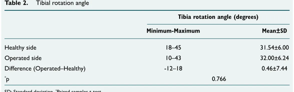
All measurements were calculated by independent radio-graphic reviewer who was blinded to the study. Observer assessed all radiographs twice at an interval of 1 week, and mean of the 2 values was accepted as rotation degree of each tibia. Reviewer had ample time to evaluate the radiographs, as no time limit was imposed on review. Measurements were taken using PACS System (Novard Corp., American Fork, UT, USA). Internal rotation deformity was assigned negative value and external rotation was given positive value. Rotational dif-ference >10° was defined as malrotation. Operated knees were evaluated with 0.5-mm, fine-cut, 3-dimensional CT scans (Aquilion 64; Toshiba Medical Systems Corp., Otawara, Japan) in cooperation with radiology department. Patients were scanned in supine position with adjustable supports to prevent movement of lower extremities. Scanning included multiple 5-mm cuts at the proximal tibia and distal tibia. Axial plane images were used. Proximally, reference line was the tangent to the posterior tibial ridges at level just superior to the fibular head. Tibial torsion angle was calculated using difference between angles of the proximal and transmalleolar axis just proximal to the tibial plafond with horizontal ref-erence line (Fig. 1c). Side-to side diffref-erence in tibial torsion angle >10° was considered significant degree of malrotation. Normal value for adults quoted in the literature is 30° to 40° of external rotation, with range of ±10°.[8,9]
Statistical Methodology
Statistical analyses were performed using NCSS 2007 (NCSS, LLC, Kaysville, UT, USA) software. Descriptive statistical methods were used (mean, SD, frequency, percentage, mini-mum, maximum) were used to evaluate study data. Paired samples t-test was used to compare group of variables that demonstrated normal distribution. Pearson correlation analy-sis and Spearman correlation analyanaly-sis were used to analyze relationships between variables. Statistical significance was determined at p<0.05.
RESULTS
Intraobserver reliability was calculated as 0.92. Mean follow-up period was 20.00±9.46 months (range: 18–51 months). Mean body mass index of the patients was 22.04±1.55 kg/ m2 (range: 20-25 kg/m2). Mean time to union was 15.42±1.67
weeks (range: 13–19 weeks). Mean VAS score was 2.58±0.83 (range: 1–4) and mean AOFAS score was 87.50±4.05 (range: 78–93). (Table 1).
(4.2%), 42B1in 3 (12.5%), 42B2 in 2 (8.3%), 42B3 in 6 (25%), 42C1 in 2 (8.3%), 42C2 in 1 (4.2%), 43A1 in 1 (4.2%), 43A3 in 4 (16.7%) and 43C2 in 1 (4.2%).
Tibial rotation angle was determined as mean 31.54±6.00° (range: 18–45°) on healthy side and mean 32.00±6.24° (range: 10–43°) on operated side.
No statistically significant difference was found between healthy side and operated side with respect to rotation angle (p>0.05) (Table 2). No statistically significant relationship was determined between VAS and AOFAS scores and difference between operated side rotation angle and healthy side ro-tation angle (p>0.05) (Table 3). In 1 patient, implants were removed due to soft tissue irritation 1 year after surgery.
Table 1. Distribution of descriptive characteristics
n % Minimum–Maximum Mean±SD
Gender
Male 11 45.8
Female 13 54.2
Age (years) 19–55 33.67±10.72 Body mass index (kg/m2) 20–25 22.04±1.55
Follow-up period (months) 18–51 20.00±9.46 Time to union (weeks) 13–19 15.42±1.67 Visual analogue scale score 1–4 2.58±0.83 American Orthopaedic Foot and Ankle Society 78–93 87.50±4.05
SD: Standard deviation.
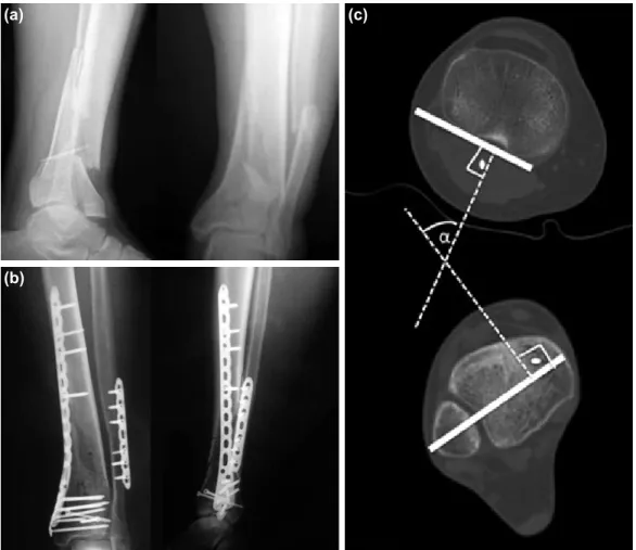
Figure 1. (a) Preoperative radiographs of a 41-year-old patient with distal tibial fracture accompanied by fibular
fracture. Tibial fracture extends to the ankle joint. (b) Early postoperative radiographs of the same patient.
Intra-articular fragments were fixed with minimal soft tissue dissection. (c) Measurement of tibial torsion.
(a) (c)
DISCUSSION
This study has demonstrated that rotational profile of the distal tibia can be observed accurately with standard postop-erative CT scans, even when standard intraoppostop-erative fluoros-copy was used to reduce malrotation of distal tibial fractures treated with MIPO technique. CT scanning has become the gold standard for assessment of limb rotational alignment.
[8–11] Test is accurate and has good inter- and intra-observer
reliability and repeatability.[12] The present study had good
intraobserver reliability, as recommended in the literature. The results of this study confirmed that using intraoperative fluoroscopy, cable technique, and uninjured extremity as a reference can decrease incidence of rotational malalignment of distal tibial fractures treated with MIPO.
With the popularity of biological fixation, MIPO is now be-ing used more frequently at trauma centers. MIPO with indirect fracture reduction can lead to higher rates of limb malalignment.[13–15] Rotational deformity in the lower
extrem-ity has been shown to influence articular cartilage shearing and development of joint arthrosis, leading to clinically rel-evant degenerative changes.[3,4,16,17] Quality of reduction and
limb alignment with use of MIPO technique has not been well documented. The few studies that have documented post-reduction limb rotation following MIPO cite incidence of malrotation >10° in 0% to 9% of limbs based on clinical examination and side-to-side comparison with the uninjured extremity.[18–22] In the present study, there were no cases with
tibial malrotation compared with uninjured side. This was
considered to be related to accurate use of intraoperative fluoroscopy and the experience of the surgical team, who were certified in trauma.
In the present study, the uninjured limb was prepared to as-sess rotation profile of the extremity. Inclusion of the unin-jured limb in surgical preparation has been recommended to benefit reduction assessment, and previous studies that have used CT scans to examine uninjured lower limbs for rotation have reported average side-to-side difference in rotation of only 3° to 4°.[11] Tornetta has described technique of using
distal femoral traction pin and fluoroscopy of both normal and injured extremities to better judge rotation for femoral shaft fractures.[23] In the current study, there was no
statis-tically significant difference between injured and uninjured extremities with respect to rotational alignment (p=0.766). Preparing the uninjured limb as reference for normal rotation and intraoperative cable technique with guidance of fluoros-copy were considered to be beneficial in judging rotation of the injured extremity. However, no convenient and reliable method of assessing intraoperative lower limb rotation has been developed and used consistently. This is an area for fur-ther research.
One study concluded that fibular plate fixation increased initial rotational stability of distal third tibia and fibula shaft fractures compared with intramedullary nailing of the tibia alone.[24] Prasad et al. stated that average rotation at the ankle
was less, with 7.67° of external rotation in group with fibula fixed, compared with 10.68° of external rotation in group with non-fixed fibula (p=0.01).[25] In the current study, only 3
fibulae were stabilized with open reduction internal fixation technique. There was no statistically significant difference be-tween these 3 cases and non-fixed fibula fractures (p>0.05). However, strength of recommendation is low due to small sample size in each group. Prospective randomized studies could be designed to evaluate difference in rotational ma-lalignment of both groups (fixed and non-fixed fibula). Soft tissue coverage is weak in the distal tibia metaphyseal region and in the distal diaphysis. Various studies have re-ported on postoperative wound healing and infection risk in this area. Guo et al. reported wound problems at rate of Sönmez et al. Minimal invasive fixation of distal tibial fractures does not result in rotational malalignment
Table 2. Tibial rotation angle
Tibia rotation angle (degrees)
Minimum-Maximum Mean±SD
Healthy side 18–45 31.54±6.00 Operated side 10–43 32.00±6.24 Difference (Operated–Healthy) -12–18 0.46±7.44
*p 0.766
SD: Standard deviation. *Paired samples t-test.
Table 3. Relationship between TRA and VAS and AOFAS
scores
TRA difference (operated–healthy) r p
VAS b-0.199 0.350
AOFAS c-0.138 0.521 bSpearman correlation coefficient. cPearson correlation coefficient. TRA: Tibia rotation angles; VAS: Visual analogue scale; AOFAS: American Orthopaedic Foot and Ankle Society.
14.6%[26] and Lau et al. reported late stage infection following
MIPO at 15%.[27] In the current study, implant was removed in
1 patient (3.3%) due to implant irritation.
Limitations of the Study
Primary limitations of this study were small number of cases and short duration of follow-up. Studies with larger patient population may demonstrate real extent of malalignment af-ter such surgery. In addition, long-af-term follow-up results may reveal useful results regarding such deformities.
In conclusion, the best method to avoid malrotation during MIPO technique is comparison with the opposite leg and use of intraoperative fluoroscopy and cable technique. It may be appropriate to prepare the normal leg for comparison. Malro-tation is best identified by clinical inspection and best quanti-fied by limited CT scan.
Conflict of interest: None declared.
REFERENCES
1. Hazarika S, Chakravarthy J, Cooper J. Minimally invasive locking plate osteosynthesis for fractures of the distal tibia--results in 20 patients. In-jury 2006;37:877–87. Crossref
2. Ronga M, Longo UG, Maffulli N. Minimally invasive locked plat-ing of distal tibia fractures is safe and effective. Clin Orthop Relat Res 2010;468:975–82. Crossref
3. Gugenheim JJ, Probe RA, Brinker MR. The effects of femoral shaft mal-rotation on lower extremity anatomy. J Orthop Trauma 2004;18:658–64. 4. Netz P, Olsson E, Ringertz H, Stark A. Functional restitution after
lower leg fractures. A long-term follow-up. Arch Orthop Trauma Surg 1991;110:238–41. Crossref
5. Suero EM, Hawi N, Westphal R, Sabbagh Y, Citak M, Wahl FM, et al. The effect of distal tibial rotation during high tibial osteotomy on the con-tact pressures in the knee and ankle joints. Knee Surg Sports Traumatol Arthrosc 2015 Mar 6.
6. Richmond J, Colleran K, Borens O, Kloen P, Helfet DL. Nonunions of the distal tibia treated by reamed intramedullary nailing. J Orthop Trau-ma 2004;18:603–10. Crossref
7. van der Schoot DK, Den Outer AJ, Bode PJ, Obermann WR, van Vugt AB. Degenerative changes at the knee and ankle related to malunion of tibial fractures. 15-year follow-up of 88 patients. J Bone Joint Surg Br 1996;78:722–5.
8. Jakob RP, Haertel M, Stüssi E. Tibial torsion calculated by computerised tomography and compared to other methods of measurement. J Bone Joint Surg Br 1980;62:238–42.
9. Jend H, Heller M, Dallek M, Schoettle H. Measurement of tibial torsion by computer tomography. Acta Radiol 1981;22:271–5. Crossref
10. Murphy SB, Simon SR, Kijewski PK, Wilkinson RH, Griscom NT. Femoral anteversion. J Bone Joint Surg Am 1987;69:1169–76. Crossref
11. Strecker W, Keppler P, Gebhard F, Kinzl L. Length and torsion of the lower limb. J Bone Joint Surg Br 1997;79:1019–23. Crossref
12. Puloski S, Romano C, Buckley R, Powell J. Rotational malalignment of the tibia following reamed intramedullary nail fixation. J Orthop Trauma 2004;18:397–402. Crossref
13. Neer CS, Grantham SA, Shelton ML. Supracondylar fracture of the adult femur. A study of one hundred and ten cases. J Bone Joint Surg Am 1967;49:591–613. Crossref
14. Schatzker J, Home G, Waddell J. The Toronto experience with the supra-condylar fracture of the femur, 1966-72. Injury 1974;6:113–28. Crossref
15. Williams J, Gibbons M, Trundle H, Murray D, Worlock P. Complica-tions of nailing in closed tibial fractures. J Orthop Trauma 1995;9:476– 81. Crossref
16. Kettelkamp DB, Hillberry BM, Murrish DE, Heck DA. Degenerative arthritis of the knee secondary to fracture malunion. Clin Orthop Relat Res 1988;234:159–69. Crossref
17. van der Schoot DK, Den Outer AJ, Bode PJ, Obermann WR, van Vugt AB. Degenerative changes at the knee and ankle related to malunion of tibial fractures. 15-year follow-up of 88 patients. J Bone Joint Surg Br 1996;78:722–5.
18. Cole PA, Zlowodzki M, Kregor PJ. Treatment of proximal tibia fractures using the less invasive stabilization system: surgical experience and early clinical results in 77 fractures. J Orthop Trauma 2004;18:528–35. Crossref
19. Kregor PJ, Stannard JA, Zlowodzki M, Cole PA. Treatment of dis-tal femur fractures using the less invasive stabilization system: surgical experience and early clinical results in 103 fractures. J Orthop Trauma 2004;18:509–20. Crossref
20. Ricci WM, Rudzki JR, Borrelli J Jr. Treatment of complex proximal tibia fractures with the less invasive skeletal stabilization system. J Orthop Trauma 2004;18:521–7. Crossref
21. Stannard JP, Wilson TC, Volgas DA, Alonso JE. The less invasive stabili-zation system in the treatment of complex fractures of the tibial plateau: short-term results. J Orthop Trauma 2004;18:552–8. Crossref
22. Weight M, Collinge C. Early results of the less invasive stabilization sys-tem for mechanically unstable fractures of the distal femur (AO/OTA types A2, A3, C2, and C3). J Orthop Trauma 2004;18:503–8. Crossref
23. Tornetta P, Ritz G, Kantor A. Femoral torsion after interlocked nailing of unstable femoral fractures. J Trauma 1995;38:213–9. Crossref
24. Morrison KM, Ebraheim NA, Southworth SR, Sabin JJ, Jackson WT. Plating of the fibula. Its potential value as an adjunct to external fixation of the tibia. Clin Orthop Relat Res 1991;266:209–13.
25. Prasad M, Yadav S, Sud A, Arora NC, Kumar N, Singh S. Assess-ment of the role of fibular fixation in distal-third tibia-fibula fractures and its significance in decreasing malrotation and malalignment. Injury 2013;44:1885–91. Crossref
26. Guo JJ, Tang N, Yang HL, Tang TS. A prospective, randomised trial comparing closed intramedullary nailing with percutaneous plating in the treatment of distal metaphyseal fractures of the tibia. J Bone Joint Surg Br 2010;92:984–8. Crossref
27. Lau TW, Leung F, Chan CF, Chow SP. Wound complication of mini-mally invasive plate osteosynthesis in distal tibia fractures. Int Orthop 2008;32:697–703. Crossref
OLGU SUNUMU
Distal tibia kırıklarının minimal invaziv tespiti rotasyonel deformiteye neden olmaz:
Yirmi dört hastanın bilgisayarlı tomografi incelemesi
Dr. Mehmet Mesut Sönmez,1 Dr. Deniz Gülabi,2 Dr. Meriç Uğurlar,1 Dr. Metin Uzun,4 Dr. Sezgin Sarban,5 Dr. Ali Şeker3 1Şişli Etfal Eğitim ve Araştırma Hastanesi, Ortopedi ve Travmatoloji Kliniği, İstanbul
2Kartal Dr. Lütfi Kırdar Eğitim ve Araştırma Hastanesi, Ortopedi ve Travmatoloji Kliniği, İstanbul 3Medipol Üniversitesi Tıp Fakültesi, Ortopedi ve Travmatoloji Anabilim Dalı, İstanbul 4Acıbadem Üniversitesi Tıp Fakültesi, Ortopedi ve Travmatoloji Anabilim Dalı, İstanbul 5International Hospital, Ortopedi ve Travmatoloji Kliniği, İstanbul
AMAÇ: Tibial rotasyon prosimal eklemin transvers planda distal ekleme göre rotasyonda olmasıdır. Bu çalışmanın amacı minimal invaziv plaklama yöntemi (MİPO) ile tedavi edilen distal tibia kırıklarında, malrotasyonun bilgisayarlı tomografi (BT) ile tespit edilimesi, rotasyonel farkın klinik sonuç-lar ve Vizüel Analog Skala (VAS) skoru üzerine etkisini araştırmaktı.
GEREÇ VE YÖNTEM: 2010–2012 yılları arasında kapalı distal tibia kırığı nedeniyle MİPO yöntemiyle ameliyat edilen 24 hasta çalışmaya dahil edil-di. On dereceden fazla rotasyon farkı malrotasyon olarak kabul ediledil-di. Ameliyat edilen bacak 0.5 mm aralıklarla alınan üç boyutlu BT kesitleriyle radyoloji bölümü tarafından değerlendirildi. İki alt ekstremite arasındaki 10 dereceden fazla fark malrotasyon olarak kabul edildi. Tüm hastalar son takiplerinde klinik (VAS ve Amerikan Ortopedi Ayak ve Ayak Bileği Topluluğu [AOFAS]) ve radyolojik olarak değerlendirildi.
BULGULAR: Ortalama takip süresi 20.00±9.46 aydı (dağılım, 18–51 ay). Ortalama VAS skoru 2.58±0.83 (dağılım, 1–4) ve ortalama AOFAS skoru 87.50±4.05 (dağılım, 78–93) idi. Ortalama tibia rotasyon açısı sağlam tarafta 31.54°±6.00° (dağılım, 18°–45°) ve ameliyatlı tarafta ise 32.00°±6.24° (dağılım, 10°–43°) idi. İstatiksel olarak anlamlı fark saptanmadı (p>0.05).
TARTIŞMA: Minimal invaziv plaklama yöntemiyle distal tibia kırıklarının tedavi edilmesinde ameliyat sırasında skopi kullanılması, kablo tekniğinin ve sağlam tarafın referans olarak kullanılma yöntemi rotasyonel dizilim sorununu azaltacağı kanaatindeyiz.
Anahtar sözcükler: BT; distal tibia kırığı; malrotasyon; MİPO.
Ulus Travma Acil Cerrahi Derg 2017;23(2):144–149 doi: 10.5505/tjtes.2016.59153
ORİJİNAL ÇALIŞMA - ÖZET