The prevalence and distribution of the dental anomalies in the Turkish
population
Article · July 2019 DOI: 10.4103/JASI.JASI_31_19 CITATIONS 0 READS 118 5 authors, including: Aybüke Ersen 2PUBLICATIONS 0CITATIONS SEE PROFILE Benin DikmenIstanbul Medipol University 7PUBLICATIONS 26CITATIONS
SEE PROFILE
All content following this page was uploaded by Aybüke Ersen on 30 July 2019.
Address for correspondence:
Dr. Benin Dikmen,
TEM Avrupa Otoyolu Goztepe Cikisi, Number: 1, 34214, Bagcilar, Istanbul, Turkey. E‑mail: bdikmen@medipol. edu.tr
Access this article online
Website: www.jasi.org.in DOI:
10.4103/JASI.JASI_31_19 Quick Response Code: Abstract
Introduction: The incidence of developmental dental anomalies can provide valuable information. The aim of this study is to investigate the prevalence and distribution of dental anomalies in the Turkish population. Material and Methods: Totally 2203 patients who came to university hospital were examined clinically and radiographically for 10 dental anomalies: rotation, diastema, hypodontia, hyperdontia, microdontia, macrodontia, dilaceration, talon cusp, gemination and ectopia. Descriptive characteristics of these dental anomalies, including gender and regions of the jaw, were recorded. The data were analyzed with Chi‑square test and Yates continuity correction. Results: In 1517 patients (68.9%), tooth anomaly was detected. Rotation was found in 58.4% of patients, significantly lower in the molar region than in the anterior and premolar regions (P < 0.01). Diastema was found in 22.7% of the samples, at higher rates in the anterior region than in the premolar and molar regions (P < 0.01). Dilaceration was observed 3.8% of the patients, at significantly higher rates in the molar region (P < 0.01). Ectopia was found in 2.9% of individuals, less common in the molar region than in the anterior and premolar regions (P < 0.01). Hypodontia was seen in 3.4% of patients, significantly lower in the anterior teeth than in the premolar and molar teeth (P < 0.01). Patients having talon cusp were 2.7%, microdontia was 1%, and only 0.5% showed the presence of hyperdontia. The distribution of hyperdontia and microdontia did not significantly differ between regions (P > 0.05). Gemination was seen in only one anterior tooth and macrodontia in only one premolar tooth. Discussion and Conclusion: Rotation is the most common dental anomaly in Turkish population, followed by diastema. Knowledge of the prevalence of anomalies may help clinicians to the detection of these anomalies at early stages.
Keywords: Prevalence, radiography, tooth abnormalities
The Prevalence and Distribution of the Dental Anomalies in the Turkish
Population
Ozge Gurbuz, Aybuke Ersen, Benin Dikmen, Burak Gumustas, Mustafa Gundogar1 Departments of Restorative Dentistry and 1Endodontics,Faculty of Dentistry, Istanbul Medipol University, Istanbul, Turkey
How to cite this article: Gurbuz O, Ersen A,
Dikmen B, Gumustas B, Gundogar M. The prevalence and distribution of the dental anomalies in the Turkish population. J Anat Soc India 2019;68:46-51.
Introduction
Developmental dental anomalies can occur as a result of genetic or environmental factors or a combination of both of them and could be observed as an evolutionary trend.[1] The development of anomalies in tooth number, shape, and position results from disturbances that occur during the morphodifferentiation stage of development.[2,3] Moreover, some systemic diseases may result in dental alterations.[3,4] The incidence of developmental dental anomalies can provide valuable information for genetic studies and can help to understand the variation between populations. In addition, since these anomalies can lead to some clinical problems such as malocclusion, difficulties in mastication, poor oral hygiene, periodontal problems, increased
susceptibility to caries, and esthetic problems, the early diagnosis of dental anomalies may be important.[4,5] There are many studies that researched the prevalence of dental anomalies in different populations, and the results of these studies are too conflicting. This situation can be attributed to variation in methods of sampling and investigation and the differences of race etc.[3]
Pipi conducted a literature search for epidemiologic studies and reported that the prevalence of supernumerary teeth varied from 0.04% to 2.29%.[6] Popoola et al. reported that the prevalence of dental hard tissue anomalies varies between countries.[4] They claimed that hypodontia is the most prevalent dental anomaly found in Indian, Saudi Arabian, Turkish, and Norwegian children. In addition, it has been reported that hypodontia is more common in the permanent dentition, with frequencies ranging from 0.03% to 10.1%.[7] The
This is an open access journal, and articles are distributed under the terms of the Creative Commons Attribution-NonCommercial-ShareAlike 4.0 License, which allows others to remix, tweak, and build upon the work non-commercially, as long as appropriate credit is given and the new creations are licensed under the identical terms.
For reprints contact: [email protected]
Gurbuz, et al.: Prevalence of dental anomalies
Journal of the Anatomical Society of India ¦ Volume 68 ¦ Issue 1 ¦ January-March 2019 47
prevalence of diastema varied from 7.8% to 18.3% in the literature.[8‑11] There are only a few publications about the prevalence of dilacerations, with rates ranging from 5.6% to 16.3%.[12‑15]
The studies which analyzed the prevalence of dental anomalies found that anomalies are seen more often in some teeth than others.[16] Some studies revealed that the most frequently missing teeth in the dental arch are the third molar, followed by the mandibular and maxillary second premolar, maxillary lateral incisor, and mandibular central incisor.[16,17] Many studies found that the most common supernumerary tooth was mesiodens.[5,18,19]
While some studies claim that gender affects the prevalence of anomalies,[5,9,20] others decline this effect.[21,22] Moreover, some of them reported that the effect of gender depends on the type of anomaly.[4,23]
Because of the conflicting results of published studies about the prevalence of dental anomalies, there is a need for further research to help clarify the prevalence and distribution of dental anomalies in the Turkish population. The aim of this study is to investigate the prevalence and distribution of associated dental anomalies such as rotation, diastema, hypodontia, oligodontia, hyperdontia, microdontia, macrodontia, dilaceration, talon cusp, and ectopia in the Turkish population.
Material and Methods
Totally 2203 consecutive patients (1220 females and 983 males) aged between 12 and 93 years who attended our clinic were examined clinically and radiographically. İntraoral and radiographic examinations of these patients had been made by one examiner carefully.
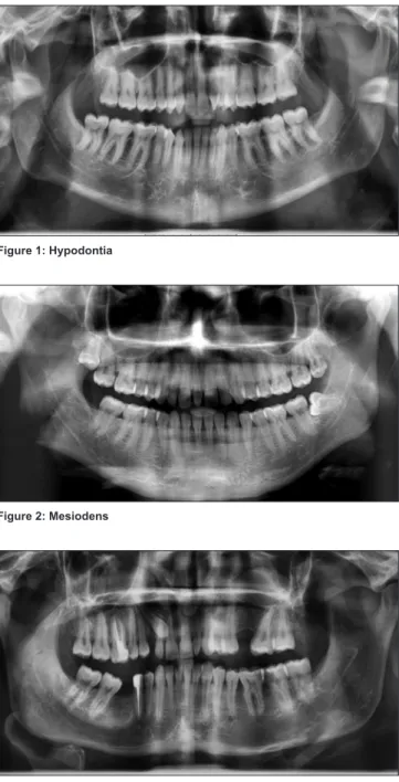
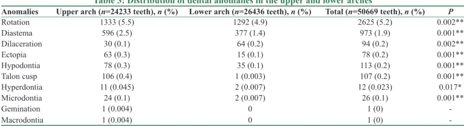
The clinical records and radiographs who attended the clinic and had been diagnosed as having rotation, diastema, hypodontia, hyperdontia, microdontia, macrodontia, root dilaceration, talon cusp, gemination and ectopia in all arches were selected for review [Figures 1‑5].
Good‑quality panoramic radiographs taken at the time of diagnosis had to be available. The radiographs were taken with a Dentsply Gendex Orthoralix 9200 panoramic unit (Dentsply Asia, Milford, US).
İndividuals were selected according to the following criteria:
A family and dental history involved questions about the primary dentition and details such as color, tooth wear, abscess formation, tooth mobility, and loss of teeth. Selection criteria of the samples included the patients who were not diagnosed with any serious childhood illnesses and systemic syndromes. Patients with no history of previous orthodontic treatment were included in this study. Patients who had metabolic diseases of
bone and radiopacities around the teeth with deep caries or large restorations were also used for exclusion from the study. Only participants of Turkish origin were selected.
The following details were recorded for each patient: • Age of patients
• Presence of associated dental anomalies such as rotation, diastema, hypodontia, oligodontia, hyperdontia, microdontia, root dilaceration, cingulum hypertropia, and ectopia
• The location on the arch of the abnormalities • Gender.
Data were pooled and analyzed for sex and side distribution.
Figure 1: Hypodontia
Figure 2: Mesiodens
IBM SPSS Statistics 22 (IBM Corp., Armonk, NY, USA) was used for statistical analysis. Obtained data were statistically analyzed with descriptive statistics and the differences between the groups were tested using Pearson’s Chi‑square test and Fisher’s exact test. P ≤ 0.05 was considered as statistically significant (P < 0.05).
The present study was approved by the Research Ethics Committee of the Istanbul Medipol University (Number: 389). In addition, an informed written consent was obtained from all the participants.
Results
The distribution of dental anomalies by gender is presented in Table 1. Patients having only one anomaly were 65.7% (997 patients), whereas only 34.3% (520 patients) showed the presence of more than one anomaly. The rate of presence of one anomaly was significantly higher than the rate of the presence of more than one anomaly (P < 0.01). The frequencies of dental anomalies and gender distribution are shown in Table 2. The distribution of dental anomalies in the upper and lower arches is presented in Table 3. The distribution of dental anomalies by region is summarized
in Table 4. The rate of rotation was significantly lower in the molar region than in the anterior and premolar regions (P < 0.01). Diastemas were observed at higher rates in the anterior region than in the premolar and molar regions (P < 0.01). The rate of the presence of diastema in the premolar region was significantly higher than the molar region (P < 0.01). Dilacerations were observed at significantly lower rates in the anterior region than in the premolar and molar regions (P < 0.01). The rate of dilaceration was significantly lower in the premolar region than in the molar region (P < 0.01). Ectopia was observed significantly less frequently in the molar region than in the anterior and premolar regions (P < 0.01). The prevalence of hypodontia showed that anterior teeth were less frequently affected than premolar and molar teeth (P < 0.01).
Discussion
The results of the present study revealed a much higher prevalence (68.9%) than other studies in which the prevalence of dental anomalies has been cited as ranging from 4.74% to 39.2%.[3,12,15,19,21,22,24‑26] This inconsistency might be explained by the diagnostic criteria used for
Figure 4: Polydiastema Figure 5: Dilaceration
Table 1: Distribution of dental anomalies by gender
Anomaly Female (n=1220 patients), n (%) Male (n=983 patients), n (%) Total (n=2203 patients), n (%) P
Present 831 (68.1) 686 (69.8) 1517 (68.9) 0.400
Absent 389 (31.9) 297 (30.2) 686 (31.1)
Chi‑square test
Table 2: Frequencies of dental anomalies and gender distribution
Anomalies Female (n=1220 patients), n (%) Male (n=983 patients), n (%) Total (n=2203 patients), n (%) P
Rotation 692 (56.7) 594 (60.4) 1286 (58.4) 0.079 Diastema 280 (23.0) 220 (22.4) 500 (22.7) 0.751 Dilaceration 47 (3.9) 37 (3.8) 84 (3.8) 0.914 Ectopia 36 (3.0) 27 (2.7) 63 (2.9) 0.775 Hypodontia 47 (3.9) 28 (2.8) 75 (3.4) 0.196 Talon cusp 30 (2.5) 29 (3.0) 59 (2.7) 0.478 Hyperdontia 4 (0.3) 8 (0.8) 12 (0.5) 0.212 Microdontia 14 (1.1) 8 (0.8) 22 (1.0) 0.570 Gemination 0 1 (0.1) 1 (0) ‑ Macrodontia 0 1 (0.1) 1 (0) ‑
Gurbuz, et al.: Prevalence of dental anomalies
Journal of the Anatomical Society of India ¦ Volume 68 ¦ Issue 1 ¦ January-March 2019 49
identifying and classifying the dental anomalies, genetic factors, racial factors, environmental differences within each population, and dietary factors.[12,18] In addition, the types of anomalies examined by studies might be another explanation for this inconsistency since each study evaluated different numbers of dental anomalies, not all of them.[5,12] Moreover, the anomalies evaluated by our studies are more common than the other anomalies generally.[1,27] In addition, a wider range of ages in our study may have an effect on these results.
The most common anomaly found in our study was rotation (58.4%). The prevalence of rotation reported by previous studies varies from 10.24% to 15.66%.[1,27,28] Primožič et al. investigated 198 individuals and reported that rotations were found in 70.20% of the patients, whereas rotations >45° were found in 15.66% of the patients.[1] They used a protractor to detect the rotations >45°. In our study, rotations were recorded by visual inspection only as a limitation of the study. This situation can explain the high prevalence of this anomaly in the present study.
The second common anomaly was diastema which was present in 500 patients (22.7%). Shenoy and Shenoy‑Panchmal conducted a cross‑sectional study among 1340 patients and assessed the prevalence of dentofacial abnormalities and orthodontic treatment need. They
found diastema in 183 patients (13.7%) and reported that females presented with higher values for diastema with insignificant differences.[9] In accordance with this study, there were no statistically significant differences between different genders in the prevalence of diastema in our study too. However, based on the numerical values, diastema also occurred more frequently in females (280 patients) than males (220 patients). Other studies reported various prevalence of diastema, ranging from 7.8% to 18.3%.[8,10,11] The literature review on the prevalence of dilaceration reveals that 5.6%–16.3% of the population have root dilacerations.[12‑15] The prevalence of root dilaceration in our study (3.8%) is lower than in other studies. This difference might be explained by the higher sample size in our study. It can be claimed that smaller samples tend to be less reliable for epidemiologic studies.
Macrodontia was seen in only one male patient in the present study which is in line with a previous study conducted by Altug‑Atac and Erdem.[2] They investigated 3043 Turkish children who had orthodontic treatment and reported only one macrodontia case. On the contrary to our study, this macrodontia case was seen in a female in that study. On the other hand, some of the studies reported a relatively higher prevalence of macrodontia, ranging from 0.4% to 1.8%.[3,5,20,22]
Table 3: Distribution of dental anomalies in the upper and lower arches
Anomalies Upper arch (n=24233 teeth), n (%) Lower arch (n=26436 teeth), n (%) Total (n=50669 teeth), n (%) P
Rotation 1333 (5.5) 1292 (4.9) 2625 (5.2) 0.002** Diastema 596 (2.5) 377 (1.4) 973 (1.9) 0.001** Dilaceration 30 (0.1) 64 (0.2) 94 (0.2) 0.002** Ectopia 63 (0.3) 15 (0.1) 78 (0.2) 0.001** Hypodontia 78 (0.3) 35 (0.1) 113 (0.2) 0.001** Talon cusp 106 (0.4) 1 (0.003) 107 (0.2) 0.001** Hyperdontia 11 (0.045) 2 (0.007) 12 (0.023) 0.017* Microdontia 24 (0.1) 2 (0.007) 26 (0.1) 0.001** Gemination 1 (0.004) 0 1 (0) ‑ Macrodontia 1 (0.004) 0 1 (0) ‑
Chi‑square test and Yates continuity correction. *P<0.05, **P<0.01
Table 4: The distribution of dental anomalies by region
Anomalies Anterior region (n=21804),
n (%) Premolar region (n=13409), n (%) Molar region (n=15456), n (%) Total (n=50669), n (%) P
Rotation 1411 (6.5) 904 (6.7) 310 (2.0) 2625 (5.2) 0.001** Diastema 802 (3.7) 124 (0.9) 47 (0.3) 973 (1.9) 0.001** Dilaceration 2 (0.009) 11 (0.1) 81 (0.5) 94 (0.2) 0.001** Ectopia 47 (0.2) 23 (0.2) 8 (0.1) 78 (0.2) 0.001** Hypodontia 28 (0.1) 33 (0.2) 52 (0.3) 113 (0.2) 0.001** Talon cusp 107 (0.5) 0 0 107 (0.2) 0.001** Hyperdontia 7 (0.03) 3 (0.02) 3 (0.02) 13 (0.03) 0.724 Microdontia 13 (0.1) 10 (0.1) 3 (0.02) 26 (0.1) 0.092 Gemination 1 (0.004) 0 0 1 (0.002) ‑ Macrodontia 0 1 (0.007) 0 1 (0.002) Chi‑square test. **P<0.01
Gemination was also seen only in one anterior tooth which is supported by previous studies.[2‑4,12,29] Low percentage of prevalence causes disregarding of this anomaly; however, the formation of gemination can cause esthetic problems, increase the risk for dental caries and periodontal disease, and complicate dental treatments.[12,29] Thus, the importance of oral hygiene should be emphasized to prevent caries for these teeth.
Primožič et al. reported that premolars and laterals are more frequently affected than other teeth in terms of tooth shape anomalies, anomalies of tooth position, and tooth number anomalies.[1] Our study supports this idea partially since rotation, diastema, ectopia, microdontia, gemination, and macrodontia were seen more commonly in the anterior or premolar region than in the molar region.
While the prevalence of hypodontia varies from 0.03% to 10.1%,[7] hyperdontia is reported to vary from 0.04% to 2.29% in various populations.[6] Our findings revealed that 3.4% of the population had hypodontia and 0.5% of the population had hyperdontia, which fit between the range limits cited above. The types of teeth reported to be missing vary in different studies. Some studies reported that mandibular second premolars are the most frequently missing teeth,[5,30‑33] while in other studies, the most commonly missing teeth were maxillary lateral incisors.[3,19,28,34] In our study, the frequency of hypodontia was most common in the molar and premolar regions, followed by the anterior region. We did not exclude the third molars in our study; this situation can explain these results. Gábris et al. found that the most frequently missing tooth in the dental arch is the third molar.[16] Hyperdontia was seen more common in the anterior region than in the premolar and molar regions with an insignificant difference in our study. This result is supported by different studies in which mesiodens was the most frequently supernumerary tooth.[5,18,19,35‑37]
In the present study, the prevalence of ectopic eruption was 2.9%, confirming the findings of some previous studies.[5,38] On the other hand, other reported prevalence of this anomaly is highly variable (0.7%–7.93%).[3,19,28,39] Ectopic eruption is a disturbance of the differential growth pattern of the individual. When the balance between growth rates and times of different tissues is disturbed, whether due to congenital factors or environmental interferences, ectopia can develop.[40] Ectopia can be diagnosed by oral examination supporting with radiography. Teeth with ectopia can be treated multidisciplinary approach including orthodontics, restorative dentistry, and oral surgery.
Hagiwara et al. investigated the prevalence and distribution of anomalies of permanent dentition in the Japanese population at high school. They found 6 talon cusps (0.06%) which were maxillary lateral incisors (5 cases) and mandibular canines (1 case).[29] This prevalence is
much lower than our finding on the prevalence of talon cusp (2.7%). The reason for this inconsistency may be due to the sample selection. They selected only Japanese students in their study whereas we conducted our study on the Turkish population agedbetween 12 and 93 years. Racial factors, environmental differences, and age range differences with these populations may also contribute the difference of the results. On the other hand, talon cusp was much more common in the upper arch in our study as an aforementioned study.
The prevalence of microdontia has been reported to be between 0.5% and 2.6%.[2,3,5,19‑22] Our result (1%) confirms these results. Microdontia can cause midline asymmetry and esthetic problems. Therefore, the need for restorative treatment becomes greater in teeth with microdontia.[22]
Conclusion
Within the limitations of the study, data provide important information about the prevalence of selected dental anomalies. Early diagnosis of dental anomalies can prevent some esthetic, orthodontic, and periodontal problems, and knowledge of the prevalence and distribution of the anomalies may help clinicians to the detection of these anomalies at early stages. Our study evaluated the prevalence of selected dental anomalies; future studies should investigate the prevalence of dental anomalies of all types.
Financial support and sponsorship
Nil.
Conflicts of interest
There are no conflicts of interest.
References
1. Primožič J, Farčnik F, Ovsenik M. Places in the dental arch that show a greater variability in tooth number, shape and position – A prevalence study. Arch Oral Biol 2012;57:744‑8. 2. Altug‑Atac AT, Erdem D. Prevalence and distribution of dental
anomalies in orthodontic patients. Am J Orthod Dentofacial Orthop 2007;131:510‑4.
3. Kazanci F, Celikoglu M, Miloglu O, Ceylan I, Kamak H. Frequency and distribution of developmental anomalies in the permanent teeth of a Turkish orthodontic patient population. J Dent Sci 2011;6:82‑9.
4. Popoola BO, Onyejaka N, Folayan MO. Prevalence of developmental dental hard‑tissue anomalies and association with caries and oral hygiene status of children in Southwestern, Nigeria. BMC Oral Health 2016;17:8.
5. Yassin SM. Prevalence and distribution of selected dental anomalies among Saudi children in Abha, Saudi Arabia. J Clin Exp Dent 2016;8:e485‑90.
6. Pippi R. Odontomas and supernumerary teeth: Is there a common origin? Int J Med Sci 2014;11:1282‑97.
7. Mattheeuws N, Dermaut L, Martens G. Has hypodontia increased in Caucasians during the 20th century? A meta‑analysis. Eur J
Gurbuz, et al.: Prevalence of dental anomalies
Journal of the Anatomical Society of India ¦ Volume 68 ¦ Issue 1 ¦ January-March 2019 51 8. Gábris K, Márton S, Madléna M. Prevalence of malocclusions in
Hungarian adolescents. Eur J Orthod 2006;28:467‑70.
9. Shenoy RP, Shenoy‑Panchmal G. Dentofacial abnormalities among adolescents: A study on the prevalence and severity. J Clin Exp Dent 2015;7:e273‑7.
10. Shivakumar KM, Chandu GN, Subba Reddy VV, Shafiulla MD. Prevalence of malocclusion and orthodontic treatment needs among middle and high school children of davangere city, İndia by using dental aesthetic index. J Indian Soc Pedod Prev Dent 2009;27:211‑8.
11. Tak M, Nagarajappa R, Sharda AJ, Asawa K, Tak A, Jalihal S,
et al. Prevalence of malocclusion and orthodontic treatment
needs among 12‑15 years old school children of Udaipur, İndia. Eur J Dent 2013;7:S45‑53.
12. Bilge NH, Yeşiltepe S, Törenek Ağırman K, Çağlayan F, Bilge OM. Investigation of prevalence of dental anomalies by using digital panoramic radiographs. Folia Morphol (Warsz) 2018;77:323‑8.
13. Dalili Z, Nemati S, Dolatabadi N, Javadzadeh AS, Mohtavipoor ST. Prevalence of developmental and acquired dental anomalies on digital panoramic radiography in patients attending the dental faculty of Rasht, Iran. J Dentomaxillofac 2012;1:24‑32.
14. Ezoddini AF, Sheikhha MH, Ahmadi H. Prevalence of dental developmental anomalies: A radiographic study. Community Dent Health 2007;24:140‑4.
15. Shokri A, Poorolajal J, Khajeh S, Faramarzi F, Kahnamoui HM. Prevalence of dental anomalies among 7‑ to 35‑year‑old people in Hamadan, İran in 2012‑2013 as observed using panoramic radiographs. Imaging Sci Dent 2014;44:7‑13.
16. Gábris K, Fábián G, Kaán M, Rózsa N, Tarján I. Prevalence of hypodontia and hyperdontia in paedodontic and orthodontic patients in Budapest. Community Dent Health 2006;23:80‑2. 17. Jorgenson RJ. Clinician’s view of hypodontia. J Am Dent Assoc
1980;101:283‑6.
18. Dang HQ, Constantine S, Anderson PJ. The prevalence of dental anomalies in an Australian population. Aust Dent J 2017;62:161‑4.
19. Patil S, Doni B, Kaswan S, Rahman F. Prevalence of dental anomalies in İndian population. J Clin Exp Dent 2013;5:e183‑6. 20. Basalamah M, Baroudi K. Prevalence of oro‑dental anomalies
among schoolchildren in Sana’a city, Yemen. East Mediterr Health J 2016;22:33‑8.
21. Aren G, Guven Y, Guney Tolgay C, Ozcan I, Bayar OF, Kose TE, et al. The prevalence of dental anomalies in a Turkish population. J Istanb Univ Fac Dent 2015;49:23‑8.
22. Fekonja A. Prevalence of dental developmental anomalies of permanent teeth in children and their influence on esthetics. J Esthet Restor Dent 2017;29:276‑83.
23. Temilola DO, Folayan MO, Fatusi O, Chukwumah NM, Onyejaka N, Oziegbe E, et al. The prevalence, pattern and clinical presentation of developmental dental hard‑tissue anomalies in children with primary and mix dentition from İle‑İfe, Nigeria. BMC Oral Health 2014;14:125.
24. Goutham B, Bhuyan L, Chinnannavar SN, Kundu M, Jha K, Behura SS. Prevalence of dental anomalies in odisha population:
A panoramic radiographic study. J Contemp Dent Pract 2017;18:549‑53.
25. Saberi EA, Ebrahimipour S. Evaluation of developmental dental anomalies in digital panoramic radiographs in Southeast İranian population. J Int Soc Prev Community Dent 2016;6:291‑5.
26. Yamunadevi A, Selvamani M, Vinitha V, Srivandhana R, Balakrithiga M, Prabhu S, et al. Clinical evaluation of nonsyndromic dental anomalies in dravidian population: A cluster sample analysis. J Pharm Bioallied Sci 2015;7:S499‑503. 27. Kathariya MD, Nikam AP, Chopra K, Patil NN, Raheja H,
Kathariya R. Prevalence of dental anomalies among school going children in İndia. J Int Oral Health 2013;5:10‑4.
28. Gupta SK, Saxena P, Jain S, Jain D. Prevalence and distribution of selected developmental dental anomalies in an İndian population. J Oral Sci 2011;53:231‑8.
29. Hagiwara Y, Uehara T, Narita T, Tsutsumi H, Nakabayashi S, Araki M. Prevalence and distribution of anomalies of permanent dentition in 9584 Japanese high school students. Odontology 2016;104:380‑9.
30. Behr M, Proff P, Leitzmann M, Pretzel M, Handel G, Schmalz G,
et al. Survey of congenitally missing teeth in orthodontic patients
in Eastern Bavaria. Eur J Orthod 2011;33:32‑6.
31. Kim YH. Investigation of hypodontia as clinically related dental anomaly: Prevalence and characteristics. ISRN Dent 2011;2011:246135.
32. Nordgarden H, Jensen JL, Storhaug K. Reported prevalence of congenitally missing teeth in two norwegian counties. Community Dent Health 2002;19:258‑61.
33. Tallón‑Walton V, Nieminen P, Arte S, Carvalho‑Lobato P, Ustrell‑Torrent JM, Manzanares‑Céspedes MC. An epidemiological study of dental agenesis in a primary health area in Spain: Estimated prevalence and associated factors. Med Oral Patol Oral Cir Bucal 2010;15:e569‑74.
34. Sisman Y, Uysal T, Gelgor IE. Hypodontia. Does the prevalence and distribution pattern differ in orthodontic patients? Eur J Dent 2007;1:167‑73.
35. Ersin NK, Candan U, Alpoz AR, Akay C. Mesiodens in primary, mixed and permanent dentitions: A clinical and radiographic study. J Clin Pediatr Dent 2004;28:295‑8.
36. Lara TS, Lancia M, da Silva Filho OG, Garib DG, Ozawa TO. Prevalence of mesiodens in orthodontic patients with deciduous and mixed dentition and its association with other dental anomalies. Dental Press J Orthod 2013;18:93‑9.
37. Primosch RE. Anterior supernumerary teeth – assessment and surgical intervention in children. Pediatr Dent 1981;3:204‑15. 38. Mucedero M, Rozzi M, Cardoni G, Ricchiuti MR, Cozza P.
Dentoskeletal features in individuals with ectopic eruption of the permanent maxillary first molar. Korean J Orthod 2015;45:190‑7.
39. Afify AR, Zawawi KH. The prevalence of dental anomalies in the Western region of Saudi Arabia. ISRN Dent 2012;2012:837270.
40. Yaseen SM, Naik S, Uloopi KS. Ectopic eruption – A review and case report. Contemp Clin Dent 2011;2:3‑7.
View publication stats View publication stats