The Journal of Experimental Medicine
A RT I C L E
JEM © The Rockefeller University Press $15.00 831
Frequent and specifi c immunity to
the embryonal stem cell–associated
antigen SOX2 in patients with
monoclonal gammopathy
Radek Spisek,
1Anjli Kukreja,
1Lin-Chi Chen,
2Phillip Matthews,
1Amitabha Mazumder,
4David Vesole,
4Sundar Jagannath,
4Henry A. Zebroski,
9Andrew J.G. Simpson,
3Gerd Ritter,
3Brian Durie,
6John Crowley,
5John D. Shaughnessy Jr.,
7Matthew J. Scanlan,
3Ali O. Gure,
3,8Bart Barlogie,
7and Madhav V. Dhodapkar
11Laboratory of Tumor Immunology and Immunotherapy, The Rockefeller University, New York, NY 10021 2Department of Medicine, Memorial Sloan-Kettering Cancer Center, New York, NY 10021
3New York Branch of Human Cancer Immunology, Ludwig Institute for Cancer Research, New York, NY 10021 4St. Vincent’s Comprehensive Cancer Center, New York, NY 10011
5Southwest Oncology Group Statistical Center, Seattle, WA 98109 6Cedars-Sinai Medical Center, Los Angeles, CA 90048
7Myeloma Institute for Research and Therapy, University of Arkansas for Medical Sciences, Little Rock, AR 72205 8Department of Molecular Biology and Genetics, Bilkent University, 06800 Ankara, Turkey
9Proteomics Resource Center, The Rockefeller University, New York, NY 10021
Specifi c targets of cellular immunity in human premalignancy are largely unknown.
Mono-clonal gammopathy of undetermined signifi cance (MGUS) represents a precursor lesion to
myeloma (MM). We show that antigenic targets of spontaneous immunity in MGUS differ
from MM. MGUS patients frequently mount a humoral and cellular immune response against
SOX2, a gene critical for self-renewal in embryonal stem cells. Intranuclear expression of
SOX2 marks the clonogenic CD138
−compartment in MGUS. SOX2 expression is also
de-tected in a proportion of CD138
+cells in MM patients. However, these patients lack
anti-SOX2 immunity. Cellular immunity to anti-SOX2 inhibits the clonogenic growth of MGUS cells
in vitro. Detection of anti-SOX2 T cells predicts favorable clinical outcome in patients with
asymptomatic plasmaproliferative disorders. Harnessing immunity to antigens expressed by
tumor progenitor cells may be critical for prevention and therapy of human cancer.
The immune system has long been debated as a
potential barrier to carcinogenesis and may
provide a valuable approach to early detection
and prevention of cancer (1). Studies have
doc-umented the ability of the immune system to
respond to antigens expressed by tumor cells in
cancer patients (2). However, the specifi c nature
of antigenic targets of T cell immunity in
hu-man premalignancy is largely unknown (3, 4).
Understanding the specifi c targets of immune
recognition of the earliest human tumors and
their precursors directly in patients is therefore
a critical fi rst step for harnessing the immune
system to detect and prevent human cancer.
Monoclonal gammopathy of undetermined
sig-nifi cance (MGUS) occurs in 3% of the
popula-tion
>50 yr of age and represents a precursor
lesion to myeloma (MM) (5). Tumor cells in
MGUS carry most of the known cytogenetic
and genomic abnormalities found in MM (6),
but only a small proportion transform into
clin-ical malignancy, suggesting a role for additional
events, including those involving the host, in
regulating malignant transformation.
Studies have recently provided experimental
evidence for the concept that the growth of
several human tumors may depend on a small
proportion of clonogenic or “cancer stem cells”
(7). Although the bulk tumor in MM consists
of plasma cells that express syndecan-1 (CD138),
recent studies have suggested that the clonogenic
growth may be enriched in a fraction missing
this marker (8, 9). However, specifi c markers to
CORRESPONDENCE Madhav V. Dhodapkar: [email protected] Abbreviations used: AMM, asymptomatic MM; BMMNC, bone marrow MNC; EBNA, Epstein-Barr nuclear antigen; IP-10, IFN-γ–inducible protein 10; MGUS, monoclonal gammopathy of undetermined signifi -cance; MM, myeloma; MNC, mononuclear cell; SADA, serum antibody detection array; SEREX, serological expression of cDNA expression libraries.
Dr. Scanlan died on 12 March 2004.
The online version of this article contains supplemental material.
on August 29, 2017
jem.rupress.org
Downloaded from
http://doi.org/10.1084/jem.20062387
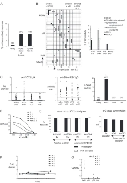
Figure 1. Antibody responses in patients with monoclonal gammo-pathies. (A) Preabsorbed serum samples from patients with MGUS, AMM, and MM were evaluated by SADA for the presence of IgG anti-bodies against a panel of 83 SEREX-defi ned antigens. The frequency of antibody responses within the cohorts of patients with asymptom-atic plasma cell disease and multiple MM is shown. Numbers indicate the patients with positive antibody response out of absolute numbers of patients evaluated by SADA for each group. (B, left) Patterns of antigenic reactivity in patients with MGUS, MM, and AMM. Rows depict individual patients according to their diagnosis, and columns show antibody reactivity against 23 tumor antigens with seropositivity in this cohort. Specifi c antigens in each column correspond numerically to antigens 1–23 in Table S2. (right) Shown are specific antigens inducing differential antibody response between subgroups of mono-clonal gammopathies and the frequency of antibody reactivity as
detected by SADA. *, P < 0.05. PRPF31, pre-mRNA–processing factor 31 homologue; RNPC1, RNA-binding region (RNP1, RRM)–containing 1. (C) ELISA for detection of SOX2-specifi c (left) and EBNA EBV–specifi c (middle) IgG antibodies in sera of MGUS, AMM, and MM patients. The distribution of antibody titers within specifi c groups is shown. (right) Overall SOX2 reactivity. Dotted lines represent the cutoff values for seropositivity. *, P < 0.05. (D) Titers of SOX2 antibodies in serial dilutions of sera from SOX2-reactive patients. (E) Sera from SOX2-positive patients were absorbed on SOX2-coated plates (or NY-ESO1–coated plates as irrelevant controls) and evaluated for anti-SOX2 IgG (or anti-EBNA IgG as a control) antibodies by ELISA (left) or detection of monoclonal Ig (by serum protein electrophoresis). Prior absorption on SOX2-abrogated anti-SOX2 reactivity without affecting anti-EBNA IgG reactivity (left) and mono clonal paraprotein concentration (right) are shown. Absorption on NY-ESO1 protein was
on August 29, 2017
A RT I C L E
identify this population are lacking. Whether the immune
system has the capacity to specifi cally target antigens
ex-pressed on cancer stem cells in humans is also not known.
In this paper, we show that the expression of an
embryo-nal stem cell marker, SOX2, specifi cally marks the clonogenic
CD138
−compartment in MGUS patients, and these patients
frequently mount humoral and cellular immunity to this
antigen. These data demonstrate the capacity of the human
immune system to spontaneously target antigens expressed on
tumor progenitors and the association of spontaneous
immu-nity against this target with an improved clinical outcome.
RESULTS
Detection of anti-SOX2 IgG antibodies in MGUS but not
MM patients or healthy donors
In prior studies, we have shown that the immune system is
capable of recognizing the preneoplastic lesions in MGUS
(10). To begin a systematic analysis of antigenic targets of
antitumor immunity in MGUS/MM, we initially analyzed
sera from patients with MM (n
= 35), MGUS (n = 28), and
asymptomatic MM (AMM; n
= 14) for the presence of IgG
antibodies against a panel of 83 serological expression of
cDNA expression libraries (SEREX)–defi ned tumor antigens
using a serum antibody detection array (SADA; Fig. 1 A and
Table S1, available at http://www.jem.org/cgi/content/full/
jem.20062387). Reactivity against 23 of the antigens in this
panel was detected in the sera from MGUS, AMM, or MM
patients but only in 1 out of 27 sera from normal blood donors
with this assay. Interestingly, the pattern of antigenic reactivity
diff ered between these cohorts (Fig. 1 B and Table S2).
Im-mune responses to Sry-HMG-box 2 (SOX2) protein were
seen only in MGUS, whereas antibodies against certain other
antigens (DNA methyltransferase 3, synaptonemal complex
protein 1, and kinesin family member 15) were only detected
in AMM (Fig. 1 B). To validate and quantify the presence of
anti-SOX2 antibodies in MGUS, we reanalyzed a larger
cohort of patients and age-matched healthy controls with an
ELISA-based assay (Fig. 1 C). Overall, SOX2 IgG
anti-bodies were detected in 12 out of 52 (23%) MGUS patients
but in none of the AMM (n
= 23) and MM (n = 40) patients
and in only 1 out of 92 healthy donors tested (P
< 0.001). As
controls, immune responses to Epstein-Barr nuclear antigen 1
(EBNA-1; Fig. 1 C) and tetanus toxoid (not depicted) were
comparably detectable in all cohorts. Anti-SOX2 antibodies
were present at a high titer and were detectable at a dilution
of
≥1:400 (Fig. 1 D). No anti-SOX2 IgM antibodies were
detected in any cohort (unpublished data). Anti-SOX2 IgG
antibodies were of both
κ and λ light chain specifi city and
were detected in both IgG and non-IgG gammopathies
(unpublished data). Preabsorption of sera with recombinant
SOX2 protein abrogated anti-SOX2 reactivity without
af-fecting the monoclonal paraprotein (Fig. 1 E). Therefore, the
observed reactivity is not caused by the monoclonal Ig found
in these patients. For six MGUS patients with high titers of
anti-SOX2 antibodies, follow-up samples over 2 yr revealed
that the antibody titers, as well as the clinical status, remain
stable over time (Fig. 1 F). In all SOX2-reactive MGUS
sera, antibodies were predominantly of the IgG1 subclass
(Fig. 1 G). Collectively, these data demonstrate the presence
of anti-SOX2 IgG1 antibodies in a substantial proportion of
MGUS patients.
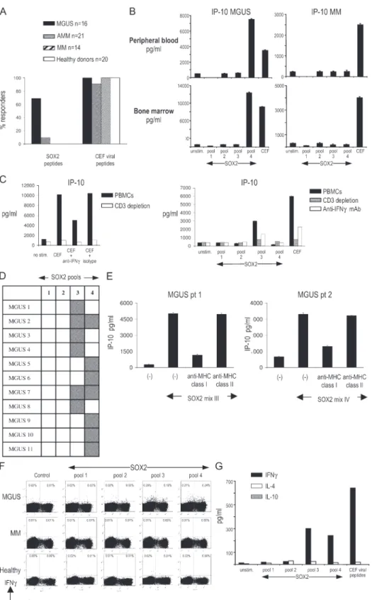
Detection of anti-SOX2 T cell responses in MGUS
but not MM patients or healthy donors
We examined if SOX2 was also a target of antitumor T cell
response in these patients. Freshly isolated PBMCs were
stimulated with a library of overlapping 15-mer peptides
spanning the entire SOX2 protein (Table S3, available at
http://www.jem.org/cgi/content/full/jem.20062387/DC1).
Peptide-reactive chemokine production (IFN-
γ–inducible
protein 10 [IP-10]) was monitored by Luminex analysis.
Using this assay, SOX2-specifi c T cells were detected in fresh
PBMCs from 11 out of 16 MGUS patients tested but in none
of the MM patients (n
= 14) or healthy donors (n = 20; P <
0.05; Fig. 2, A and B). Anti-SOX2–specifi c T cells were also
detected in 2 out of 21 patients with AMM. T cell reactivity
against a pool of MHC class I–restricted viral antigen peptides
(CEF, derived from CMV, EBV, and infl uenza virus) was
comparable in all four cohorts. SOX2-specifi c T cells were
detected in both blood and bone marrow in four patients
tested (Fig. 2 B). In this assay, the production of IP-10, a
po-tent antiangiogenic chemokine, in response to viral or SOX2
peptides is a sensitive readout for T cell–derived IFN-
γ, as it
is abrogated by prior depletion of CD3
+T cells and
sub-stantially reduced in the presence of neutralizing anti–IFN-
γ
mAb (Fig. 2 C). SOX2-specifi c reactivity in MGUS patients
targeted peptides in pools 3 or 4 of the SOX2 peptide library.
In two patients, SOX2-specifi c T cells were detected in
response to both pools 3 and 4 (Fig. 2 D). SOX2-reactive
T cells detected in this assay were predominantly MHC class I
restricted, as the response was inhibited by anti–MHC class I
blocking antibody (Fig. 2 E). To further analyze the nature of
this T cell response, PBMCs from MGUS patients were
stimulated for 2 wk with autologous DCs loaded with the
SOX2 peptide library and analyzed for the presence of
SOX2-specifi c T cells by intracellular IFN-
γ fl ow cytometry.
These experiments demonstrated that SOX2-reactive T cells
included both CD4
+and CD8
+T cells (Fig. 2 F). In
con-trast, SOX2-specifi c T cells could not be detected from MM
patients or healthy donors, even after four restimulations with
peptide-pulsed DCs (Fig. 2 F and not depicted). Expanded
SOX2-reactive CD4
+T cells were of the Th1 cell phenotype,
performed as a control. Data represent the mean ± SEM. (F) Long-term persistence of anti-SOX2 IgG response in follow-up samples. Titers of anti-SOX2–specific IgG antibodies were detected in follow-up
samples from patients with MGUS (patients 1–6). (G) IgG subclass– specific analysis of SOX2-specific antibody response in patients with MGUS.
on August 29, 2017
jem.rupress.org
Figure 2. Analysis of SOX2-specifi c T cell responses. (A) Overall analysis of frequency of anti-SOX2 T cell responses in patients with MGUS, AMM, and MM and healthy donors. (B) Analysis of anti-SOX2 T cell reactivity in freshly isolated PBMCs and BMMNCs. Freshly isolated MNCs were stimulated with peptide pools derived from the SOX2 peptide library for 48 h, and supernatants were analyzed for IP-10 production. Data rep-resent the mean ± SEM. (C) Depletion of CD3+ T cells or neutralization of
IFN-γ decreases antigen-specifi c production of IP-10 in response to viral peptides (left) or SOX2-derived peptides (right). (left) PBMCs were stimu-lated with a cocktail of peptides derived from viral antigens (CEF) with or without prior depletion of CD3+ T cells or prior treatment with 10 μg/ml
anti–IFN-γ blocking antibody or isotype control mAb. (right) PBMCs from a MGUS patient were similarly stimulated with four pools of derived peptides. Supernatants were analyzed for IP-10 production by
on August 29, 2017
A RT I C L E
as they mainly produced IFN-
γ, but not IL-4 or IL-10, upon
SOX2 stimulation (Fig. 2 G). SOX2-specifi c T cells in
MGUS were detected in seven out of nine patients with
positive anti-SOX2 antibodies tested, as well as in four of
seven patients without detectable anti-SOX2 antibodies.
Therefore, SOX2 is a frequent target for specifi c T cell
im-munity in MGUS patients but not in MM or healthy donors,
and the detection of specifi c T cell responses may be more
sensitive than current assays in detecting humoral responses.
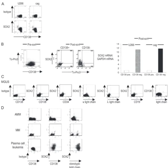
Intranuclear SOX2 marks the clonogenic compartment
in MGUS
Expression of SOX2 is restricted to embryonal and neural
stem cells, wherein it plays a critical role in regulating the
self-renewal and pluripotency of stem cells (11–15). Recent
studies in MM and MGUS have suggested that a minor
CD138
−subpopulation of both MM cell lines and primary
cells is enriched in clonogenic progenitors, capable of growth
in methylcellulose as well as in immune-defi cient mice (8, 9).
Analysis of intranuclear SOX2 expression in two MM cell
lines by fl ow cytometry, as well as SOX2 mRNA by
Taq-Man, revealed that SOX2 indeed specifi cally marks this
CD138
−subpopulation (Fig. 3, A and B). Analysis of SOX2
expression in marrow from MGUS patients revealed that
SOX2
+cells were restricted for tumor-associated Ig light
chain but lacked the expression of the terminal plasma cell
diff erentiation marker CD138 and the hematopoietic stem
cell marker CD34 (Fig. 3 C). These cells expressed lower
levels of Ig light chain compared with CD138
+plasma cells
and lacked expression of CD19, a B cell marker. Overall,
SOX2
+cells in the MGUS marrow accounted for only 0.5–
1.5% of mononuclear cells (MNCs) and had a phenotype
consistent with preplasma cells (CD138
−CD19
−IgL
lo) (16).
Interestingly, in patients with active MM, SOX2
+cells were
also observed in the more diff erentiated CD138
+IgL
highcom-partment in fi ve out of fi ve patients tested (Fig. 3 D). The
SOX2 expression pattern in patients with AMM was
inter-mediate, with three out of fi ve patients resembling the
stain-ing in MGUS (Fig. 3 D), whereas the other two were more
like MM. Circulating tumor cells in a patient with advanced
MM in leukemic phase showed higher reactivity, suggesting
the acquisition of this marker by more diff erentiated cells in
some patients with more aggressive disease (Fig. 3 D).
Targeting SOX2 immunity inhibits the clonogenic growth
of tumors
T cells from HLA-A2
+MGUS patients were capable of IP-10
secretion in response to a CD138
−compartment of A2
+U266 cells (wherein the SOX2 expression is limited; Fig. 3 A),
Luminex. (D) Patterns of anti-SOX2 T cell reactivity in MGUS patients. Gray squares represent a positive T cell response against a corresponding pool of the SOX2 peptide library. (F) Analysis of anti-SOX2 T cell reactivity in PBMCs from two patients with SOX2-specifi c T cells against SOX2. Stimu-lation with SOX2 peptides was performed in the presence of anti–MHC class I blocking antibody and supernatants analyzed for IP-10 production.
Data represent the mean ± SEM. (F) Expansion of SOX2-specifi c CD4 and CD8 T cells in MGUS after two stimulations with SOX2 peptide library– loaded DCs evaluated by intracellular staining for IFN-γ. Representative results of one experiment out of three with similar results are shown. (G) ELISA for IFN-γ, IL-4, and IL-10 in the supernatants from cultures of SOX2-stimulated T lymphocytes.
consistent with the recognition of endogenously presented
antigen (Fig. 4 A). Although SOX2 is expressed only by a
proportion of the bulk tumor, immunity against this antigen
could still be important if this subpopulation or antigen was
important for the clonogenic growth of tumors. To directly
test this, marrow MNCs from MGUS patients were
stimu-lated with the SOX2 peptide library, and SOX2
responsive-ness was documented by the production of IP-10 (Fig. 4 B).
Marrow MNCs (CD34
−CD138
−) stimulated under these
conditions were plated in clonogenic assays (9). Cultures
stimulated with the SOX2 peptide library demonstrated
sub-stantially inhibited clonogenic growth in three out of three
MGUS patients tested (Fig. 4 B and not depicted). As noted
earlier, stimulation of marrow MNCs from MM with SOX2
peptides did not lead to any detectable reactivity (Fig. 2, A
and F), and consistent with this, prestimulation with the
SOX2 peptide library did not lead to inhibition of
clono-genic growth in MM (Fig. 4 C).
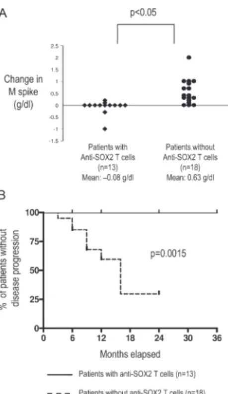
Detection of anti-SOX2 T cells predicts a favorable outcome
in patients with asymptomatic plasmaproliferative disorders
Most studies evaluating the clinical signifi cance of anti tumor
immunity in humans are based on retrospective data. Several
of the patients analyzed in this study were enrolled in an
observational trial of patients with asymptomatic
plasmapro-liferative disorders, performed under the auspices of the
South-west Oncology Group. This provided a unique opportunity to
prospectively evaluate whether T cell im munity to a single
antigen could predict tumor progression. With a median
follow up of 24 mo, patients with anti-SOX2 T cells had a
signifi cantly lower net change in the level of tumor-derived
monoclonal Ig (M spike) over time compared with those
lacking anti-SOX2 T cells (mean increase in M spike
= 0.08
g/dl vs. 0.63 g/dl, respectively; P
< 0.05; Fig. 5 A). The
pa-tients with anti-SOX2 T cells also had a signifi cantly lower
likelihood of disease progression, with a 2-yr progression-free
survival rate of 100 versus 30% compared with patients lacking
anti-SOX2 T cells. (P
< 0.01; Fig. 5 B). Therefore, immunity to
SOX2 predicts the clinical outcome in patients with
asymp-tomatic plasmapro liferative disorders.
DISCUSSION
A growing body of evidence points to the capacity of the
hu-man immune system to recognize preneoplastic lesions (3, 4,
10, 17, 18). However, the nature of specifi c antigens
recog-nized by T cells in the preneoplastic stage of human cancer is
largely unknown. The data in this paper suggest that the pattern
of antigens spontaneously recognized by the immune system
in preneoplastic lesions may diff er from that in clinical cancer.
on August 29, 2017
jem.rupress.org
Therefore, these data have implications for harnessing the
immune system for the early detection and prevention of
cancer in humans (19, 20).
The fi nding (originating from an unbiased search) that
immunity to SOX2 (a gene critical for the self-renewal and
pluripotency of embryonic stem cells) (14, 15) predicts
clin-ical outcome supports the importance of stem cell genes and
self-renewal pathways in cancer biology. These data also
show that intranuclear SOX2 specifi cally marks the putative
MM progenitors (8). SOX2 has also been recently found
to be expressed in other cancer stem cells and implicated in
intestinal metaplasia in gastric cancer (12, 13). Identifi cation
of a specifi c marker for the putative progenitor population
in MM should facilitate understanding of myelomagenesis,
as well as the development of specifi c therapies targeting this
population. A subpopulation of CD138
+cells also acquires
the expression of SOX2 in patients with progressive MM.
Thus, the progression from MGUS to MM may involve the
acquisition of self-renewal properties by the diff erentiated
compartment of the clone. In other words, there may be a
fundamental diff erence in the nature of the self-renewing
compartment in MGUS versus MM. Further studies are
Figure 3. Phenotype of SOX2-expressing cells in MM cell lines and in patients with MGUS, AMM, and MM. (A) Intranuclear staining for SOX2 expression in multiple MM cell lines U266 and cag. Data shown are gated on live cells based on scatter properties, and SOX2 expression was analyzed by intranuclear staining. The percentage of cells in each quadrant is indicated. (B) Analysis of SOX2 mRNA and protein in sorted CD138+ and CD138− MM cells. FACS data shown are gated for live cells based on scatter properties. Cells were stained with anti-CD138 and To-Pro-3 (to further discriminate dying cells) before the cell sort. Live (To-Pro-3–negative) cells were then sorted into CD138− and CD138+
populations by fl ow sorting. Each population was stained for the expres-sion of intranuclear SOX2 expresexpres-sion and analyzed for the expresexpres-sion of
SOX2 mRNA by TaqMan. The relative quantity of SOX2 transcripts (nor-malized to GAPDH) is shown. The percentage of cells in each quadrant is indicated. (C) SOX2 expression in BMMNCs in a patient with IgG κ MGUS. BMMNCs were stained with mAbs against SOX2 in combination with other markers. Representative results of one out of eight MGUS patients are shown. (D) SOX2 expression patterns in tumor cells from patients with AMM, MM, and plasma cell leukemia. Data are representative of fi ve pa-tients tested with MM and of seven papa-tients with AMM. Three out of fi ve AMM patients had SOX2 expression patterns similar to MGUS patients (AMM). In two out of fi ve patients with AMM, the pattern was similar to that in MM. Clonotypic light chain refers to the light chain of the tumor-derived monoclonal Ig.
on August 29, 2017
A RT I C L E
needed to evaluate this possibility. Very similar correlations
have recently been described in patients with chronic
my-elogenous leukemia, wherein blast transformation is
associ-ated with the acquisition of self-renewal genes by the more
diff erentiated compartment of the clone (21, 22).
In earlier experiments, we had shown that the tumor bed
in MGUS is enriched for T cells reactive against autologous
CD138
+preneoplastic cells (10). The data in the current
paper extend these fi ndings to show that MGUS patients also
mount an immune response against antigens expressed in the
CD138
−compartment of the clone, thought to be enriched
in clonogenic tumor progenitors. The frequency of these
T cells is low (relative to an antiviral responses during acute
infection), and they target an antigen expressed only in a
pro-portion of the clone. However, stimulation of the marrow
MNCs with this antigen, without prior in vitro expansion,
was suffi
cient to inhibit the clonogenic growth of MGUS
cells in vitro. One possibility is that the immune response
focused against progenitors or self-renewal genes may be
more effi
cient in suppressing clonogenic growth than in
immunity against bulk tumor. Interestingly, spontaneous
immunity to SOX2 was not detectable in MM patients, who
also carry SOX2-expressing cells. Further studies are needed
to understand the absence of spontaneous SOX2 immunity
in MM patients. Importantly, MGUS patients with anti-SOX2
Figure 4. Relevance of SOX2-specifi c T cells for the recognition of tumor cells and inhibition of clonogenic tumor growth. (A) Produc-tion of IP-10 in HLA-A2+ MGUS patients in response to MACS-sorted CD138− and CD138+ subsets of U266 cell line (HLA-A2+). Representative
results for two patients are shown. (B) Inhibition of clonogenic growth of primary tumor cells after stimulation with the SOX2 peptide library in MGUS (left). BMMNCs were depleted of CD138+ and CD34+ cells, incu-bated with SOX2 or control peptides, and plated with 5 × 10⁵
monocyte-derived DCs/ml at a ratio of 1:2 in Methocult. Colonies were counted by microscopy 2–3 wk after plating. Effective prestimulation with SOX2 pep-tides was documented by IP-10 production 48 h after stimulation (right). The relative growth of tumor colonies for two patients with SOX2-reac-tive T cells is shown. (C) Clonogenic growth of primary tumor cells after stimulation with the SOX2 peptide library in MM. BMMNCs were depleted of CD138+ and CD34+ cells, incubated with SOX2 or left unstimulated, and plated with 5 × 10⁵ monocyte-derived DCs/ml at a ratio of 1:2 in
Methocult. Colonies were counted by microscopy 2–3 wk after plating. Relative growth of tumor colonies for two patients with SOX2-reactive T cells is shown. Data represent the mean ± SEM.
Figure 5. Correlation of detectable SOX2-reactive T cell immunity with clinical outcome in patients with asymptomatic plasmaprolif-erative disorders. (A) Net change in levels of tumor-derived monoclonal Ig (M spike) in patients with asymptomatic plasmaproliferative diseases with or without detectable SOX2-specifi c T cells. (B) Comparison of time-to-progression Kaplan-Meier’s curves constructed for the cohorts with
and without SOX2-specifi c T cells (P < 0.01 using the log-rank test). on August 29, 2017
jem.rupress.org
The potential role of the immune system in regulating
cancer development has been extensively debated (1, 23). It
is likely that distinct components of the immune system have
the capacity to both promote as well as suppress cancer. It is
therefore of interest that T cell immunity to SOX2 correlates
with a favorable outcome. Antibodies against SOX2 have
also been detected in some patients with small cell lung
cancer and impart a favorable prognosis, although
SOX2-specifi c T cells have not yet been studied in these patients
(24, 25). However, the correlation of SOX2 immunity with
a favorable outcome does not establish a causal relationship.
For example, the detection of SOX2-specifi c T cells may be
refl ective of altered biology of tumor progenitors in MGUS
versus MM. Therefore, clinical studies to enhance SOX2
immunity are needed to directly assess whether immunity to
SOX2 or other antigens on tumor progenitors can induce
tumor regressions in patients with MM and other cancers.
These data should also encourage a systematic search for
spe-cifi c targets of spontaneous T cell immunity in other human
preneoplastic states.
These data also have several clinical implications. Current
management of patients with asymptomatic
plasmaprolifera-tive tumors is a challenge, as it is often diffi
cult to predict
dis-ease progression and the need for therapy in these patients
(26). These data suggest that in addition to changes in tumor
cells, the nature of tumor-specifi c host immune response may
also provide a novel approach for predicting an outcome.
Most studies of immunotherapy of human cancer to date
have focused on trying to target antigens expressed by bulk
tumors. This approach has led to generally low rates of
clini-cal regressions, often in spite of high frequencies of immunity
to vaccine antigens. These data suggest the possibility that
targeting drugs or the immune system against targets critical
to the biology of tumor progenitors may be needed for the
eff ective control of cancer.
MATERIALS AND METHODS
Patient samples. Bone marrow and peripheral blood samples used in this study were obtained from patients with a diagnosis of MGUS, AMM, and MM based on standard clinical criteria (27). All patients signed an informed consent approved by the institutional review board. Several samples were obtained under the auspices of a prospective multicenter Southwest Oncol-ogy Group observational clinical trial (S0120) of patients with asymptomatic plasmaproliferative disorders.
Cell lines and media. Multiple MM cells lines U266 and cag have been previously described (28). For DC and T cell cultures, 5% pooled human serum (Labquip) was used.
SADA for analyzing serum reactivity. Preabsorbed serum samples from 28 patients with MGUS, 14 patients with AMM, 35 patients with MM, and 27 healthy blood donors were evaluated by SADA for the presence of IgG antibody to a panel of 83 SEREX-defi ned antigens, as previously described (29). In brief, precut nitrocellulose membranes (80 × 120 mm) were pre-coated with a layer (≈0.2 mm) of growth media and placed on a reservoir layer of media in Omni Tray (86 × 128 mm; Nalge Nunc International
on the precoated nitrocellulose membranes. 30 SEREX-defi ned antigens were spotted in duplicate on each nitrocellulose membrane. Membranes were incubated for 15 h at 37°C and processed as per the standard SEREX protocol (29). In brief, membranes were blocked in 0.5% nonfat dried milk, incubated in 10 ml of a 1:200 dilution of sera at room temperature for 15 h, and incu-bated in a 1:3,000 dilution of alkaline phosphatase–conjugated, Fc fragment– specifi c, goat anti–human IgG (Jackson ImmunoResearch Laboratories). Serum IgG reactivity was detected with the alkaline phosphatase substrate 4-nitro blue tetrazolium chloride/5-bromo-4-chloro-3-indolyl-phosphate (Biosource International). Positive assays were repeated in a blinded manner to confi rm reactivity.
ELISA for the detection of anti-SOX2, EBV (EBNA-1) antibodies. Sera were screened for the presence of anti-SOX2 antibodies by standard ELISA. A 96-well microtiter polystyrene half-area immunoassay plate (Corning) was coated in PBS with 1 μg/ml of recombinant human SOX2 protein overnight at 4°C. Plates were washed with PBS and blocked with 5% nonfat dry milk in PBS for 2 h. After washing, serum samples at 1:100 and 1:400 dilutions in the blocking solution were added and incubated for 1 h at room temperature. Plates were extensively washed with 0.05% Tween 20 in PBS, and the secondary antibody, horseradish peroxidase–labeled goat anti–human IgG, IgG1, IgG2, IgG3, or IgG4 (Southern Biotechnology As-sociates, Inc.), in blocking solution was added. Plates were incubated for 1 h at room temperature and washed, and SOX2 reactivity was revealed by the addition of substrate solution (Biosource International). After 30 min of in-cubation in the dark, the reaction was ended with Stop solution (Biosource International), and the absorbance at 450 nm was measured using an ELISA plate reader (MultiSkan Plus; Thermo Fisher Scientifi c). The threshold for seropositivity for SOX2 antibodies was determined as ODN450 at 0.11, based on the mean background for no antigen controls plus 4 SD. Sero-positivity for anti–EBNA-1 IgG was determined using a commercial kit (SCIMEDX) according to the manufacturer’s instructions. Samples with an antibody index ≥1 were considered to be seropositive for EBNA IgG. Detection of cytokines by ELISA. Commercial ELISA kits (Biosource International) were used to measure IFN-γ, IL-4, and IL-10 concen-trations in the cell-culture supernatants, according to the manufac-turer’s recommendations.
Preabsorption of anti-SOX2–positive samples. Before ELISA analysis for SOX2 reactivity, samples with anti-SOX2 IgGs were preabsorbed over-night at 4°C in 96-well plates previously coated with 30 μg/ml of recombi-nant SOX2 protein and blocked with 5% nonfat dry milk in PBS.
Synthesis of the SOX-2 peptide library. Peptides were synthesized in collaboration with the Proteomics Resource Center at the Rockefeller Uni-versity. Overlapping sequences from the Sox-2 protein were determined and optimized for synthesis by using the epitope library fragment generation pro-gram PeptGen, developed by Los Alamos National Laboratories, as part of the HIV Immunology Database (available at http://www.hiv.lanl.gov). All peptides were created in a microtiter plate (96 well) format using a parallel peptide synthesizer/spotter (MultiPep; Intavis) on resin (TentaGel R RAM; Rapp Polymere) loaded at 5 μm per well, using Fmoc-protected amino acids (Anaspec). Deprotection of the amine was accomplished with 20% piperidine (Sigma-Aldrich) in N-methylpyrrolidinone (NMP; EMD Bio-sciences, Inc.). Repetitive coupling reactions were conducted using 0.3 M HATU/ HOBt and 0.4 M NMM using NMP as the primary solvent. Simul-taneous resin cleavage and side-chain deprotection were achieved by treatment with 0.8 ml/well of concentrated, sequencing grade trifl uoroacetic acid (Fisher Scientifi c) with triisopropylsilane, water, and DODT in a ratio of 95:2:2:1 for 2 h. After vacuum fi ltration to a collection plate, centrifugal evaporation (Genevac) was used to remove TFA from the plate containing
on August 29, 2017
jem.rupress.org
A RT I C L E
the soluble peptides. Peptides were treated with 8 M acetic acid, and the acidic mixture was evaporated and redissolved in 20% acetonitrile and HPLC (Waters Chromatography)-grade water and dried twice more. All crude products were subsequently analyzed by reversed-phase HPLC using a C18 column (Chromolith Performance; Merck). Individual peptide integrity was verifi ed by matrix-assisted laser desorption/ionization (MALDI) mass spectro-metry using a delayed extraction spectrometer system (Voyager; PerSeptive/ Applied Biosystems).
Screening for SOX2-reactive T cells in fresh PBMCs. MNCs from blood or bone marrow were separated by density gradient centrifugation us-ing Ficoll-Hypaque (GE Healthcare). 2 × 10⁵ PBMCs or BMMCs in 200 μl of media were cultured in the presence of 2.5 μg/ml of peptide pools de-rived from the SOX2 peptide library or with a mixture of MHC class I– restricted peptides derived from CMV, EBV, and infl uenza virus (CEF mix) (30). The composition of the SOX2 peptide library pools is noted in Table S3. After 48 h, supernatants were collected and assayed for the production of IP-10 by Luminex, using the manufacturer’s directions (Upstate Biotech-nology), and analyzed by Beadview software (Upstate Biotechnology). In some experiments, CD3 T cells were depleted by negative selection, or PBMC stimulation by specifi c antigens was performed in the presence of IFN-γ or anti–MHC class I blocking mAbs (Biolegend) to confi rm the specifi city of IP-10 production. For some experiments, CD138+ and CD138− fractions of U266 (HLA-A2+ cell line) were also used for the stim-ulation of fresh PBMCs from HLA-A2+ MGUS patients with documented anti-SOX2 T cell reactivity, and IP-10 production was analyzed by Luminex 48 h later. A twofold or greater increase in IP-10 production relative to con-trol was considered as positive for the presence of antigen-specifi c T cells. DC generation and expansion of SOX2-specifi c T cells. MNCs from blood or bone marrow were separated by density gradient centrifugation us-ing Ficoll-Hypaque. DCs were generated from monocytes isolated by CD14 magnetic beads (Miltenyi Biotec) and cultured for 5 d in the presence of GM-CSF (Immunex) and IL-4 (R&D Systems), as previously described (30). Day-5 DCs were matured overnight with 100 ng/ml LPS (Sigma-Aldrich) and pulsed for 2 h with peptide pools derived from the SOX2 peptide library (15-mer peptides overlapping by 11 aa) at 2 μg/ml. A CD14− T cell–enriched fraction was added to a U-bottom 96-well plate at 2 × 10⁵ cells/well in 200 μl of medium and stimulated with mature peptide-pulsed DCs at a ratio of 1 DC per 20 CD14− cells. Recombinant IL-2 was added at 15 U/ml every third day. Antigen-specifi c T cells were restimulated with the same antigen-pulsed DCs every week and tested for IFN-γ production at the time of restimulation by intracellular cytokine fl ow cytometry. Flow cytometry for the detection of intracellular cytokines. Anti-gen-specifi c cells were analyzed by a fl ow cytometry–based assay for the de-tection of intracellular cytokines, as described previously (31). In brief, blood or bone marrow T cells were cultured for 12 h with autologous unpulsed mature DCs, or DCs loaded with specifi c peptide mixtures, in the presence of Golgistop (Cytofi x/CytoPerm Plus Kit; BD Biosciences). Cells were fi xed and permeabilized in 100 μl Cytofi x/Cytoperm solution using the manufacturer’s instructions and stained for intracellular cytokines (IFN-γ) and surface markers (CD3 and CD8).
Intranuclear SOX2 staining. 5 × 10⁵ cells were fi xed in Cytofi x/Cyto-perm overnight at 4°C. Cells were x/Cyto-permeabilized in Cytox/Cyto-perm solution, refi xed in Cytofi x/Cytoperm for 5 min on ice, and treated with 300 μg/ml DNase I for 45 min at 37°C. Cells were stained with PE-SOX2 mAb (R&D Systems) for 20 min at room temperature, washed, and stained for surface or intracellular markers. Samples were acquired on an instrument (FACSCali-bur; BD Biosciences) using CellQuest software (BD Biosciences) and ana-lyzed with FlowJo software (TreeStar Inc.). Typically, 1–5 × 10⁵ events were collected per sample.
Cell sorting and analysis of SOX2 mRNA by TaqMan. CD138− and CD138+ fractions of the U266 and cag cell lines were sorted on a
FACSVan-tage (BD Biosciences). The purity of sorted populations used in real-time PCR experiments exceeded 95%. RNA was isolated with the RNeasy Mini Kit (QIAGEN), and RT-PCR was conducted with the Assays-on-Demand (Applied Biosystems) primer probe for SOX2 using a sequence detection system (ABI PRISM 7700; Applied Biosystems). Expression of GAPDH was monitored as a housekeeping gene. Reactions were set up in triplicates using EZ PCR Core Reagents (Applied Biosystems), according to the manufac-turer’s instructions, with 20 ng of total RNA. The relative expression of target genes was calculated using the comparative threshold cycle method. Clonogenic assays on primary tumor cells. Bone marrow MNCs (BMMNCs) were isolated from marrow samples using density gradient cen-trifugation. CD138+ and CD138– fractions were isolated using CD138 microbeads (Miltenyi Biotec), and the CD138– fraction was further depleted of normal hematopoietic progenitors using CD34 microbeads (Miltenyi Bio-tec). CD138−CD34− cells were incubated overnight with SOX2 or control peptides and plated (5 × 10⁵ cells/ml) with monocyte-derived DCs at a ratio of 1:2 in methylcellulose containing 5% leukocyte-conditioned media (Methocult; Stem Cell Technologies, Inc.), as described previously (9). Cells were plated in 35-mm2 tissue culture dishes in triplicates and incubated at 37°C and 5% CO2. Colonies consisting of >40 cells were counted by microscopy 2–3 wk after plating.
Statistical analysis. Diff erences in frequencies were assessed with Fisher’s exact test for two groups and the χ2 test for three or more groups. Diff erences in change in M protein over time between those with and without SOX2 immunity were tested using the Wilcoxon test. The criteria for disease pro-gression required an increase in M protein ≥0.5 g/dl, an increase in marrow plasmacytosis ≥10%, or a development of symptomatic disease requiring ini-tiation of therapy (32). Progression-free survival curves were constructed us-ing the Kaplan-Meier method (33) and tested usus-ing the log-rank test (34). Online supplemental material. Table S1 provides a list of tumor antigens tested in the SADA. Table S2 shows a list of tumor antigens inducing IgG antibody responses in patients with plasma cell diseases. Table S3 provides the sequences of peptides used in the SOX2 peptide library. Online supplemental material is available at http://www.jem .org/cgi/content/full/jem.20062387/DC1.
This paper is dedicated to our patients for their encouragement of this work and to the loving memory of Matt Scanlan.
The authors thank Dr. R.M. Steinman for critical reading of the manuscript; Drs. L.J. Old and K.L. Calame for helpful discussions; J. Krasovsky, A. Hutchinson, and A. Murray for excellent technical assistance; A. Hurley for help with clinical aspects; and J. Adams for help with illustrations.
This work was supported in part by funds from the National Institutes of Health (grants CA106802, CA109465, P50-AT02779, MO1-RR00102, and PO1-CA55819), an Eli Lilly-Damon Runyon Clinical Investigator Award, the Dana Foundation, the Irma T. Hirschl Foundation, the Fund to Cure Myeloma, and the Southwest Oncology Group.
The authors have no confl icting fi nancial interests. Submitted: 14 November 2006
Accepted: 28 February 2007
REFERENCES
1. Dunn, G.P., A.T. Bruce, H. Ikeda, L.J. Old, and R.D. Schreiber. 2002. Cancer immunoediting: from immunosurveillance to tumor escape.
Nat. Immunol. 3:991–998.
2. Blattman, J.N., and P.D. Greenberg. 2004. Cancer immunotherapy: a treatment for the masses. Science. 305:200–205.
3. Finn, O.J. 2003. Premalignant lesions as targets for cancer vaccines.
J. Exp. Med. 198:1623–1626.
4. Dhodapkar, M.V. 2005. Harnessing host immune responses to preneoplasia: promise and challenges. Cancer Immunol. Immunother. 54:409–413.
on August 29, 2017
jem.rupress.org
N. Engl. J. Med. 354:1362–1369.
6. Fonseca, R., B. Barlogie, R. Bataille, C. Bastard, P.L. Bergsagel, M. Chesi, F.E. Davies, J. Drach, P.R. Greipp, I.R. Kirsch, et al. 2004. Genetics and cytogenetics of multiple myeloma: a workshop report.
Cancer Res. 64:1546–1558.
7. Reya, T., S.J. Morrison, M.F. Clarke, and I.L. Weissman. 2001. Stem cells, cancer, and cancer stem cells. Nature. 414:105–111.
8. Matsui, W., C.A. Huff , Q. Wang, M.T. Malehorn, J. Barber, Y. Tanhehco, B.D. Smith, C.I. Civin, and R.J. Jones. 2004. Characterization of clonogenic multiple myeloma cells. Blood. 103:2332–2336. 9. Kukreja, A., A. Hutchinson, K. Dhodapkar, A. Mazumder, D. Vesole,
R. Angitapalli, S. Jagannath, and M.V. Dhodapkar. 2006. Enhancement of clonogenicity of human multiple myeloma by dendritic cells. J. Exp.
Med. 203:1859–1865.
10. Dhodapkar, M.V., J. Krasovsky, K. Osman, and M.D. Geller. 2003. Vigorous premalignancy-specifi c eff ector T cell response in the bone marrow of patients with monoclonal gammopathy. J. Exp. Med. 198:1753–1757.
11. Wegner, M., and C.C. Stolt. 2005. From stem cells to neurons and glia: a Soxist’s view of neural development. Trends Neurosci. 28:583–588. 12. Tatematsu, M., T. Tsukamoto, and K. Inada. 2003. Stem cells and
gastric cancer: role of gastric and intestinal mixed intestinal metaplasia.
Cancer Sci. 94:135–141.
13. Hemmati, H.D., I. Nakano, J.A. Lazareff , M. Masterman-Smith, D.H. Geschwind, M. Bronner-Fraser, and H.I. Kornblum. 2003. Cancerous stem cells can arise from pediatric brain tumors. Proc. Natl. Acad. Sci.
USA. 100:15178–15183.
14. Boyer, L.A., T.I. Lee, M.F. Cole, S.E. Johnstone, S.S. Levine, J.P. Zucker, M.G. Guenther, R.M. Kumar, H.L. Murray, R.G. Jenner, et al. 2005. Core transcriptional regulatory circuitry in human embryonic stem cells. Cell. 122:947–956.
15. Takahashi, K., and S. Yamanaka. 2006. Induction of pluripotent stem cells from mouse embryonic and adult fi broblast cultures by defi ned factors. Cell. 126:663–676.
16. McHeyzer-Williams, L.J., and M.G. McHeyzer-Williams. 2005. Antigen-specifi c memory B cell development. Annu. Rev. Immunol. 23:487–513.
17. Suzuki, H., D.F. Graziano, J. McKolanis, and O.J. Finn. 2005. T cell-dependent antibody responses against aberrantly expressed cyclin B1 protein in patients with cancer and premalignant disease. Clin. Cancer
Res. 11:1521–1526.
18. Comtesse, N., A. Zippel, S. Walle, D. Monz, C. Backes, U. Fischer, J. Mayer, N. Ludwig, A. Hildebrandt, A. Keller, et al. 2005. Complex humoral immune response against a benign tumor: frequent antibody response against specifi c antigens as diagnostic targets. Proc. Natl. Acad.
Sci. USA. 102:9601–9606.
19. Spisek, R., and M.V. Dhodapkar. 2006. Immunoprevention of cancer.
Hematol. Oncol. Clin. North Am. 20:735–750.
20. Forni, G., P.L. Lollini, P. Musiani, and M.P. Colombo. 2000. Immuno-prevention of cancer: is the time ripe? Cancer Res. 60:2571–2575.
in blast-crisis CML. N. Engl. J. Med. 351:657–667.
22. Jamieson, C.H., I.L. Weissman, and E. Passegue. 2004. Chronic versus acute myelogenous leukemia: a question of self-renewal. Cancer Cell. 6:531–533.
23. de Visser, K.E., A. Eichten, and L.M. Coussens. 2006. Paradoxical roles of the immune system during cancer development. Nat. Rev. Cancer. 6:24–37.
24. Gure, A.O., E. Stockert, M.J. Scanlan, R.S. Keresztes, D. Jager, N.K. Altorki, L.J. Old, and Y.T. Chen. 2000. Serological identifi cation of embryonic neural proteins as highly immunogenic tumor antigens in small cell lung cancer. Proc. Natl. Acad. Sci. USA. 97:4198–4203. 25. Vural, B., L.C. Chen, P. Saip, Y.T. Chen, Z. Ustuner, M. Gonen, A.J.
Simpson, L.J. Old, U. Ozbek, and A.O. Gure. 2005. Frequency of SOX Group B (SOX1, 2, 3) and ZIC2 antibodies in Turkish patients with small cell lung carcinoma and their correlation with clinical parameters.
Cancer. 103:2575–2583.
26. Kyle, R.A. 2004. New Strategies for MGUS and Smoldering Multiple Myeloma. Clin. Adv. Hematol. Oncol. 2:507–509.
27. Durie, B.G., R.A. Kyle, A. Belch, W. Bensinger, J. Blade, M. Boccadoro, J.A. Child, R. Comenzo, B. Djulbegovic, D. Fantl, et al. 2003. Myeloma management guidelines: a consensus report from the Scientifi c Advisors of the International Myeloma Foundation. Hematol. J. 4:379–398.
28. Dhodapkar, K., J. Krasovsky, B. Williamson, and M. Dhodapkar. 2002. Antitumor monoclonal antibodies enhance cross-presentation of cellular antigens and the generation of tumor-specifi c killer T cells by dendritic cells. J. Exp. Med. 195:125–133.
29. Chen, Y.T., A.O. Gure, and M.J. Scanlan. 2005. Serological analysis of expression cDNA libraries (SEREX): an immunoscreening tech-nique for identifying immunogenic tumor antigens. Methods Mol. Med. 103:207–216.
30. Dhodapkar, K.M., J.L. Kaufman, M. Ehlers, D.K. Banerjee, E. Bonvini, S. Koenig, R.M. Steinman, J.V. Ravetch, and M.V. Dhodapkar. 2005. Selective blockade of inhibitory Fcgamma receptor enables human dendritic cell maturation with IL-12p70 production and immunity to antibody-coated tumor cells. Proc. Natl. Acad. Sci.
USA. 102:2910–2915.
31. Chang, D.H., K. Osman, J. Connolly, A. Kukreja, J. Krasovsky, M. Pack, A. Hutchinson, M. Geller, N. Liu, R. Annable, et al. 2005. Sustained expansion of NKT cells and antigen-specifi c T cells after injection of α-galactosyl–ceramide–loaded mature dendritic cells in cancer patients. J. Exp. Med. 201:1503–1517.
32. Durie, B.G., J.L. Harousseau, J.S. Miguel, J. Blade, B. Barlogie, K. Anderson, M. Gertz, M. Dimopoulos, J. Westin, P. Sonneveld, et al. 2006. International uniform response criteria for multiple myeloma.
Leukemia. 20:1467–1473.
33. Kaplan, E.L., and P. Meier. 1958. Nonparametric estimation from incomplete observations. J. Am. Stat. Assoc. 53:457–481.
34. Mantel, N. 1966. Evaluation of survival data and two new rank order statistics arising in its evaluation. Cancer Chemother. Rep. 50:163–170.
on August 29, 2017
jem.rupress.org